Comparative Study
. 2006 Nov 8;26(45):11562-74.
doi: 10.1523/JNEUROSCI.3092-06.2006.

Cell migration along the lateral cortical stream to the developing basal telencephalic limbic system

Affiliations
Comparative Study

Cell migration along the lateral cortical stream to the developing basal telencephalic limbic system

Rosalind S E Carney et al. J Neurosci. .

Abstract

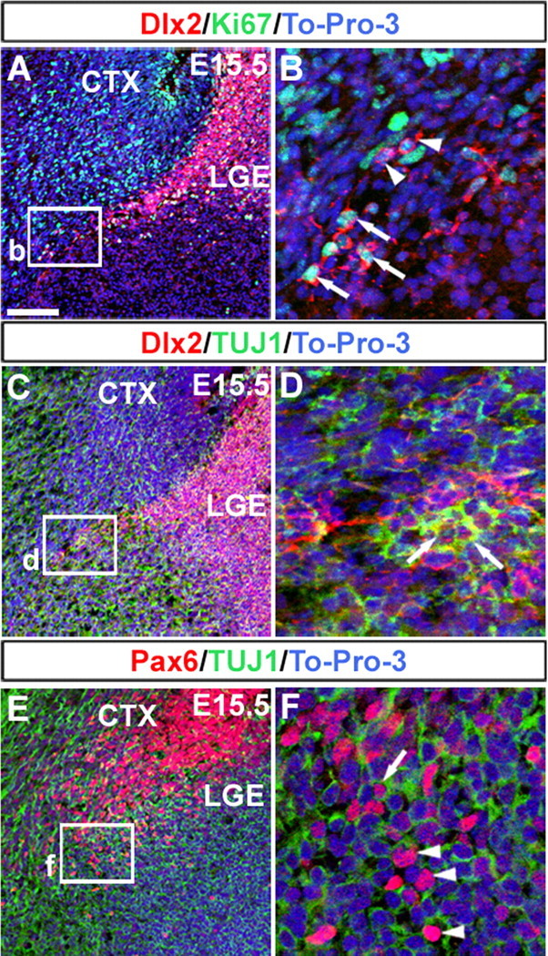
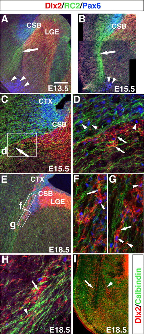
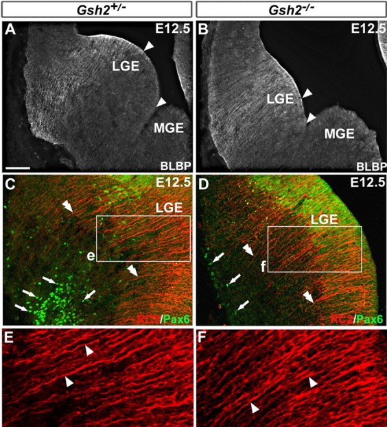
During embryogenesis, the lateral cortical stream (LCS) emerges from the corticostriatal border (CSB), the boundary between the developing cerebral cortex and striatum. The LCS is comprised of a mix of pallial- and subpallial-derived neural progenitor cells that migrate to the developing structures of the basal telencephalon, most notably the piriform cortex and amygdala. Using a combination of in vitro and in vivo approaches, we analyzed the timing, composition, migratory modes, origin, and requirement of the homeodomain-containing transcription factor Gsh2 (genomic screened homeobox 2) in the development of this prominent migratory stream. We reveal that Pax6 (paired box gene 6)-positive pallial-derived and Dlx2 (distal-less homeobox 2)-positive subpallial-derived subpopulations of LCS cells are generated in distinct temporal windows during embryogenesis. Furthermore, our data indicate the CSB border not only is comprised of separate populations of pallial- and subpallial-derived progenitors that contribute to the LCS but also a subpopulation of cells coexpressing Pax6 and Dlx2. Moreover, despite migrating along a route outlined by a cascade of radial glia, the Dlx2-positive population appears to migrate primarily in an apparent chain-like manner, with LCS migratory cells being generated locally at the CSB with little contribution from other subpallial structures such as the medial, lateral, or caudal ganglionic eminences. We further demonstrate that the generation of the LCS is dependent on the homeodomain-containing gene Gsh2, revealing a novel requirement for Gsh2 in telencephalic development.


Figures

Figure 1.
Figure 1.
Dlx2+ and Pax6+ cells of the LCS during embryogenesis. The LCS consists of two subsets of cells: a pallial-derived population that is Pax6+ (A–C) and a subpallial-derived population that is Dlx2+ (D–F). At E11.5, Pax6 is highly expressed in the pallial and CSB ventricular zone (A). At this age, a few Pax6+ cells were observed along the putative route of the LCS (A, arrowheads) and at the pial surface (double arrowheads). At E15.5, Pax6+ cells were observed along the route of the LCS (B, arrowheads) and in the developing basolateral complex of the amygdala (B). By E18.5, multiple Pax6+ cells were observed in distinct developing emerging amygdala nuclei (C), including the cortical, basomedial nuclei, and medial nuclei. At E13.5, the first Dlx2+ cells emanated from the CSB (D, arrowhead). By E15.5, Dlx2+ cell migration along the LCS was highly prominent (E, arrow), with Dlx2+ cells found in the prospective basolateral complex and cortical nuclei (arrowheads) of the developing amygdala. By E18.5, Dlx2+ cells continued to migrate along the LCS (arrow) and accumulated in the piriform cortex (black line) and cortical amygdala nucleus (F, arrowhead). AMY, Amygdala; BLC, basolateral complex; CTX, cerebral cortex; PIR, piriform cortex; STR, striatum. Scale bar: A, 50 μm; B, D, E, 100 μm; C, F, 150 μm.
Figure 2.
Figure 2.
Dlx2 and Pax6 expression at the corticostriatal border. At E13.5, Pax6+ cells (red) are shown emanating from the border with Dlx2+ cells (green) and migrating along the LCS (arrowheads) to the developing piriform cortex (A). Higher magnification of the boxed region in (A) revealed numerous Pax6+ cells (arrowheads) in the Dlx2+ subpallium (B). Many of these Pax6+ cells also expressed Dlx2 (arrow), in which a Pax6+ nucleus was surrounded by a Dlx2+ cell body as shown in C. At E15.5, the LCS consisted of both Dlx2+ and Pax6+ cells (arrow) (D). Higher magnification of the boxed region in D also revealed numerous Pax6+ cells in the subpallium (arrowheads), many of which expressed Dlx2+ (F, arrows). At E18.5, Pax6+ and Dlx2+ cells were intermingled in both the pallial and subpallial aspects of the border (G). High magnification of the boxed regions in G revealed numerous Pax6+ and Dlx2+ cells intermingled throughout the border region (H, I, arrows). In each case, To-Pro-3 iodide (To-Pro-3; blue) staining was used to label individual cell nuclei. Scale bar: A, 200 μm; B, 50 μm; C, F, H, I, 20 μm; D, 100 μm; E, 35 μm; G, 80 μm. CTX, Cerebral cortex.
Figure 3.
Figure 3.
Expression of TUJ1 and Ki67 in the lateral cortical stream. At E15.5, Dlx2+ cells are shown migrating along the LCS (red) through a region of Ki67+ proliferating cells (green, A). Higher magnification of boxed area in A revealed that some Dlx2+ cells were also Ki67+, as indicated by a Dlx2+ cell body surrounding a Ki67+ nucleus (arrows), whereas other Dlx2+ cells were Ki67 negative (arrowheads, B). At this same age, Dlx2+ (red) cells of the LCS migrated into a high TUJ1 (green) expressing region (C). Higher magnification of boxed area in C also revealed that some Dlx2+ cells coexpressed the neuronal marker TUJ1 (D, arrows). At E15.5, Pax6+ cells (red) were also observed migrating along the LCS into a TUJ1+ (green) region (E). Higher magnification of the boxed region in E revealed that some of these Pax6+ cells were also TUJ1+ (arrows), whereas others were TUJ1 negative (arrowheads, F). Scale bar: A, C, E, 100 μm; B, D, F, 20 μm. CTX, Cerebral cortex.
Figure 4.
Figure 4.
Route of Dlx2+ and Pax6+ cell migration along the LCS. As shown at E13.5 (A) and E15.5 (B), the route of the LCS is marked by a prominent cascade of RC2+ (green) radial glia (arrows). At E13.5 and E15.5, a pool of Pax6+ cells (blue) was observed in the ventrolateral telencephalon (A, B, arrowheads). At E15.5, migrating Dlx2+ cells (red) were observed along the proximal aspect of the LCS (C, arrow). High magnification of boxed region in C revealed distinct cellular distributions of Pax6+ cells (blue, arrowheads) compared with Dlx2+ cells (arrows), with Pax6+ cells dispersed widely and Dlx2+ cells more tightly packed (D). This pattern of Dlx2+ cell dispersion along the LCS was also observed at E18.5 (E–H, arrows). However, some Dlx2+ cells were also more closely associated with radial glia (F–H, arrows) and not in packed, chain-like configurations. Additionally, a subset of cells along the LCS appeared to coexpress Pax6 and Dlx2 (F, G, double arrowheads), with some of these cells in chain-like configurations. By E18.5, the Dlx2+ stream of the LCS split into lateral (arrow) and medial (arrowhead) routes (H). These Dlx2+ (red) cells were in proximity to calbindin+ cells (green). CTX, Cerebral cortex. Scale bar: A, E, 100 μm; B, 200 μm; C, 80 μm; D, F–H, 30 μm; I, 350 μm.
Figure 5.
Figure 5.
Dlx2+ cells of the LCS morphologically resemble chain migration in vivo. Sagittal (A–C) and coronal (D, E) views of E15.5 LacZ-stained Dlx2+/tauLacZ brains revealed tightly packed, chain-like cellular clusters along the LCS. In the sagittal view in A, two prominent Dlx2+ migratory populations are shown; those migrating from the CGE toward the developing cortex (arrowhead) and those migrating ventrally along the LCS toward the basal telencephalon (boxed region). Higher magnification of the boxed region in A shows cascades of chain-like Dlx2+ cells emanating from the CSB region and migrating more ventrally (B, arrows). High magnification of the same region from a different brain also revealed similar Dlx2+ chain-like configurations of cells migrating ventrolaterally (C, arrows). Similar distribution patterns of Dlx2+ cells (arrows) were also observed in coronal views of the LCS at low- (D) and high- (E) power views. Also note the difference in the configuration of the Dlx2+ cells of the LCS (arrow) versus Dlx2+ cells migrating to the cerebral cortex (D, arrowheads). F, Electron micrographs of the LCS revealed some migrating Dlx2+ cells (asterisks) also in close apposition to each other. G, In contrast, some Dlx2+ cells were also observed to migrate in isolation and more closely associated with radial glia. Arrowheads in F and G show perinuclear LacZ precipitate marking Dlx2+ cells. CTX, Cerebral cortex; RG, radial glia. Scale bars: A, 900 μm; B, 350 μm; C, 150 μm; D, 100 μm; E, 25 μm; F, G, 4 μm.
Figure 6.
Figure 6.
Dlx2+ cells of the LCS migrate as chains in vitro. β-Galactosidase immunostaining of Dlx2+/tauLacZ coronal telencephalic sections at E16.5 also showed that Dlx2+ cells of the LCS (A, arrow) appeared to migrate in a manner distinct from that of Dlx2+ cells tangentially migrating to the cerebral cortex (A, arrowheads). In B–F, explants from the corticostriatal border region, LGE, MGE, and CGE were dissected from Dlx2+/tauLacZ animals at E13.5 and cultured in Matrigel for 3–4 d. CSB explants stained for LacZ revealed cells that migrated away from the CSB in tightly packed configurations (arrow in B and a higher magnification shown in C). In contrast, explants from the LGE did not display significant migration (D), whereas cells from the MGE (E) and CGE (F) migrated as loose chains (E, F, arrowheads), similar to that observed for pallial-directed migrating Dlx2+ cells as shown in A (and shown in Fig. 5D). CTX, Cerebral cortex. Scale bar: A, 100 μm; B, D, 250 μm; C, E, F, 75 μm.
Figure 7.
Figure 7.
The corticostriatal border is the primary source of cells of the LCS. GFP (B, C) or PLAP (D–I) cells were homotopically transplanted at E15.5 to the CSB (B, C) or at E13.5 to the MGE, CGE, or LGE (D–I) with the aid of ultrasound-guided microscopy (A, asterisk indicates needle placement at CSB) and analyzed 2–4 d after transplantation. CSB transplanted cells migrated along the LCS (B, C, arrowheads). In contrast, although MGE transplanted cells were also observed along the route of the LCS (D, E), many of these cells possessed leading processes directed toward the developing cerebral cortex (E, M, arrowheads). Both CGE-derived (F, G) and LGE-derived (H, I) cells did not migrate along the LCS. In vitro, explants from GFP+ donors were homotopically transplanted at E13.5 into wild-type coronal telencephalic organotypic slice cultures and cultured for 2 DIV. CSB transplants (asterisk shows region of transplant) revealed robust migration along the LCS (J), with many cells extending a leading process in the direction of the ventrolateral telencephalon (K, arrowheads). Homotopically transplanted GFP+ MGE cells were also observed along the region of the LCS (L), but most of cells displayed dorsally directed leading processes (M, arrowheads). Quantification of process orientation (cells were placed into 1 of 4 bins based on the direction of the leading process) revealed that 88% (134 of 153) of MGE cells had processes directed dorsally (bins 1, 4), whereas 62% (60 of 97) of LCS cells had processes oriented ventrally (bins 2, 3) (N). Breakdown of percentages of cells found in each bin for MGE and LCS cells is shown graphically in O. For MGE grafts, a total of 153 cells were counted, and for CSB grafts, a total of 97 cells were counted. The numbers of cells whose process orientation fell into each bin were as follows: MGE–MGE grafts, bin 1 (47 cells), bin 2 (8 cells), bin 3 (11 cells), bin 4 (87 cells); LCS–LCS grafts, bin 1 (12 cells), bin 2 (18 cells), bin 3 (42 cells), bin 4 (25 cells). CTX, Cerebral cortex; GE, ganglion eminence; STR, striatum; Scale bar: B, D, F–I, 250 μm; C, J, L, 300 μm; E, 125 μm; K, M, 40 μm.
Figure 8.
Figure 8.
The LCS is significantly affected in Gsh2−/− mutants. Dlx2+ cell migration along the LCS was reduced in Gsh2−/−;Dlx2+/tauLacZ mutant mice (A, C, E) compared with controls (B, D, F). As shown at both E15.5 (A, B) and E18.5 (C, D), only a few cells were observed leaving the corticostriatal border in mutant mice compared with controls (arrows). A higher magnification of the Dlx2 expression in the CSB region in C and D is shown in c′ and d′. At E18.5, the lateral and medial branches of Dlx2+ cells that extend from the LCS into the basal telencephalon in controls (E, arrows) were not observed in Gsh2−/−;Dlx2+/tauLacZ mutant mice (F). AMY, Amygdala; CTX, cerebral cortex. Scale bar: A–F, 150 μm.
Figure 9.
Figure 9.
Status of radial glia in Gsh2−/− mutants. At E12.5, the domain of BLBP+ radial glia appeared to be shifted more ventral in Gsh2−/− mutants (A, B, arrowheads mark distance between lower limit of BLBP expression in the LGE and the LGE/MGE sulcus). As revealed by Pax6 immunostaining, a reduction in the number of Pax6+ cells (green, arrows) in the ventrolateral telencephalon was observed in Gsh2−/− mutants compared with controls (C, D). However, in Gsh2−/− mutants, RC2+ radial glial morphology (red, double arrowheads C, D and boxed regions shown in higher magnification in E, F) appeared normal. Scale bar (in A): A, B, 150 μm; C, D, 50 μm; E, F, 20 μm.
Figure 10.
Figure 10.
The defect in Dlx2+ migration in Gsh2−/− mutants along the LCS is not rescued in vitro. Telencephalic explants of the CSB region were dissected from Gsh2−/−;Dlx2+/tauLacZ and control animals at E13.5 and were cultured on Matrigel for 3–4 d, fixed, and subsequently LacZ stained. In control brains (A–D), robust, Dlx2+ chain-like migration was observed along the presumptive LCS and RMS. Boxed regions in A and B are shown at higher magnification in C and D, respectively. Arrows in C show tight chains of putative RMS migrating Dlx2+ cells. Arrows in D show chains of Dlx2+ putative LCS cells. A reduced number of explants from Gsh2−/−;Dlx2+/tauLacZ mice (E, F), displayed migration along the LCS. The asterisk indicates region of expected LCS migration in mutant explants. In controls (n = 22 animals, 77 explants), 79.2% of explants displayed migration along both RMS and LCS, whereas in Gsh2−/−;Dlx2+/tauLacZ mutants (n = 7 animals, 27 explants), only 22.2% of explants displayed migration along both RMS and LCS. Scale bar: A, B, E, F, 250 μm; C, D, 100 μm.

References

    1. Alheid GF. Extended amygdala and basal forebrain. Ann NY Acad Sci. 2003;985:185–205. - PubMed
    1. Alvarez-Buylla A, Garcia-Verdugo JM. Neurogenesis in adult subventricular zone. J Neurosci. 2002;22:629–634. - PMC - PubMed
    1. Anthony TE, Klein C, Fishell G, Heintz N. Radial glia serve as neuronal progenitors in all regions of the central nervous system. Neuron. 2004;41:881–890. - PubMed
    1. Assimacopoulos S, Grove EA, Ragsdale CW. Identification of a Pax6-dependent epidermal growth factor family signaling source at the lateral edge of the embryonic cerebral cortex. J Neurosci. 2003;23:6399–6403. - PMC - PubMed
    1. Bayer S, Altman J. Neocortical development. New York: Raven; 1991.

Publication types

MeSH terms

LinkOut - more resources