Comparative Study
. 2006 Nov 8;26(45):11682-94.
doi: 10.1523/JNEUROSCI.3656-06.2006.

Different types of cerebellar GABAergic interneurons originate from a common pool of multipotent progenitor cells

Affiliations
Comparative Study

Different types of cerebellar GABAergic interneurons originate from a common pool of multipotent progenitor cells

Ketty Leto et al. J Neurosci. .

Abstract

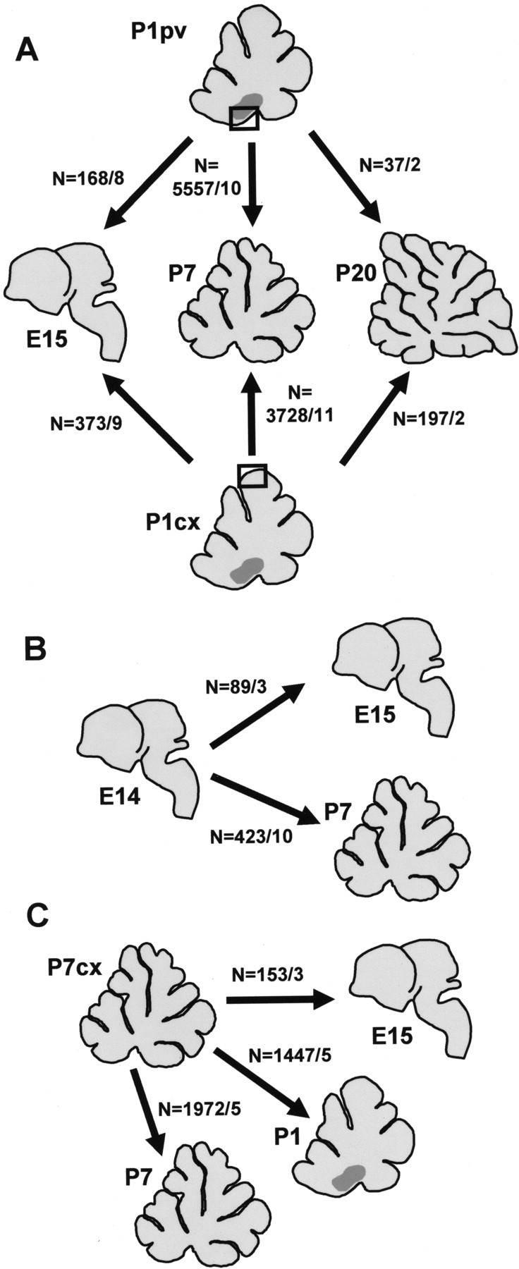
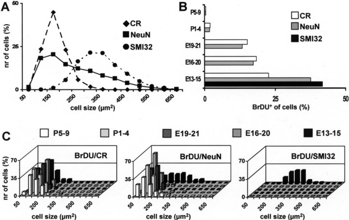
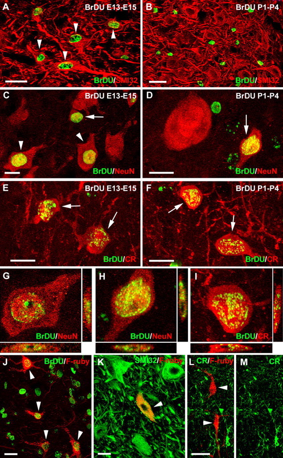
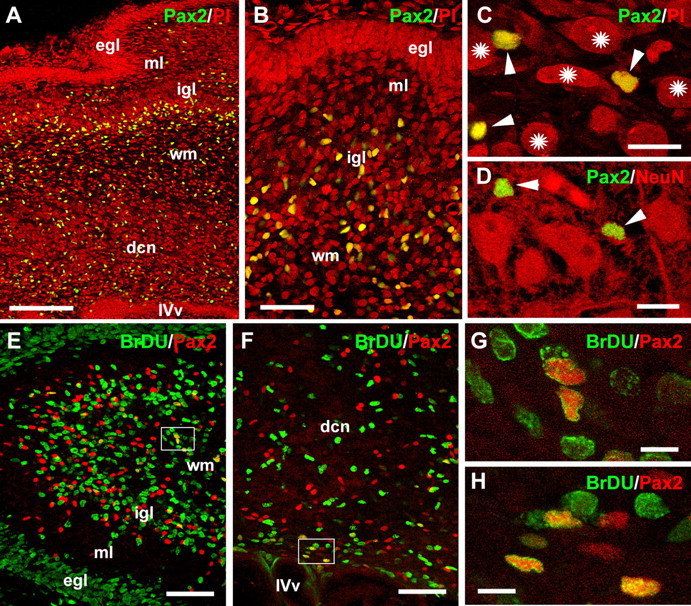
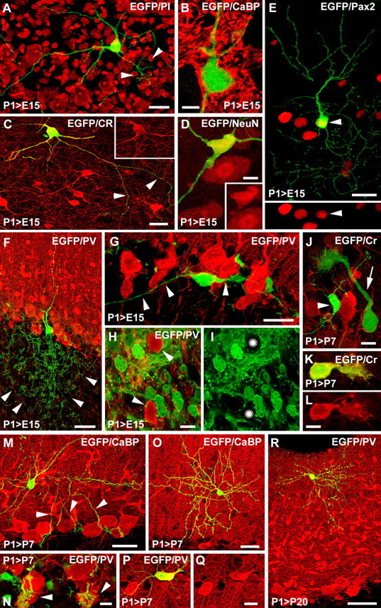
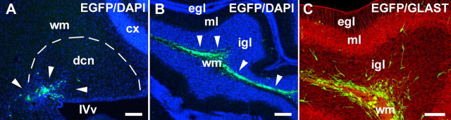
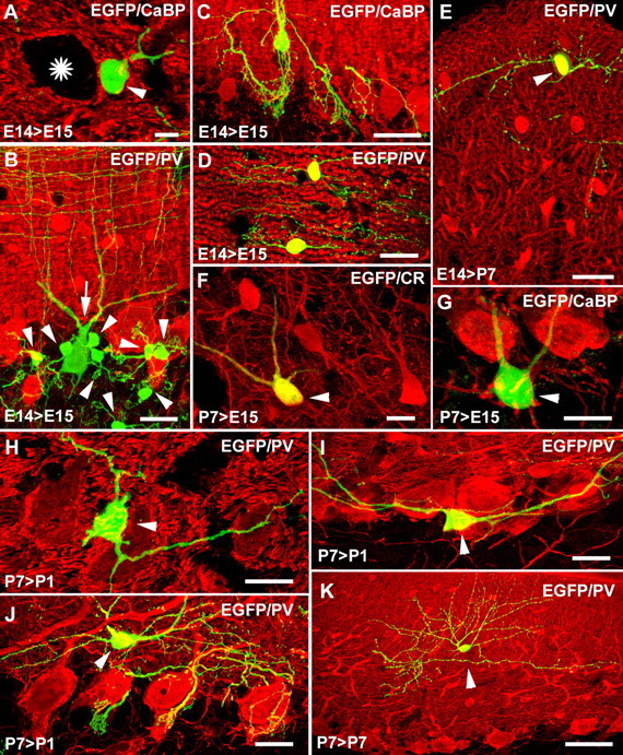
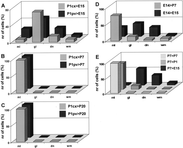
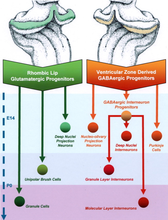
Different cerebellar phenotypes are generated according to a precise spatiotemporal schedule, in which projection neurons precede local interneurons. Glutamatergic neurons develop from the rhombic lip, whereas GABAergic neurons originate from the ventricular neuroepithelium. Progenitors in these germinal layers are committed toward specific phenotypes already at early ontogenetic stages. GABAergic interneurons are thought to derive from a subset of ventricular zone cells, which migrate in the white matter and proliferate up to postnatal life. During this period, different interneuron categories are produced according to an inside-out sequence, from the deep nuclei to the molecular layer (we show here that nuclear interneurons are also born during late embryonic and early postnatal days, after glutamatergic and GABAergic projection neurons). To ask whether distinct interneuron phenotypes share common precursors or derive from multiple fate-restricted progenitors, we examined the behavior of embryonic and postnatal rat cerebellar cells heterotopically/heterochronically transplanted to syngenic hosts. In all conditions, donor cells achieved a high degree of integration in the cerebellar cortex and deep nuclei and acquired GABAergic interneuron phenotypes appropriate for the host age and engraftment site. Therefore, contrary to other cerebellar types, which derive from dedicated precursors, GABAergic interneurons are produced by a common pool of progenitors, which maintain their full developmental potentialities up to late ontogenetic stages and adopt mature identities in response to local instructive cues. In this way, the numbers and types of inhibitory interneurons can be set by spatiotemporally patterned signals to match the functional requirements of developing cerebellar circuits.


Figures

Figure 1.
Figure 1.
Schematic representation of the different transplantation paradigms applied in the study. A, In a first set of experiments, cerebellar cells were isolated from the periventricular region (P1pv, boxed area) or the cortex (P1cx, boxed area) of P1 rat cerebella and grafted to E15, P7, or P20 cerebella. B, E14 donors were transplanted to either age-matched embryos or P7 cerebella. C, Finally, P7 cortical cells (P7cx) were grafted to E15 embryos or to P1 or P7 pups. For each condition, represented by an arrow linking donor to host, the total number of donor-derived interneurons and the number of examined cases are reported.
Figure 2.
Figure 2.
Birthdating of deep nuclei neuron subtypes. A shows the cell size distribution of adult deep nuclear neurons identified by the different markers applied in the study: NeuN (squares) labels cells along the whole range of perikaryal dimensions, whereas calretinin (CR, diamonds) and SMI-32 (circles) stain two distinct subsets of small and large neurons, respectively. B illustrates the percentage of BrdU-immunoreactive nuclear neurons that are colabeled by NeuN (gray bars), SMI32 (black bars), or calretinin (white bars), after application of the nucleotide analog at different time windows during embryonic and postnatal life. BrdU/SMI32-positive neurons are exclusively present between E13 and E15, whereas NeuN- or calretinin-immunoreactive cells incorporate BrdU at all ages. The histograms in C report the cell size distribution of deep nuclear neurons double labeled for BrdU and calretinin (CR), NeuN, or SMI32, after administration of BrdU at different embryonic and postnatal ages (indicated by the different shades of gray). The large NeuN/SMI32-positive neurons are exclusively generated between E13 and E15, whereas smaller cells, stained by NeuN and calretinin, are also born at later developmental stages.
Figure 3.
Figure 3.
A–M, Birthdating of deep nuclei neuron subtypes. A–F, Immunolabeling of deep nuclei neurons after administration of BrdU at E13–E15 (A, C, E) or P1–P4 (B, D, F). Double immunostaining for BrdU (green) and SMI32 (red, A, B), NeuN (red, C, D), or calretinin (CR, red, E, F). Large SMI32/NeuN-immunoreactive neurons with BrdU-positive nuclei (pointed by arrowheads in A, C) are exclusively present after BrdU application at E13–E15. In contrast, small NeuN/calretinin-immunolabeled cells incorporate BrdU at both ages (arrows in C–F). G–I, Multiplane views demonstrate the nuclear labeling of some of the neurons depicted in the above micrographs. Nucleo-olivary projection neurons, stained in red by retrograde Fluoro Ruby (F-ruby) tracing from the inferior olive, are indicated by arrowheads in J: these neurons incorporated BrdU (green) administered between E13 and E15. K and L,M show that the Fluoro Ruby-traced nucleo-olivary neurons (red, arrowheads in K, L) are double labeled for SMI32 (green in K) but not for calretinin (green in L, M). Scale bars: A, B, J, L, 20 μm; C–F, K, 10 μm.
Figure 4.
Figure 4.
A–H, Distribution and proliferation of Pax-2-expressing cells in P1 cerebellum. At this age, Pax-2-positive cells, which appear yellow in A–C because of double labeling with propidium iodide (PI, red), are present in both the cortex and the region of the nuclei (dcn), being more numerous at the border between the white matter (wm) and the nascent internal granular layer (igl). The distribution of these cells in the cortex can be also appreciated by the higher-magnification picture B. Arrowheads in C point to small Pax-2-expressing cells intermingled with large deep nuclei neurons (some are indicated by asterisks). Some of such cells are also double labeled with anti-NeuN antibodies (arrowheads in D). E–H show double labeling for BrdU (green) and Pax-2 (red) in the cortex (E, G) or periventricular region (F, H) after a pulse of BrdU. Note that double-labeled cells are most frequent in the cortical white matter (wm, E) and in the region bordering the fourth ventricle (IVv, F). The higher-magnification pictures G and H show the double-labeled nuclei contained in the insets in E and F, respectively. egl, External granular layer, ml, molecular layer; igl, internal granular layer; wm, white matter; dcn, deep cerebellar nuclei; IVv, fourth ventricle. Scale bars: A, 200 μm; B, E, F, 100 μm; C, D, 20 μm; G, H, 10 μm.
Figure 5.
Figure 5.
A–R, GABAergic interneurons generated by P1 cortical or periventricular progenitors grafted embryonic (A–I) or postnatal cerebella (J–R). The micrographs show representative examples of the phenotypes developed by donor cells. Interneurons of the deep nuclei (A–D) are characterized by small round-like perikarya (A) covered by calbindin-immunolabeled Purkinje axon terminals (B), long slender dendrites, and a short axon branching close to the cell body (arrowheads in A, C). These cells are also labeled by anti-calretinin (C, inset) and anti-NeuN (D, inset) antibodies. Golgi cells (E, F) are located at various depth in the granular layer; they display highly branched axonal arbors in the granular layer (arrowheads in F) and dendrites elongating into the molecular layer. They are also stained by anti-Pax2 antibodies (arrowhead in E, inset). G shows an EGFP-positive Lugaro cell, whose dendrites are typically extended along the Purkinje cell layer. H and I show a cluster of donor granule cells that remained ectopically positioned in the deep white matter of the recipient cerebellum. Arrowheads in H point to two parvalbumin-immunopositive interneurons of host origin (asterisks in I indicate their position, showing that they do not express EGFP) intermingled with the donor cells. The arrow in J points to a unipolar brush cell derived from a P1 donor grafted to a P7 host; arrowhead indicates an EGFP-positive granule neuron. A fraction of the donor unipolar brush cells was also immunostained for calretinin (K, L). M shows a basket cell with several axon branches (arrowheads) impinging on Purkinje cell bodies. The higher-magnification picture N illustrates the correct integration of donor basket axons in the recipient pinceaux (arrowheads) at the basal pole of Purkinje cell perikarya. A typical stellate cell is displayed in O. P and Q show that donor molecular layer interneurons also express parvalbumin. R depicts another stellate cell in the uppermost region of the molecular layer of a P20 host. P1>E15, P1 donor to E15 host; P1>P7, P1 donor to P7 host; P1>P20, P1 donor to P20 host. A–C, E, H, I, K–M, P, Q, R, Cortical donor cells; D, F, G, J, N, O, periventricular donor cells. PI, Propidium iodide; CaBP, calbindin; PV, parvalbumin; CR, calretinin. Scale bars: A, C, E, G, M–Q, 30 μm; B, D, H–L, 10 μm; F, R, 50 μm.
Figure 6.
Figure 6.
A–C, Initial placement of P1 donor cells transplanted to P7 cerebella. The survey micrographs show the position of donor cerebellar cells 1 d (B, C) or 2 d (A) after transplantation. Arrowheads in A point to EGFP-positive cells placed close to the deep cerebellar nuclei (dcn, outlined by the dashed line). When placed in cortical folia (B), donor cells (arrowheads) were typically found along the axial white matter, from which they spread into the adjacent cortex. The migration of donor cells (arrowheads in C) across the cortical layers can be better appreciated in the higher-magnification micrograph C, double labeled for EGFP (green) and anti-GLAST antibodies (red). cx, Cerebellar cortex; egl, external granular layer; ml, molecular layer; igl, internal granular layer; wm, white matter; dcn, deep cerebellar nuclei; IVv, fourth ventricle. DAPI, Blue in A and B. Scale bars: A, B, 100 μm; C, 50 μm.
Figure 7.
Figure 7.
A–K, GABAergic interneurons generated by E14 (A–E) or P7 (F, G) donor progenitors. Embryonic cerebellar cells grafted to embryonic recipients (A–D) generated all types of interneurons, including deep nuclei interneurons (arrowhead in A; asterisk points to a presumptive host projection neuron highlighted by Purkinje axon terminals), Golgi (arrow in B; arrowheads indicate granule cells), basket (C), and stellate (D) cells. When transplanted to P7 hosts (E), the same donors produced a vast majority of molecular layer interneurons (arrowhead indicates a parvalbumin-immunopositive stellate cell). F and D show that cerebellar cells isolated at P7 and transplanted to E15 recipients are still able to adopt early-generated identities, such as calretinin-immunopositive interneurons in the deep nuclei (arrowhead in F) or Golgi cells (arrowhead in G). The same cells transplanted to P1 hosts (H–J) still generate interneurons in the deep nuclei (arrowhead in H), granular layer (arrowhead in I points to a Lugaro cell), and molecular layer (arrowhead in J indicates a basket cell). In contrast, when homochronically transplanted to P7 cerebella (K), P7 progenitors almost exclusively acquire the phenotypes of molecular layer interneurons (arrowhead points to a stellate cell). E14>E15, E14 donor to E15 host; E14>P7, E14 donor to P7 host; P7>E15, P7 donor to E15 host; P7>P1, P7 donor to P1 host; P7>P7, P7 donor to P7 host. CaBP, Calbindin; PV, parvalbumin; CR, calretinin. Scale bars: A, F, 10 μm; B, D, E, 30 μm; C, K, 50 μm; G–J, 20 μm.
Figure 8.
Figure 8.
Relative frequencies of the different GABAergic interneuron phenotypes generated by donor cells in the different transplantation experiments. Histograms A–C show the phenotypic repertoires produced by P1 cortical (P1cx, light gray bars) or periventricular (P1pv, dark gray bars) cells grafted to E15 (A), P7 (B), or P20 (C) cerebella. D illustrates the distribution of interneuron types produced by E14 donors grafted to P7 (light gray bars) or E15 (dark gray bars) hosts. E reports the distribution obtained with P7 cortical donors transplanted to E15 recipients in utero or to postnatal cerebella at P1 or P7. ml, Molecular layer interneurons (basket and stellate cells); gl, granular layer interneurons (Golgi and Lugaro cells); dn, deep nuclei interneurons; wm, ectopic interneurons in white matter. In all graphs, the frequency of the different phenotypes is represented as percentage of the total number of interneurons observed in all of the cases belonging to the same experimental group (see Fig. 1).
Figure 9.
Figure 9.
The origins of GABAergic and glutamatergic neurons in the cerebellum. The dumbbell-shaped rhombic lip of the cerebellar anlage (highlighted in green) is the birthplace for progenitors that generate all cerebellar glutamatergic neurons (green arrows). The cerebellar ventricular zone (highlighted in orange) is the birthplace for progenitors that generate all cerebellar GABAergic neurons. These progenitors differentiate into Purkinje cells and likely nucleo-olivary projection neurons (orange arrows). In addition, a subpopulation of progenitors is formed, termed GABAergic interneuron progenitors, that exclusively produces all the GABAergic cerebellar interneurons (red arrows), including deep nuclei interneurons and Golgi and Lugaro cells (granular layer interneurons), as well as basket and stellate cells (molecular layer interneurons), according to the precise spatiotemporal schedule of the developing cerebellum. The timeline on the left of the figure (dashed arrow) depicts the course of cerebellar development.

Similar articles

Cited by

References

    1. Alder J, Cho NK, Hatten ME. Embryonic precursor cells from the rhombic lip are specified to a cerebellar granule neuron identity. Neuron. 1996;17:389–399. - PubMed
    1. Altman J, Bayer SA. Prenatal development of the cerebellar system in the rat. I. Cytogenesis and histogenesis of the deep nuclei and the cortex of the cerebellum. J Comp Neurol. 1978;179:23–48. - PubMed
    1. Altman J, Bayer SA. Development of the cerebellar system in relation to its evolution, structures and functions. New York: CRC; 1997.
    1. Baader S, Bergmann M, Mertz K, Fox PA, Gerdes J, Oberdick J, Schilling K. The differentiation of cerebellar interneurons is independent of their mitotic history. Neuroscience. 1999;90:1243–1254. - PubMed
    1. Barbe MF, Levitt P. The early commitment of fetal neurons to the limbic cortex. J Neurosci. 1991;11:519–533. - PMC - PubMed

Publication types

MeSH terms