. 2007 Jan;97(1):837-48.
doi: 10.1152/jn.00873.2006. Epub 2006 Nov 8.

Adenosine inhibits activity of hypocretin/orexin neurons by the A1 receptor in the lateral hypothalamus: a possible sleep-promoting effect

Affiliations

Adenosine inhibits activity of hypocretin/orexin neurons by the A1 receptor in the lateral hypothalamus: a possible sleep-promoting effect

Zhong-Wu Liu et al. J Neurophysiol. 2007 Jan.

Abstract

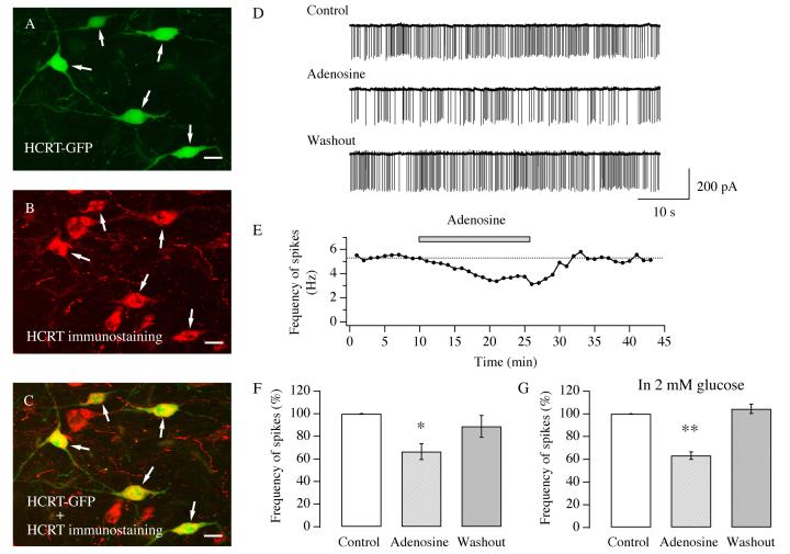
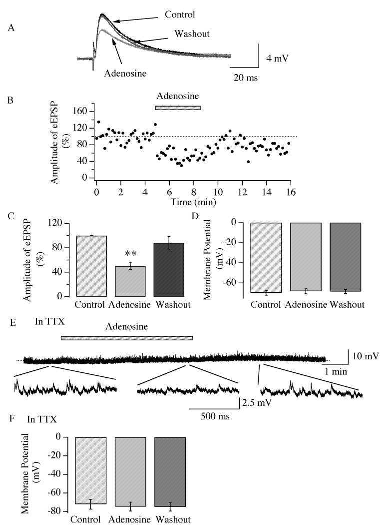
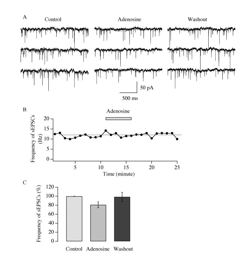
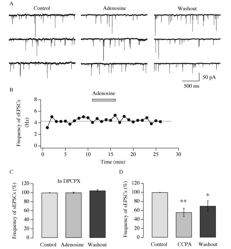
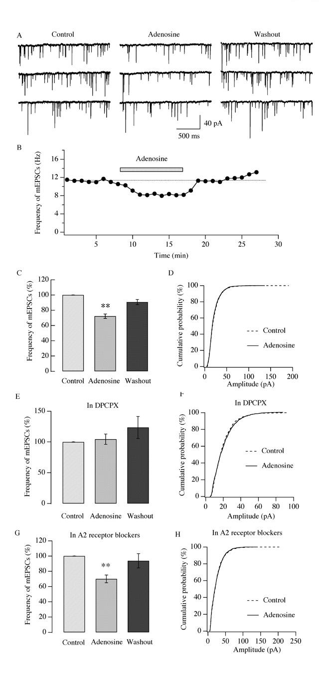
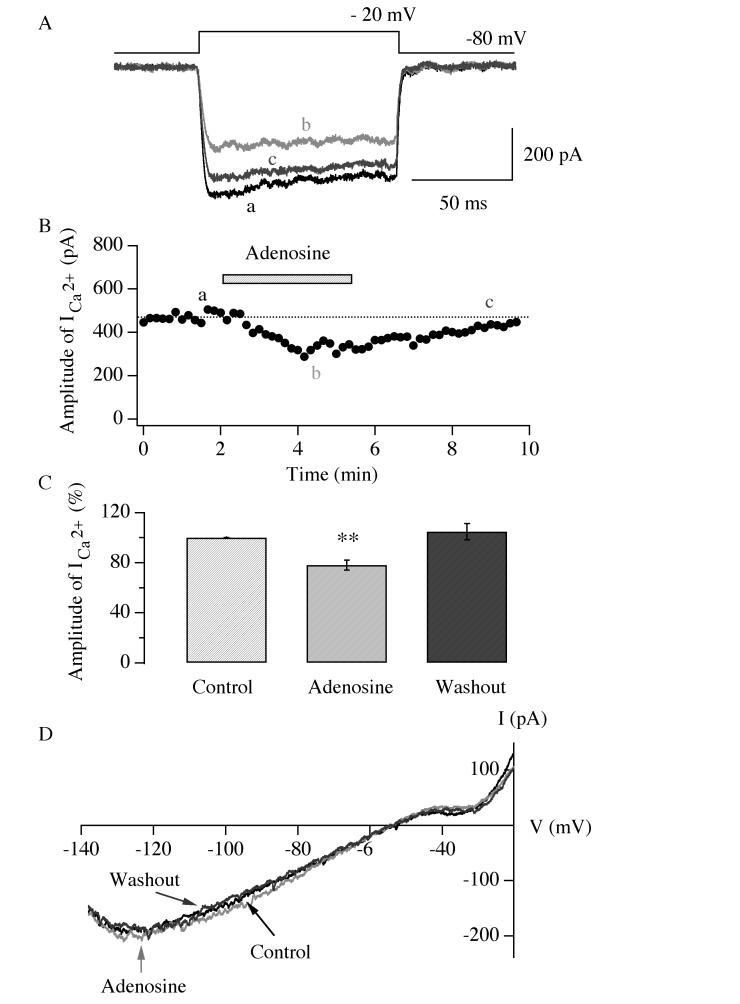
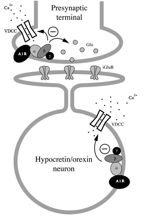
Neurons in the lateral hypothalamus (LH) that contain hypocretin/orexin have been established as important promoters of arousal. Deficiencies in the hypocretin/orexin system lead to narcolepsy. The inhibition of hypocretin/orexin neurons by sleep-promoting neurotransmitters has been suggested as one part of the sleep regulation machinery. Adenosine has been identified as a sleep promoter and its role in sleep regulation in the basal forebrain has been well documented. However, the effect of adenosine on arousal-promoting hypocretin/orexin neurons has not been addressed, despite recent evidence that immunocytochemical visualization of adenosine receptors was detected in these neurons. In this study, we examined the hypothesis that adenosine inhibits the activity of hypocretin/orexin neurons by using electrophysiological methods in brain slices from mice expressing green fluorescent protein in hypocretin/orexin neurons. We found that adenosine significantly attenuated the frequency of action potentials without a change in membrane potential in hypocretin/orexin neurons. The adenosine-mediated inhibition arises from depression of excitatory synaptic transmission to hypocretin/orexin neurons because adenosine depresses the amplitude of evoked excitatory postsynaptic potential and the frequency of spontaneous and miniature excitatory postsynaptic currents in these neurons. At the cell body of the hypocretin/orexin neurons, adenosine inhibits voltage-dependent calcium currents without the induction of GIRK current. The inhibitory effect of adenosine is dose dependent, pertussis toxin sensitive, and mediated by A1 receptors. In summary, our data suggest that in addition to its effect in the basal forebrain, adenosine exerts its sleep-promoting effect in the LH by inhibition of hypocretin/orexin neurons.


Figures

Fig.1
Fig.1
Adenosine depresses the frequency of action potentials in hypocretin/orexin neurons. A-C, double labeling of hypocretin/orexin neurons in brain slices. A, expression of GFP in hypocretin/orexin neurons under the control of a specific hypocretin/orexin promoter. B, immunocytochemical staining of hypocretin/orexin immunoreactive neurons (red). C, green and red signals overlap in hypocretin/orexin neurons indicated by arrows. All GFP-expressing neurons are hypocretin/orexin positive. Scale bar: AC, 15 μm. D-G, loose patch recordings of spikes (action currents) in visually identified hypocretin/orexin neurons were performed in slices from P14-21 mice expressing GFP in hypocretin/orexin neurons. D, sample traces of action currents recorded at various stages of our experiments under voltage clamp with loose patch recordings are presented. E, the time course of a typical experiment shows that adenosine (100 μM) applied to the recorded neuron attenuated the frequency of spikes. This attenuation was reversible upon the removal of adenosine. Application of adenosine is indicated by the filled bar. F, pooled data for frequency of spikes from all hypocretin/orexin neurons examined in our experiments are presented. An ANOVA was used to examine the significance of difference among all three groups. An asterisk “*” indicates P<0.05. G, pooled data for frequency of spikes from all 6 hypocretin/orexin neurons examined in ACSF containing 2 mM glucose are presented. An ANOVA was used to examine the significance of difference among all three groups. A double asterisk “**” indicates P<0.01.
Fig.2
Fig.2
Adenosine decreases the frequency of sEPSCs in hypocretin/orexin neurons. Experiments were performed in the presence of bicuculline (30 μM) in all solutions and sEPSCs were recorded in hypocretin/orexin neurons held at −60 mV under voltage clamp. The frequency of sEPSCs decreased rapidly in the presence of adenosine (100 μM). Sample traces recorded in our experiments at various stages are presented in A and the time course of a typical experiment is plotted in B. Application of adenosine is indicated by the filled bar in B. C, pooled data from all tested hypocretin/orexin neurons at different concentrations of adenosine show that the inhibitory effect of adenosine on the frequency of sEPSCs is dose-dependent. The symbol “**” indicates P<0.01 (ANOVA test). D, frequency of sEPSCs obtained from all hypocretin/orexin neurons (n=8) examined in ACSF containing 2 mM glucose demonstrated that adenosine depressed the frequency of sEPSCs in hypocretin/orexin neurons (**, P<0.01, ANOVA). Inset, sample traces demonstrated that sEPSCs were completely blocked by ionotropic glutamate receptor antagonists AP5 and CNQX (n=7), suggesting a tone of glutamatergic transmission.
Fig.3
Fig.3
Adenosine attenuates the amplitude of evoked EPSPs in hypocretin/orexin neurons. Whole-cell patch clamp experiments were performed in the presence of bicuculline (30 μM) in all solutions and the stimulating electrode was placed on the medial forebrain bundle (MFB). Evoked EPSPs (eEPSPs) were recorded in hypocretin/orexin neurons under current clamp and presented in A. In order to obtain a stable recording of eEPSP without triggering action potentials, hyperpolarizing current was injected to the recorded neurons to maintain the membrane potential between −60 to −70 mV. The amplitude of eEPSPs declined in the presence of adenosine and recovered after its removal. The time course of a typical experiment is plotted in B. C, pooled data from all experiments show that the amplitude of eEPSPs is significantly decreased by the application of adenosine (**, p<0.01, ANOVA). D, pooled data from all experiments show that the membrane potential of hypocretin/orexin neurons does not change (P>0.05, ANOVA). E-F, the membrane potential of hypocretin/orexin neurons recorded in the presence of TTX (1 μM). E, a sample trace from our experiments is presented (M.P., −68.0 mV). The application of adenosine is indicated with the filled bar and the dotted line represents the baseline of the recording. F, pooled data from all experiments show that adenosine does not cause any changes in membrane potential in the presence of TTX (P>0.05, ANOVA).
Fig.4
Fig.4
Adenosine-mediated inhibition of frequency of sEPSCs was abolished in the presence of pertussis toxin (PTX). All experiments were performed in slices treated with PTX (500 ng/ml) for two hours in advance. sEPSCs were recorded in hypocretin/orexin neurons under voltage clamp in the presence of bicuculline (30 μM) in all solutions. A, sample traces of sEPSCs recorded at various stages of our experiment. B, the time course of a typical experiment is shown. Application of adenosine is indicated by the filled bar. C, pooled data from all tested neurons showing that the frequency of sEPSCs does not significantly decrease in the presence of adenosine in slices pre-treated with PTX (P>0.05, ANOVA).
Fig.5
Fig.5
A1 adenosine receptor is responsible for the effect of adenosine in hypocretin/orexin neurons. Whole-cell recordings were performed in hypocretin/orexin neurons held at −60 mV under voltage clamp. Bicuculline (30 μM) was present in all solutions. In the presence of DPCPX (3 μM), a selective A1 receptor antagonist, adenosine (100 μM) was applied to recorded neurons after a stable recording of sEPSCs was obtained and its effect on the frequency of sEPSCs was monitored. Sample traces are presented in A and the time course of a typical experiment is shown in B. Application of adenosine is indicated by the filled bar above the time course plotting. C, pooled data from all neurons examined in our experiments show that adenosine does not inhibit the frequency of sEPSCs in the presence of DPCPX (P>0.05, ANOVA). D, experiments were performed in ACSF containing bicuculline (30 μM). After a stable recording of sEPSCs was obtained, a selective A1 receptor agonist, CCPA was applied to the recorded neurons. Data from all tested hypocretin/orexin neurons are pooled and presented here. The frequency of sEPSCs significantly declined in the presence of CCPA and after its removal (**, p<0.01; *, P<0.05, ANOVA).
Fig.6
Fig.6
A2 adenosine receptors are not responsible for the effect of adenosine in hypocretin/orexin neurons. Whole-cell recordings were performed in hypocretin/orexin neurons held at −60 mV under voltage clamp. Bicuculline (30 μM) was present in all solutions. Selective A2 receptor antagonists, MRS 1706 and SCH 58261, were applied to the recorded neurons. After a stable recording of sEPSCs were obtained, in the presence of MRS 1706 and SCH 58261, adenosine (100 μM) was applied and its effect on the frequency of sEPSCs was monitored. Sample traces are presented in A and the time course of a typical experiment is shown in B. In the presence of MRS 1706 and SCH 58261, the frequency of sEPSCs declined in the presence of adenosine (100 μM) and recovered after its withdrawal. Application of adenosine is indicated by the filled bar. C, pooled data from all neurons examined in our experiments show that the inhibitory effect of adenosine was intact in the presence of selective A2 receptor antagonists (**, P<0.01, ANOVA).
Fig.7
Fig.7
Adenosine inhibits the frequency but not the amplitude of miniature EPSCs. mEPSCs were recorded in hypocretin/orexin neurons held at −60mV under voltage clamp in the presence of bicuculline and TTX in all solutions. A-C, after a stable recording of mEPSCs was obtained, adenosine (100 μM) was applied to the recorded neuron via bath solution. Sample traces of mEPSCs recorded at various stages of our experiments are shown in A and the time course of a typical experiment is presented in B. The filled bar above the trace in B indicates the application of adenosine. C, pooled data of frequency of mEPSCs from all tested neurons indicate that the frequency of mEPSCs significantly declines in the presence of adenosine and recovers after its withdrawal (**, P<0.01, ANOVA). D, accumulative probability curves for the amplitude of mEPSC events detected before and during application of adenosine are generated and plotted. There is no significant difference between events in control and adenosine groups as indicated by the Kolmogorov-Smirnov test. Solid line: control, 2770 events; dotted line: plus adenosine, 1937 events. E-F, the effect of adenosine on the frequency and amplitude of mEPSCs was examined in the presence of DPCPX, a selective A1 receptor antagonist. Adenosine does not inhibit the frequency and amplitude of mEPSCs in the presence of DPCPX as suggested by ANOVA (P>0.05, E) and the Kolmogorov-Smirnov test (P>0.05, F). In F, solid line: control, 1228 events; dotted line: plus adenosine, 1170 events. G-H, experiments were performed as described above in the presence of A2 receptor antagonists (SCH 58261 and MRS 1706). In their presence, adenosine significantly depresses the frequency (G, **, P<0.01, ANOVA) but not the amplitude of mEPSCs (H, P>0.05, Kolmogorov-Smirnov test). In H, solid line: control, 1450 events; dotted line: plus adenosine, 974 events.
Fig.8
Fig.8
Adenosine inhibits voltage-dependent calcium currents in hypocretin/orexin neurons. A, sample traces of whole-cell voltage-dependent calcium currents recorded at various stages in our experiments are shown. Voltage-dependent calcium currents were induced in hypocretin/orexin neurons held at −80 mV under voltage clamp with a voltage step from −80 mV to −20 mV. B, the time course of a typical experiment is shown. The application of adenosine (30 μM) is indicated as the filled bar above the time course curve. Letters a, b and c indicate time points when sample traces were recorded. C, pooled data from all tested neurons were analyzed and plotted, which demonstrate that the amplitude of voltage-dependent calcium currents significantly decreases in the presence of adenosine (30 μM) (**, P<0.01, ANOVA). D, the I-V relationship of membrane currents induced by a ramp pulse (from −140 mV to −20 mV, duration=600 ms) before, during and after the application of adenosine is shown. There is no significant GIRK current in the presence of adenosine.
Fig.9
Fig.9
A schematic graph illustrates pathways mediating the effect of adenosine in hypocretin/orexin neurons. Activation of A1 receptor occurs both at presynaptic terminals innervating hypocretin/orexin neurons and at cell bodies of these neurons. At presynaptic terminals, adenosine might depress voltage-dependent calcium channels which leads to a reduction in calcium-dependent glutamate release. Adenosine might also directly inhibit calcium-independent exocytosis of vesicles containing glutamate. At the soma of hypocretin/orexin neurons, adenosine inhibits voltage-dependent calcium channels but does not induce a GIRK current. A1R: A1 adenosine receptor; VDCC: voltage-dependent calcium channel; iGluR: ionotropic glutamate receptor.

References

    1. Abel T, Nguyen PV, Barad M, Deuel TA, Kandel ER, Bourthouladze R. Genetic demonstration of a role for PKA in the late phase of LTP and in hippocampus-based longterm memory. Cell. 1997;88:615–626. - PubMed
    1. Alam MN, Szymusiak R, Gong H, King J, McGinty D. Adenosinergic modulation of rat basal forebrain neurons during sleep and waking: neuronal recording with microdialysis. J Physiol. 1999;521:679–690. - PMC - PubMed
    1. Arrigoni E, Rainnie DG, McCarley RW, Greene RW. Adenosine-mediated presynaptic modulation of glutamatergic transmission in the laterodorsal tegmentum. J Neurosci. 2001;21:1076–1085. - PMC - PubMed
    1. Arrigoni E, Chamberlin NL, Saper CB, McCarley RW. Adenosine inhibits basal forebrain cholinergic and noncholinergic neurons in vitro. Neuroscience. 2006;140:403–413. - PubMed
    1. Bading H, Ginty DD, Greenberg ME. Regulation of gene expression in hippocampal neurons by distinct calcium signaling pathways. Science. 1993;260:181–186. - PubMed

Publication types

MeSH terms

LinkOut - more resources