. 2007 Feb;81(3):1148-61.
doi: 10.1128/JVI.01812-06. Epub 2006 Nov 8.

Packaging of the virion host shutoff (Vhs) protein of herpes simplex virus: two forms of the Vhs polypeptide are associated with intranuclear B and C capsids, but only one is associated with enveloped virions

Affiliations

Packaging of the virion host shutoff (Vhs) protein of herpes simplex virus: two forms of the Vhs polypeptide are associated with intranuclear B and C capsids, but only one is associated with enveloped virions

G Sullivan Read et al. J Virol. 2007 Feb.

Abstract

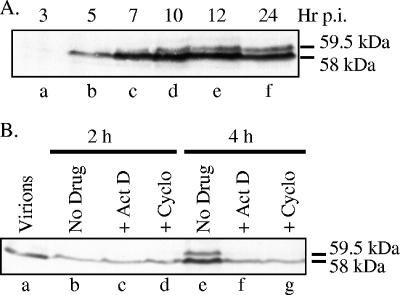
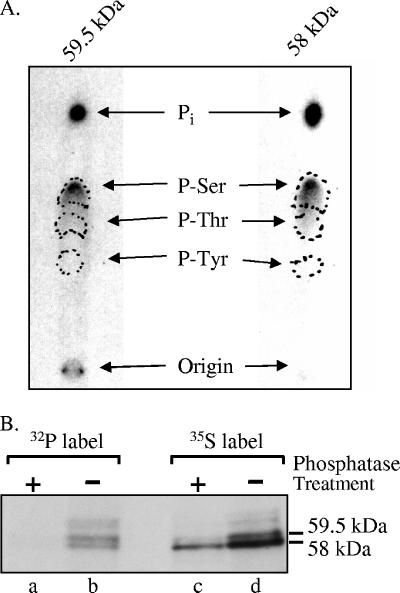
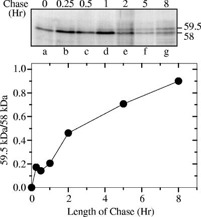
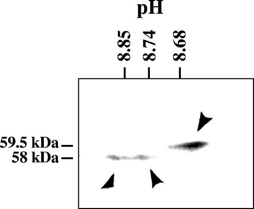
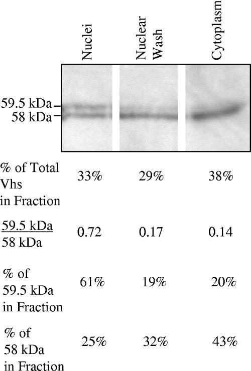
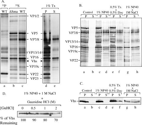
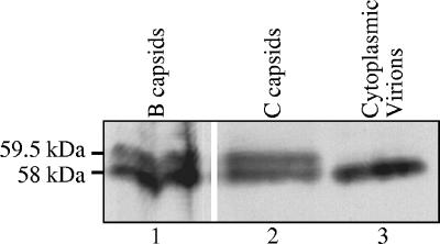
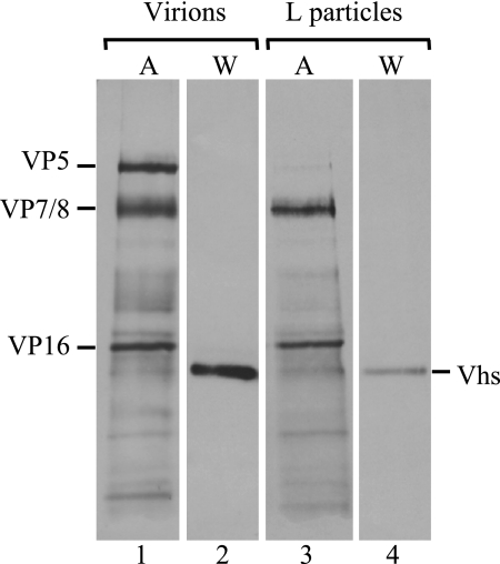
The virion host shutoff (Vhs) protein (UL41) is a minor component of herpes simplex virus virions which, following penetration, accelerates turnover of host and viral mRNAs. Infected cells contain 58-kDa and 59.5-kDa forms of Vhs, which differ in the extent of phosphorylation, yet only a 58-kDa polypeptide is incorporated into virions. In pulse-chase experiments, the primary Vhs translation product comigrated in sodium dodecyl sulfate-polyacrylamide gel electrophoresis with the 58-kDa virion polypeptide, and could be chased to 59.5 kDa. While both 59.5-kDa and 58-kDa forms were found in nuclear and cytoplasmic fractions, the 59.5-kDa form was significantly enriched in the nucleus. Both forms were associated with intranuclear B and C capsids, yet only the 58-kDa polypeptide was found in enveloped cytoplasmic virions. A 58-kDa form, but not the 59.5-kDa form, was found in L particles, noninfectious particles that contain an envelope and tegument but no capsid. The data suggest that virions contain two populations of Vhs that are packaged by different pathways. In the first pathway, the primary translation product is processed to 59.5 kDa, is transported to the nucleus, binds intranuclear capsids, and is converted to 58 kDa at some stage prior to final envelopment. The second pathway does not involve the 59.5-kDa form or interactions between Vhs and capsids. Instead, the primary translation product is phosphorylated to the 58-kDa virion form and packaged through interactions with other tegument proteins in the cytoplasm or viral envelope proteins at the site of final envelopment.


Figures

FIG. 1.
FIG. 1.
Appearance of the 59.5-kDa Vhs form requires de novo protein synthesis. (A) Time course of Vhs accumulation. Vero cells were infected with 20 PFU/cell of wild-type HSV-1 (KOS). Whole-cell lysates were prepared at the indicated times postinfection (p.i.) and analyzed by SDS-PAGE and Western blotting using antiserum raised against a UL41-LacZ fusion protein (58). The 58- and 59.5-kDa forms of Vhs are indicated to the right of lane f. (B) Vero cells were infected with 50 PFU/cell of wild-type HSV-1 in the absence of drugs (lanes b and e) or in the presence of 5 μg/ml of actinomycin D (ActD) (lanes c and f) or 50 μg/ml of cycloheximide (Cyclo) (lanes d and g). Whole-cell lysates were prepared at the indicated times and analyzed by SDS-PAGE and Western blotting using antiserum raised against a UL41-LacZ fusion protein. Lane a contains an aliquot of the infecting virions equivalent to the number used to infect the number of cells in each of the other samples. The 58- and 59.5-kDa forms of Vhs are indicated to the right of lane g.
FIG. 2.
FIG. 2.
Phosphoamino acid analysis of Vhs. (A) The 58-kDa and 59.5-kDa forms of Vhs are phosphorylated predominantly on serines. Vero cells were infected with 20 PFU/cell of wild-type HSV-1 and labeled from 4 to 18 h after infection by incubation in medium containing [32P]orthophosphate. After preparation of whole-cell lysates, the 58-kDa and 59.5-kDa forms of Vhs were immunoprecipitated and purified by preparative SDS-PAGE. Both forms were acid hydrolyzed and mixed with unlabeled phosphoamino acid standards, and the phosphoamino acids were analyzed by 1-D high-voltage electrophoresis on cellulose thin-layer plates. Unlabeled phosphoamino acid standards were localized by staining the plate with ninhydrin, while 32P-labeled amino acids were visualized by autoradiography. The locations of phosphoserine, phosphothreonine, and phosphotyrosine standards are indicated by the dashed circles. (B) The difference in electrophoretic mobility of the 58-kDa and 59-kDa Vhs forms is due primarily to differences in the extent of phosphorylation. Vero cells were infected with 20 PFU/cell of wild-type HSV-1 and labeled from 3.5 to 19 h after infection by incubation in medium containing either [35S]methionine (lanes c and d) or [32P]orthophosphate (lanes a and b). After preparation of whole-cell lysates, the Vhs polypeptides were immunoprecipitated and treated with potato acid phosphatase (lanes a and c) or left untreated (lanes b and d). The products were analyzed by SDS-PAGE and autoradiography. The locations of the 58-kDa and 59.5-kDa forms of the Vhs polypeptide are indicated to the right of lane d.
FIG. 3.
FIG. 3.
Pulse-chase analysis of the Vhs polypeptides. Vero cells were infected with 20 PFU/cell of wild-type HSV-1 (KOS) and pulse-labeled for 15 min with [35S]methionine beginning 8 h after infection. The label was removed, and the cells were washed and incubated for the indicated chase intervals in medium containing an excess of unlabeled methionine. Whole-cell lysates were prepared and the Vhs polypeptides immunoprecipitated and analyzed by SDS-PAGE and autoradiography. The upper panel shows an autoradiogram of the resulting gel. The locations of the 58-kDa and 59.5-kDa polypeptides are shown to the right of lane g. For each time point, the amounts of 58-kDa and 59.5-kDa polypeptides were quantified by densitometric scanning of the autoradiogram. The ratio of the amounts of 59.5-kDa and 58-kDa polypeptides (59.5 kDa/58 kDa) is plotted in the lower panel.
FIG. 4.
FIG. 4.
2-D gel electrophoresis of the Vhs polypeptides. Vero cells were harvested 24 h after infection with 10 PFU/cell of wild-type HSV-1, and solubilized in 2-D gel sample buffer. The lysate was analyzed by 2-D gel electrophoresis, involving isoelectric focusing in the first dimension and SDS-PAGE in the second. Following electrophoresis, the proteins were transferred to an Immobilon P membrane and detected by immunoblotting using rabbit antiserum raised against a UL41-lacZ fusion protein. The three predominant Vhs polypeptides are indicated by arrowheads. The positions of the 58-kDa and 59.5-kDa polypeptides are indicated to the left of the gel, while the pH values at different positions in the isoelectric focusing gel are indicated at the top.
FIG. 5.
FIG. 5.
The 59.5-kDa form of Vhs is enriched in the nucleus. Vero cells were harvested 8 h after infection with 20 PFU/cell of wild-type HSV-1 and separated into nuclear, nuclear wash, and cytoplasmic fractions. Aliquots of fractions from equal numbers of cells were analyzed by SDS-PAGE and Western blotting to detect the 59.5-kDa and 58-kDa forms of Vhs. The relative amounts of the 59.5-kDa and 58-kDa polypeptides were quantified for each fraction by densitometric scanning of the film resulting from the Western blot, and the results are shown in the four lines beneath the gel. Line 1 shows the percentage of the total amount (59.5 kDa plus 58 kDa) of Vhs present in each fraction. Line 2 shows the ratio of the amounts of the 59.5-kDa and 58-kDa polypeptides in each fraction. Line 3 shows the percentage of the total amount of the 59.5-kDa polypeptide that was present in each fraction, and line 4 shows the percentage of the total amount of the 58-kDa polypeptide in each fraction.
FIG. 6.
FIG. 6.
Both 59.5-kDa and 58-kDa Vhs polypeptides bind intranuclear B and C capsids. Vero cells were infected with 20 PFU/cell of wild-type HSV-1 and incubated from 4 to 18 h after infection in MEM containing [35S]methionine and [3H]thymidine to label protein and DNA, respectively. A nuclear lysate was prepared and analyzed by sedimentation through a gradient of 20 to 50% sucrose prepared in TNE. Aliquots of each gradient fraction were analyzed to determine the amounts of [35S]methionine and [3H]thymidine incorporated into TCA-precipitable material (A) and by SDS-PAGE and Western blotting to detect the 59.5-kDa and 58-kDa Vhs polypeptides (B). The peak centered at fraction 11 was presumed to be B capsids, and that at fraction 14 was presumed to be C capsids. The putative B capsids were pelleted from fraction 11 of the first gradient, resuspended in TNE, and resedimented through a second 20 to 50% sucrose gradients. An aliquot from each fraction of the second gradient was analyzed by SDS-PAGE, and the resolved proteins were blotted onto an Immobilon P membrane. The membrane was subjected to autoradiography (C) to detect all 35S-labeled proteins and probed with Vhs-specific antiserum (D) to allow immunologic detection of the Vhs polypeptides. The locations of major capsid proteins are shown by dots to the right of lane 12 of panel C and labeled to the left of lane 1 of panel C. The 59.5-kDa and 58-kDa Vhs polypeptides are indicated by dots to the right of lane 12 of panel D and labeled to the left of lane 1 of panel D. In this and other figures, the smallest of the major capsid proteins, VP26, was run off the end of the gel. This was necessary in order to run the gel long enough to resolve the 58-kDa and 59.5-kDa forms of Vhs.
FIG. 7.
FIG. 7.
Both 59.5-kDa and 58-kDa Vhs polypeptides are found associated with intranuclear B and C capsids, but VP16 is not. [35S]methionine-labeled B and C capsids were purified from nuclear lysates of infected Vero cells by sedimentation through two sequential gradients of 20 to 50% sucrose as described for B capsids in the legend to Fig. 6. Proteins in the peak fractions from the second B and C capsid gradients were resolved by SDS-PAGE and blotted onto an Immobilon P membrane. The membrane was subjected to autoradiography (A) to detect all 35S-labeled proteins and probed with Vhs-specific antiserum (B) to allow immunologic detection of the Vhs polypeptides. The locations of major capsid proteins are shown to the left of panel A, and those of the 59.5-kDa and 58-kDa Vhs polypeptides are shown to the right of panel B. The proteins in B and C capsids were compared by Western blotting to those in extracellular virions purified as described in the text. Following SDS-PAGE, the proteins were blotted onto an Immobilon P membrane. The membrane was probed using UL41-specific antiserum to detect the Vhs polypeptides (C), after which it was stripped and reprobed using VP16-specific antiserum to detect the VP16 polypeptide (D).
FIG. 8.
FIG. 8.
Limited removal of the Vhs polypeptides from B capsids by washing with GuHCl. [35S]methionine-labeled B capsids were purified from nuclear lysates from infected Vero cells by sedimentation through two sequential gradients of 20 to 50% sucrose prepared in TNE. B capsids were pelleted from the appropriate fractions of the second gradient, resuspended, and washed for 30 min in TNE containing 0, 0.2 M, 0.5 M, 1 M, or 2 M GuHCl. The capsids were then pelleted through a cushion of 25% sucrose in TNE. Polypeptides that remained associated with the pelleted capsids were resolved by SDS-PAGE, transferred to an Immobilon P membrane, and analyzed by autoradiography (A) to visualize the major capsid proteins or by probing the membrane using Vhs-specific antiserum (C) for immunologic detection of the Vhs polypeptides. For comparison, lane f of panel A contains a sample of C capsids. The profile of proteins in C capsids was similar to that in B capsids, except that C capsids lack VP22a. In this and other figures, the smallest of the major capsid proteins, VP26, was run off the end of the gel. This was necessary in order to run the gel long enough to resolve the 58-kDa and 59.5-kDa forms of Vhs. The relative amounts of capsid proteins and Vhs polypeptides remaining bound to washed and unwashed capsids were determined by densitometric scanning of the autoradiogram in panel A or the Western blot in panel C and are plotted in panel B.
FIG. 9.
FIG. 9.
Extraction of virions with detergents, salt, and GuHCl. (A) [32P]orthophosphate-labeled wild-type (WT) virions (lane a) and [35S]methionine-labeled wild-type (lanes c to e) and Vhs-ΔSma (lane b) virions were extracted with 1% Triton X-100 (Tx) and separated into pellet (P) (lane d) and supernatant (S) (lane e) fractions as described in the text or were left untreated (lanes a to c). The various virions and fractions were analyzed by SDS-PAGE and autoradiography. Various virion polypeptides are labeled between lanes c and d. The wild-type Vhs polypeptide is indicated by arrowheads to the right of lane c and left of lane d. The Vhs polypeptide is missing from Vhs-ΔSma virions. (B) [35S]methionine-labeled wild-type virions were extracted for 1 h with the following buffers as described in the text: (i) 10 mM Tris (pH 7.5) (control), (ii) 1% NP-40, (iii) 0.5% Triton X-100 plus 0.5% sodium deoxycholate (Doc), and (iv) 1% NP-40 plus 1 M NaCl. The extracted virions were separated by centrifugation into a capsid-containing pellet and a supernatant. Proteins in the various fractions were resolved by SDS-PAGE, transferred to an Immobilon P membrane, and visualized by autoradiography to detect all virion proteins. (C) The same membrane was then probed using UL41-specific antiserum to detect the Vhs polypeptide. (D) Wild-type virions were extracted with 1% NP-40 plus 1 M NaCl, after which the capsids were pelleted. The capsids, retaining approximately 50% of the Vhs protein, were washed further with TNE containing 0 M, 0.5 M, 1 M, or 2 M GuHCl. The washed capsids were pelleted through a cushion of 25% sucrose, resuspended, and analyzed by SDS-PAGE and Western blotting to detect the Vhs polypeptide. The amount of Vhs remaining bound to capsids washed with various concentrations of GuHCl was quantified by densitometric scanning and expressed as a percentage of the amount bound to TNE-washed capsids.
FIG. 10.
FIG. 10.
Enveloped cytoplasmic virions contain only the 58-kDa form of Vhs. Enveloped cytoplasmic virions (lane 3), and intranuclear B (lane 1) and C (lane 2) capsids were purified as described in the text and analyzed by SDS-PAGE and Western blotting using Vhs-specific antiserum. The locations of the 59.5-kDa and 58-kDa forms of Vhs are indicated to the left of lane 1.
FIG. 11.
FIG. 11.
L particles contain a 58-kDa, but not the 59.5-kDa, form of Vhs. [35S]methionine-labeled virions (lanes 1 and 2) and L particles (lanes 3 and 4) were purified from the extracellular medium from cultures of BHK-21 cells infected with wild-type HSV-1. Proteins were resolved by SDS-PAGE and transferred to an Immobilon P membrane, after which the membrane was subjected to autoradiography (A) (lanes 1 and 3) to detect all labeled viral proteins or probed using a UL41-specific antiserum (W) (lanes 2 and 4) for immunologic detection of the Vhs polypeptides.
FIG. 12.
FIG. 12.
Indirect immunofluorescence localization of Vhs in infected and transfected cells. Vero cells were infected with 2.5 PFU/cell of wild-type HSV-1 (A) or the Vhs mutant N237 (B) or were transfected with the wild-type Vhs expression plasmid pKOSamp (C). At 11.5 h after infection or 24 h after transfection, the cells were fixed and stained for indirect immunofluorescence using rabbit antiserum raised against a LacZ-Vhs fusion protein containing amino acids 239 through 489 of wild-type Vhs and were examined using a Zeiss LSM confocal microscope.

References

    1. Bowzard, J. B., R. J. Visalli, C. B. Wilson, J. S. Loomis, E. M. Callahan, R. J. Courtney, and J. W. Wills. 2000. Membrane targeting properties of a herpesvirus tegument protein-retrovirus Gag chimera. J. Virol. 74:8692-8699. - PMC - PubMed
    1. Boyle, W. J., P. Van Der Geer, and T. Hunter. 1991. Phosphopeptide mapping and phosphoamino acid analysis by two-dimensional separation on thin-layer cellulose plates, p. 110-149. In T. Hunter and B. M. Sefton (ed.), Protein phosphorylation, part B. Academic Press, Inc., San Diego, CA. - PubMed
    1. Brengues, M., D. Teixeira, and R. Parker. 2005. Movement of eukaryotic mRNAs between polysomes and cytoplasmic processing bodies. Science 310:486-489. - PMC - PubMed
    1. Brignati, M. J., J. S. Loomis, J. W. Wills, and R. J. Courtney. 2003. Membrane association of VP22, a herpes simplex virus type 1 tegument protein. J. Virol. 77:4888-4898. - PMC - PubMed
    1. Browne, H., S. Bell, T. Minson, and D. W. Wilson. 1996. An endoplasmic reticulum-retained herpes simplex virus glycoprotein H is absent from secreted virions: evidence for reenvelopment during egress. J. Virol. 70:4311-4316. - PMC - PubMed

Publication types

LinkOut - more resources