. 2006 Nov 29;25(23):5591-602.
doi: 10.1038/sj.emboj.7601414. Epub 2006 Nov 9.

Dynamic state of DNA topology is essential for genome condensation in bacteria

Affiliations

Dynamic state of DNA topology is essential for genome condensation in bacteria

Ryosuke L Ohniwa et al. EMBO J. .

Abstract

In bacteria, Dps is one of the critical proteins to build up a condensed nucleoid in response to the environmental stresses. In this study, we found that the expression of Dps and the nucleoid condensation was not simply correlated in Escherichia coli, and that Fis, which is an E. coli (gamma-Proteobacteria)-specific nucleoid protein, interfered with the Dps-dependent nucleoid condensation. Atomic force microscopy and Northern blot analyses indicated that the inhibitory effect of Fis was due to the repression of the expression of Topoismerase I (Topo I) and DNA gyrase. In the Deltafis strain, both topA and gyrA/B genes were found to be upregulated. Overexpression of Topo I and DNA gyrase enhanced the nucleoid condensation in the presence of Dps. DNA-topology assays using the cell extract showed that the extracts from the Deltafis and Topo I-/DNA gyrase-overexpressing strains, but not the wild-type extract, shifted the population toward relaxed forms. These results indicate that the topology of DNA is dynamically transmutable and that the topology control is important for Dps-induced nucleoid condensation.


Figures

Figure 1
Figure 1
Dps expression under different growth conditions. (A) Schematic representation of the promoter region of the dps gene in E. coli. The IHF and OxyR binding sites exist upstream of the –35/–10 sequences (promoter of dps gene). (B) Western blot analyses against Dps in the wt (W3110) and the W3110 derived Δdps, ΔoxyR and ΔhimD strains, under oxidative stress (2 mM H2O2 treatment) in the log phase and in the stationary phase.
Figure 2
Figure 2
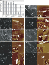
The lysis efficiencies and the nucleoid architectures of the wt, Δdps and ΔoxyR strains under three different conditions. (A) The efficiency of lysis. The number of lysed cells that dispersed fibers was divided by the total cell number and indicated as percent. The cells were observed by DAPI staining (B, D, F, H, J, L, N, P, R) or by AFM (C, E, G, I, K, M, O, Q, S). The numbers of the cells examined are following (each value separated by slash in parentheses represents the number of cells counted at each independent experiment); wt in log phase (218/154/64/34/54), wt under 2 mM H2O2 (235/77/51/135), wt in stationary phase (72/43/109), Δdps strain in the log phase (73/88/17/26), Δdps strain under 2 mM H2O2 (102/91/52), Δdps strain in the stationary phase (37/62/75), ΔoxyRs strain in the log phase (77/47/51/50), Δdps strain under 2 mM H2O2 (74/49/30/53/39), Δdps strain in the stationary phase (74/138/98/165/106). (B, C) wt in the log phase, (D, E) wt in the log phase treated with 2 mM H2O2, (F, G) wt in the stationary phase, (H, I) Δdps strain in the log phase, (J, K) Δdps strain in the log phase treated with 2 mM H2O2, (L, M) Δdps strain in the stationary phase, (N, O) ΔoxyR strain in the log phase, (P, Q) ΔoxyR strain in the log phase treated with 2 mM H2O2, (R, S) ΔoxyR strain in the stationary phase. Scale bars: 10 μm (B, D, F, H, J, L, N, P, R) and 500 nm (C, E, G, I, K, M, O, Q, S).
Figure 3
Figure 3
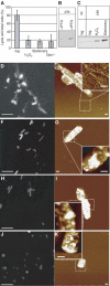
The lysis efficiency and the nucleoid architecture of the Δfis strain. (A) The efficiency of lysis. The number of lysed cells that dispersed fibers was divided by the total cell number and indicated as percent. The numbers of the cells examined are following (each value separated by slash in parentheses represents the number of cells counted at each independent experiment); the log phase (58/39/45/43), under 2 mM H2O2 (56/21/45/35), the stationary phase (125/59/77), the overexpression (76/50/74/40/29). (B) Overexpression of Dps by IPTG in the Δfis strain. The expression was detected by antibody against Dps. (C) Western blot against Dps in the Δfis strain under three different conditions. (D) DAPI image of the Δfis strain in the log phase. (E) AFM image of the Δdps strain in the log phase. (F) DAPI image of the Δfis strain under overexpression of Dps in the log phase. (G) AFM image of the Δfis strain under overexpression of DPS in the log phase. (H) DAPI image of the Δfis strain in the log phase treated with 2 mM H2O2, (I) AFM image of the Δfis strain in the log phase treated with 2 mM H2O2, (J) DAPI image of the Δfis strain in the stationary phase, (K) AFM image of the Δfis strain in the stationary phase. Scale bars: 10 μm (D, F, H, J) and 500 nm (E, G, I, K).
Figure 4
Figure 4
Northern blot analyses of top A-, gyrA-, gyrB- and dps-mRNAs. The levels of (A) topA-, (B) gyrA- and (C) gyrB-mRNAs in the wt and Δfis strains. (D) The levels of dps-mRNAs in the wt cells in the log phase and in the log phase treated with 2 mM H2O2. (A–C) do not show tight bands, whereas (D) shows a single tight band. In bacteria, many mRNAs have been detected as broad smears by Northern blot analyses (Maruyama et al, 2003).
Figure 5
Figure 5
Nucleoid dynamics in Topo I- or DNA gyrase-overexpressing cells. The expression of His-tagged Topo I (A) or His-tagged DNA gyrase (B) was detected by an antibody against His-tag. (C) AFM image of Topo I-overexpressing cell in the log phase, (D–F) AFM images of Topo I-overexpressing cells treated with 2 mM H2O2, (G) AFM image of DNA gyrase-overexpressing cell in the log phase, (H–J) AFM image of DNA gyrase-overexpressing cells treated with 2 mM H2O2. Scale bars: 500 nm.
Figure 6
Figure 6
Northern blot analyses of topA-, gyrA-, gyrB- and fis-mRNAs under the overexpression of Topo I and DNA gyrase. The levels of (A) topA-mRNA in DNA gyrase++, (B, C) gyrA- and gyrB-mRNAs in Topo I++, and (D) fis-mRNA in Topo I++ or DNA gyrase++.
Figure 7
Figure 7
DNA topology assay using Δfis, Topo I++ and Gyrase ++ cell extracts. (A) The plasmid DNA (pBSII SK) was incubated with cell extracts prepared from wt, fis-deletion cells (Δfis), Topo I-overexpressed cells (Topo I ++) and DNA gyrase-overexpressed cells (Gyrase ++) for 15 min at 37°C, and then loaded onto agarose gels containing 2.5 mg/ml chloroquine. (B) The extract from Gyrase++ was added to the extract from Topo I++, and was incubated with pBSII for 15 min at 37°C. The electrophoresis was performed without chloroquine. M represents the marker DNA (λ DNA digested by HindIII (BioLabs)), and the numbers above the gels indicate the amounts of proteins in each cell extract (ng). The graphs represent microdensitometric tracings corresponding to the lanes.
Figure 8
Figure 8
A model of the topology control of genome DNA required for the Dps-dependent nucleoid condensation. (A) In the presence of Fis, DNA topology is maintained statically by the repression of Topo I and DNA gyrase expressions, and also by the physical blocking against the topological changes of DNA topoisomers. Therefore, the expression of Dps does not lead to a nucleoid condensation. (B) In the absence of Fis, Topo I and DNA gyrase are induced and may easily change the DNA topology. Thus DNA topology is dynamic. This status allows the nucleoid to be condensed under the expression of Dps (Topo I and DNA gyrase coexist). (C) Overexpression of Topo I or DNA gyrase may overcome the barrier of Fis, cause the DNA topology dynamic, and facilitate the nucleoid condensation as in (B).

References

    1. Almiron M, Link AJ, Furlong D, Kolter R (1992) A novel DNA-binding protein with regulatory and protective roles in starved Escherichia coli. Genes Dev 6: 2646–2654 - PubMed
    1. Altuvia S, Almiron M, Huisman G, Kolter R, Storz G (1994) The dps promoter is activated by OxyR during growth and by IHF and sigma S in stationary phase. Mol Microbiol 13: 265–272 - PubMed
    1. Azam TA, Ishihama A (1999) Twelve species of the nucleoid-associated protein from Escherichia coli. Sequence recognition specificity and DNA binding affinity. J Biol Chem 274: 33105–33113 - PubMed
    1. Azam TA, Hiraga S, Ishihama A (2000) Two types of localization of the DNA-binding proteins within the Escherichia coli nucleoid. Genes Cells 5: 613–626 - PubMed
    1. Baba T, Ara T, Hasegawa M, Takai Y, Okumura Y, Baba M, Datsenko KA, Tomita M, Wanner BL, Mori H (2006) Construction of Escherichia coli K-12 in-frame, single-gene knockout mutants: the Keio collection. Mol Systems Biol 2: 2006.0008 - PMC - PubMed

Publication types

MeSH terms

LinkOut - more resources