. 2006 Nov 10:6:96.
doi: 10.1186/1471-2180-6-96.

Structure-based discovery of inhibitors of the YycG histidine kinase: new chemical leads to combat Staphylococcus epidermidis infections

Affiliations

Structure-based discovery of inhibitors of the YycG histidine kinase: new chemical leads to combat Staphylococcus epidermidis infections

Zhiqiang Qin et al. BMC Microbiol. .

Abstract

Background: Coagulase-negative Staphylococcus epidermidis has become a major frequent cause of infections in relation to the use of implanted medical devices. The pathogenicity of S. epidermidis has been attributed to its capacity to form biofilms on surfaces of medical devices, which greatly increases its resistance to many conventional antibiotics and often results in chronic infection. It has an urgent need to design novel antibiotics against staphylococci infections, especially those can kill cells embedded in biofilm.

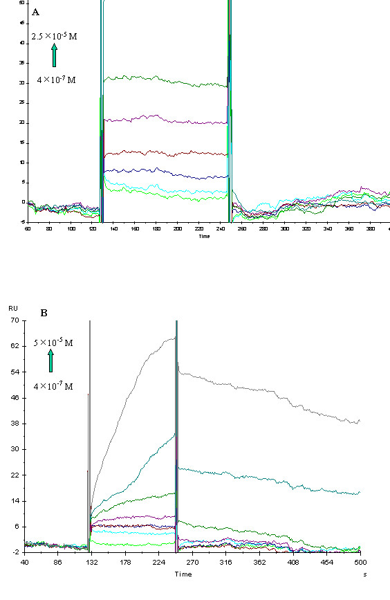
Results: In this report, a series of novel inhibitors of the histidine kinase (HK) YycG protein of S. epidermidis were discovered first using structure-based virtual screening (SBVS) from a small molecular lead-compound library, followed by experimental validation. Of the 76 candidates derived by SBVS targeting of the homolog model of the YycG HATPase_c domain of S. epidermidis, seven compounds displayed significant activity in inhibiting S. epidermidis growth. Furthermore, five of them displayed bactericidal effects on both planktonic and biofilm cells of S. epidermidis. Except for one, the compounds were found to bind to the YycG protein and to inhibit its auto-phosphorylation in vitro, indicating that they are potential inhibitors of the YycG/YycF two-component system (TCS), which is essential in S. epidermidis. Importantly, all these compounds did not affect the stability of mammalian cells nor hemolytic activities at the concentrations used in our study.

Conclusion: These novel inhibitors of YycG histidine kinase thus are of potential value as leads for developing new antibiotics against infecting staphylococci. The structure-based virtual screening (SBVS) technology can be widely used in screening potential inhibitors of other bacterial TCSs, since it is more rapid and efficacious than traditional screening technology.


Figures

Figure 1
Figure 1
Domain analysis of the YycG/YycF TCS of S. epidermidis. (A) Domain analysis of the two-component system (TCS) YycG/YycF of S. epidermidis ATCC12228. The analysis was performed based on the SMART database and the descriptions of putative functions of domains were also from SMART. HAMP: dimerization; PAS: FAD, heme, and cinnamic acid binding; HisKA: Phosphoacceptor, dimerization; HATPase_c: ATP-binding, Phosphorylation of HisKA domain, REC: phosphoacceptor, Trans_reg_C: DNA binding. The columns represent the transmembrane segment predicted by the TMHMM2 program, and the arrows indicate the start and end sites of the YycG protein fragment – YycG', as described in EXPERIMENTAL PROCEDUES. (B) The sequence alignment of the HATPase_c domain of YycG in S. epidermidis and that of EnvZ in E. coli. The height of columns below the alignment represents the similarity between two proteins. "*" denotes identical residues between two sequences, ":" means similar residues, "." means a bit different, and blank means completely different. Schematic alignment diagram was made by the program ClustalX.
Figure 2
Figure 2
The modeled structure of the YycG HATPase_c domain of S. epidermidis. (A) Structure superposition of the modeled structure of YycG HATPase_c domain of S. epidermidis (blue) with the NMR structure of the homologous domain of EnvZ in E. coli (yellow). Only backbones are shown in this picture. (B) The solid ribbon representation of the structure model of the YycG HATPase_c domain. The YycG HATPase_c domain of S. epidermidis folds into a two-layer sandwich structure. Four high conserved motifs, N-box (Asn499~Tyr507), G1-box (Ile53~Ile538), F-box (Asp544~Phe547), G2-box (Gly563~Gly567), around the catalytic domain of the HPK encompasses the active ATP-binding pocket and a long loop from Asp548 to Ala574 drifts outside the pocket. The substrate-binding site is located at the deep cleft among N, G1, F, G2 boxes. Schematic diagrams were made by the program Molscript.
Figure 3
Figure 3
Shape and surface features of the ATP-binding pocket of the YycG HATPase_c domain in S. epidermidis. (A). View from the front of the pocket of the HATPase_c domain. (B) View from the top of the pocket of the HATPase_c domain. To display the bottom of the pocket clearly, some residues which cover the top of the pocket were taken off from the surface. The ATP binding pocket is fairly large and deep. Two cavities joined by a gorge-like channel construct the whole binding pocket. The inner small cavity of the pocket is hydrophobic, composed of residues Phe498, Val501, Phe56, Ile53, and the adenine ring of natural ligand ATP may interact with this area just as in the NMR structure of EnvZ[24]. The space compressed by Asn503 and Lys542 in the middle of the pocket form the narrow channel. The outer large cavity of pocket divides into two parts (I and II) in terms of its surface property. Area I composed of residues Phe46, Thr483, Ile484, Phe485, Met493, and Leu598, exhibits hydrophobic character; Area II locates near entrance of the binding pocket and show hydrophilic character. Schematic diagrams were made with the MOLCAD program in Sybyl (see Methods).
Figure 4
Figure 4
The chemical structures of seven antibacterial compounds as potential YycG inhibitors. These compounds include three derivatives of thiazolidinone (compounds 2, 5, and 7), two derivatives of benzamide (compounds 1 and 3), one derivative of furan (compound 4) and one derivative of pyrimidinone (compound 6).
Figure 5
Figure 5
Killing biofilm cells of S. epidermidis by potential YycG inhibitors. Mature biofilm of S. epidermidis RP62A strain were formed in 12-wells polystyrene plates (see Methods). The effect of various compounds (dissolved in DMSO) on biofilm-covered cells was investigated: TSB medium (control, the first column); TSB medium with an equal volume of DMSO solution (control, the second column); TSB medium with various compounds at their MIC values (black columns); TSB medium with various compounds at their MBC values (white columns). This assay was repeated three times and the values represented the mean and SD from three experiments. The concentrations we used in this assay (MBC/MIC): C1 (200 μM/50 μM), C2 (100 μM/25 μM), C3 (100 μM/25 μM), C4 (100 μM/12.5 μM), C5 (25 μM/6.25 μM), and Van (4 μg/ml/1 μg/ml), according to the results from MBC/MIC assays with planktonic cells of S. epidermidis RP62A strain. C: compound, Van: vancomycin.
Figure 6
Figure 6
Binding affinities of the potential YycG inhibitors to the YycG' protein determined by using SPR. Real-time measurement of the interactions of compounds 1 (A) and 7 (B) to the YycG' protein was done by using the Biacore 3000 instrument. The curves represented the interaction of various concentrations of compounds (shown in the figures) with the protein. The compounds were injected for 120 s, and dissociation was monitored for more than 150s (see Methods).
Figure 7
Figure 7
Hemolytic activities on healthy human erythrocytes of 7 potential YycG inhibitors. The MICs of 7 inhibitors are compound 1 (50 μM), compound 2 (25 μM), compound 3 (12.5 μM), compound 4 (12.5 μM), compound 5 (6.25 μM), compound 6 (100 μM), and compound 7 (0.2 μM), respectively. The MICs of Tet and Cip are both 0.25 μg/ml. Each assay was performed in quadruplicate and repeated twice. The values represented the mean and SD from one separate experiment. Cells with no compounds treatment and with 1% Triton-100 treatment were for the zero and 100% hemolysis controls, respectively. Black and white columns represented the concentrations of 4×MICs and MICs of 7 inhibitors and two conventional antibiotics, respectively. Tet: tetracycline, Cip: ciprofloxacin.
Figure 8
Figure 8
Interaction models of the six potential YycG inhibitors to the HATPase_c domain of YycG protein. Six potential YycG inhibitors (compounds 15 and 7) were docked into the ATP-binding pocket of YycG protein. Surface of the pocket was made by the MOLCAD program in Sybyl. They interact with residues in the binding site by a rather similar mode. Inhibitors were figured using stick mode with different colors: compound 1 (white), compound 2 (purple), compound 3 (red), compound 4 (yellow), compound 5 (green), and compound 7 (blue).
Figure 9
Figure 9
Four-point pharmacophores model for the six potential inhibitors binding to the YycG protein. Four-point pharmacophores models of inhibitors compose of two hydrogen bond acceptors, which interact with Asn503 and Lys542, and two hydrophobic centers (orange spheres) on the both side of gorge of the binding site, which interact with hydrophobic residues, it was constructed by DISCO in Sybyl based on the six inhibitors and could provide useful information for novel inhibitor design and structural modification. A (compound 1); B (compound 2); C (compound 3); D (compound 4); E (compound 5); F (compound 7).

References

    1. Rupp ME, Archer GL. Coagulase-negative staphylococci: pathogens associated with medical progress. Clin Infect Dis. 1994;19:231–243. quiz 244-235. - PubMed
    1. Huebner J, Goldmann DA. Coagulase-negative staphylococci: role as pathogens. Annu Rev Med. 1999;50:223–236. doi: 10.1146/annurev.med.50.1.223. - DOI - PubMed
    1. Raad I, Alrahwan A, Rolston K. Staphylococcus epidermidis: emerging resistance and need for alternative agents. Clin Infect Dis. 1998;26:1182–1187. - PubMed
    1. Donlan RM, Costerton JW. Biofilms: survival mechanisms of clinically relevant microorganisms. Clin Microbiol Rev. 2002;15:167–193. doi: 10.1128/CMR.15.2.167-193.2002. - DOI - PMC - PubMed
    1. Vandecasteele SJ, Peetermans WE, Merckx R, Van Eldere J. Expression of biofilm-associated genes in Staphylococcus epidermidis during in vitro and in vivo foreign body infections. J Infect Dis. 2003;188:730–737. doi: 10.1086/377452. - DOI - PubMed

Publication types

MeSH terms

LinkOut - more resources