. 2007 Jan 19;365(3):892-9.
doi: 10.1016/j.jmb.2006.10.036. Epub 2006 Oct 14.

Cryo-electron tomography of clathrin-coated vesicles: structural implications for coat assembly

Affiliations

Cryo-electron tomography of clathrin-coated vesicles: structural implications for coat assembly

Yifan Cheng et al. J Mol Biol. .

Abstract

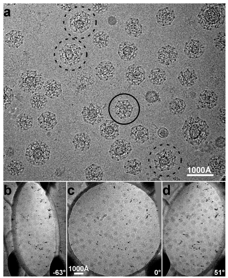
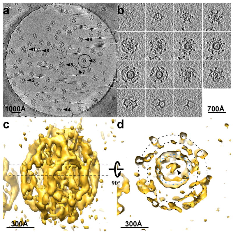
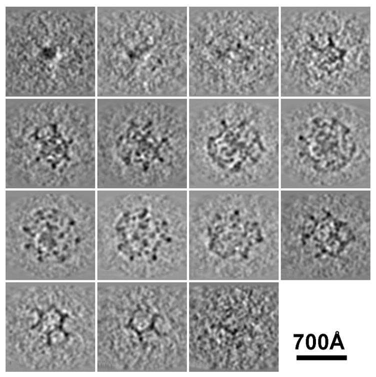
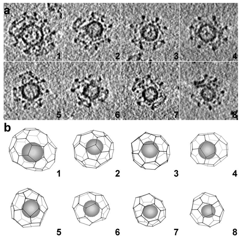
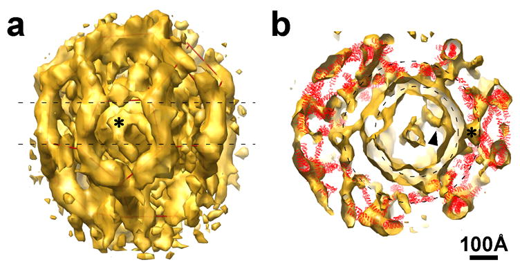
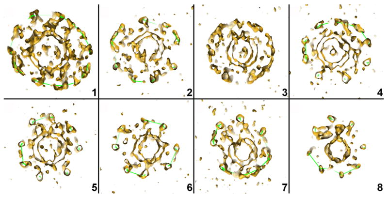
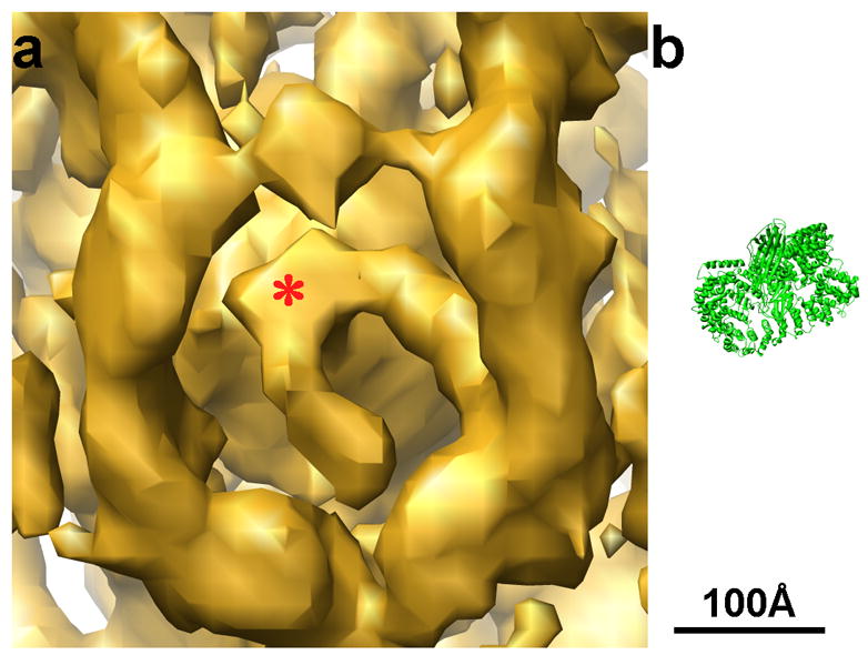
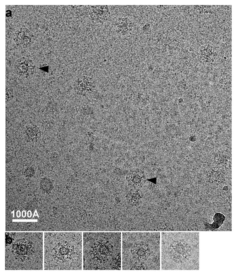
Clathrin-coated vesicles mediate vesicular traffic in cells. Three-dimensional image reconstructions of homogenous populations of in vitro assembled clathrin coats have yielded a molecular model for clathrin and its interactions with some of its partners. The intrinsic averaging required for those calculations has precluded detailed analysis of heterogeneous populations of clathrin-coated vesicles isolated from cells. We have therefore used cryo-electron tomography to study the lattice organization of individual clathrin-coated vesicles and the disposition of the captured vesicle with respect to the surrounding coat. We find a wide range of designs for the clathrin lattice, with different patterns of pentagonal, hexagonal, and occasionally heptagonal facets. Many coats, even smaller ones, enclose membrane vesicles, which are generally offset from the center of the clathrin shell. The electron density distribution between the coat and the underlying vesicle is not uniform, and the number of apparent contacts that anchor the clathrin lattice to the vesicle membrane is significantly less than the number of clathrin heavy chains in the assembly. We suggest that the eccentric position of the vesicle reflects the polarity of assembly, from initiation of coat formation to membrane pinching.


Figures

Figure 1
Figure 1
a: Cryo-electron microscopy image of clathrin coated vesicles embedded in vitreous ice. The particle marked by a continuous circle is probably the top view of a D6 barrel. From the projection image alone it is difficult to determine whether the coat contains a vesicle or not. The particles marked by dashed circles are larger and contain vesicles obviously offset from the centers of the coats. bd: Three representative raw images of a tilt series used to calculate cryo-tomograms of vitrified clathrin-coated vesicles. The tilt axis is approximately along the vertical direction.
Figure 2
Figure 2
Tomography of clathrin coated vesicles. a: Central section (155 Å thick) of an electron tomogram of coated vesicles embedded in a slab of vitreous ice over a hole in a carbon film. The diameter of the hole is 1 μm. b: Slices through a single coated vesicle (circled in A, and particle 3 in Fig. 4). The slice thickness is 62 Å. c: Isodensity surface for the coated vesicle shown in b viewed from the side. d: Central section of the same density map, corresponding to the dashed line in c, viewed from the top. The black dashed circles mark the locations of two separate layers of density that surround the vesicle; the blue dashed circle marks the membrane of the vesicle itself.
Figure 3
Figure 3
Slices through a single clathrin coat that does not contain a membrane vesicle. Coats like this are probably assembled spontaneously from clathrin triskelions during the purification process.
Figure 4
Figure 4
Outline of clathrin lattices in individual coated vesicles. a: The particles correspond to the clathrin-coated vesicles indicated in the tomogram shown in Fig. 2a. The images correspond to central sections (62 Å thick) viewed from the top. b: Clathrin lattices were determined by manual tracing along the electron density of the individual tomograms. The spheres inside the lattices represent the sizes and locations of the membrane vesicles contained within each coat. The views are presented in the same orientation as the central sections of the coated vesicles in A.
Figure 5
Figure 5
Details of coated vesicle 4 from Fig. 4. a: Surface rendered density map; the asterisk marks a feature anchored in the vesicle membrane. Red lines within the density outline the lattice. b: Central section of the same density map, with the ribbon diagrams of vertices of a clathrin lattice positioned within it. The two dashed circles represent the vesicle membrane (smaller circle) and the radial location of the N-terminal domains of the clathrin heavy chains (larger circle).
Figure 6
Figure 6
Central sections of the tomographic density maps of each of the 8 coated vesicles shown in Fig. 4, illustrating the eccentric positions of the membrane vesicles and the positions of the N-terminal domains of the clathrin heavy chains with respect to the vesicles.
Figure 7
Figure 7
a: Enlarged view of Fig. 5a, showing a feature anchored on the vesicle membrane. b: Ribbon diagram of the AP-1 core structure (pdb code: 1W63, 29) at the same scale as the density map. The size of the AP-1 core matches the size of the density anchored on the membrane in a.
Figure 8
Figure 8
a: Cryo-EM image of clathrin coated vesicles embedded in vitreous ice. The coated vesicles were purified with high ionic strength and pH to prevent recoating during purification. Vesicles within two of the coats (arrow head) are obviously eccentric. b: A gallery of selected clathrin coated vesicles boxed out from various areas of the sample.

Comment in

References

    1. Kirchhausen T. Clathrin. Annu Rev Biochem. 2000;69:699–727. - PubMed
    1. Brodsky FM, Chen CY, Knuehl C, Towler MC, Wakeham DE. Biological basket weaving: formation and function of clathrin-coated vesicles. Annu Rev Cell Dev Biol. 2001;17:517–568. - PubMed
    1. Sudhof TC. The synaptic vesicle cycle. Annu Rev Neurosci. 2004;27:509–547. - PubMed
    1. Crowther RA, Finch JT, Pearse BM. On the structure of coated vesicles. J Mol Biol. 1976;103:785–798. - PubMed
    1. Kirchhausen T, Harrison SC. Protein organization in clathrin trimers. Cell. 1981;23:755–761. - PubMed

Publication types

MeSH terms