. 2007 Feb;184(2):236-45.
doi: 10.1016/j.jmr.2006.10.008. Epub 2006 Nov 13.

Quasi Monte Carlo-based isotropic distribution of gradient directions for improved reconstruction quality of 3D EPR imaging

Affiliations

Quasi Monte Carlo-based isotropic distribution of gradient directions for improved reconstruction quality of 3D EPR imaging

Rizwan Ahmad et al. J Magn Reson. 2007 Feb.

Abstract

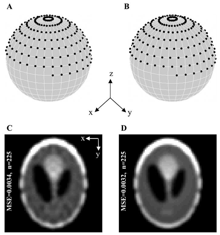
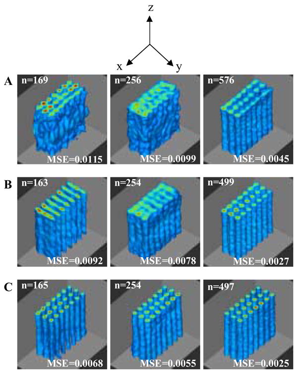
In continuous wave (CW) electron paramagnetic resonance imaging (EPRI), high quality of reconstructed image along with fast and reliable data acquisition is highly desirable for many biological applications. An accurate representation of uniform distribution of projection data is necessary to ensure high reconstruction quality. The current techniques for data acquisition suffer from nonuniformities or local anisotropies in the distribution of projection data and present a poor approximation of a true uniform and isotropic distribution. In this work, we have implemented a technique based on Quasi-Monte Carlo method to acquire projections with more uniform and isotropic distribution of data over a 3D acquisition space. The proposed technique exhibits improvements in the reconstruction quality in terms of both mean-square-error and visual judgment. The effectiveness of the suggested technique is demonstrated using computer simulations and 3D EPRI experiments. The technique is robust and exhibits consistent performance for different object configurations and orientations.


Figures

Figure 1
Figure 1
Effect of dislocating the gradient directions from a latitude-longitude grid on the reconstruction quality. A 3D Shepp-Logan phantom constructed by stacking (along z-axis) 2D Shepp-Logan phantom of size 64x64. (A) Distribution of 225 projections over a hemisphere using equal linear angle acquisition with Δθ= Δφ=π 15. All the projections corresponding to φ= 0 are identical regardless of the θ value. In order to avoid the acquisition of these identical projections, the angleφ is started from Δφ2 instead of 0. It should also be noted that the projections corresponding to φ=π 2 are only acquired for θϵ[ 0,π)since the symmetry of the projection data ensures that the other half(θϵ[ π, 2π)) is covered automatically. (B) Distribution of 225 projections after equal linear angle distribution given in A is modified by choosing a different set of azimuth angles for each iteration of mΔφ so that projection distribution does not follow the rigid latitude-longitude grid. (C) Center slice of image reconstructed using distribution given in A. (D) Center slice of image reconstructed using distribution given in B. From C to D, there is a visible reduction in the reconstruction artifacts suggesting that the isotropy of the data distribution results in an improved reconstruction quality.
Figure 2
Figure 2
Distribution of gradient directions (A) 229 projection directions distributed using equal solid angle scheme. (B) 225 projection directions distributed using the suggested QMC-based acquisition technique.
Figure 3
Figure 3
Reconstruction results of a simulated phantom using the three acquisition techniques for three orthogonal orientations of a phantom (see text for the description of the phantom). (A) Three orthogonal orientations of the simulated phantom which consists of six torus-shaped objects. (B) Reconstruction of A based on equal linear angle acquisition with 225 projections. (C) Reconstruction of A based on equal solid angle acquisition with 229 projections. (D) Reconstruction of A using the QMC-based acquisition with 225 projections. For proper display, voxels with intensity less than the 25% of the peak intensity of the reconstructed image were set to zero. In addition, all the reconstructed images were cropped from the center for better visualization.
Figure 4
Figure 4
MSE convergence for the three acquisition techniques. Number of projections vs. MSE for the phantom orientation shown in: (A) first column of Fig. 3A, (B) second column of Fig. 3A, and (C) third column of Fig. 3A. Here, ELA, ESA, and QMC stand for equal linear angle, equal solid angle, and the Quasi-Monte Carlo based acquisition, respectively.
Figure 5
Figure 5
Experimental phantom used for 3D EPRI experiment. A total of 21 capillary tubes were glued together so that they were arranged on a 7x3 grid. Each capillary tube had an inner diameter of 0.9 mm and an outer diameter of 1.4 mm. A 0.7 mM TAM solution was used to fill each capillary tube to a height of 10 mm. Sample dimensions were approximately 4.2 mm x 9.8 mm x 10 mm.
Figure 6
Figure 6
Reconstruction results for 3D EPRI of the capillary tubes phantom (shown in Fig. 5) to evaluate the performance of three different acquisition techniques. The measurements were performed on an L-band (1.2 GHz) EPRI system. Reconstruction results based on: (A) equal linear angle acquisition, (B) equal solid angle acquisition, and (C) the QMC-based acquisition for three different numbers of projections. Top 10% of the tubes were cropped for better visualization. Voxels with intensity less than the 25% of the peak intensity of the reconstructed image were set to zero.

References

    1. Fujii H, Berliner LJ. One- and two-dimensional EPR imaging studies on phantoms and plant specimens. Magn Reson Med. 1985;2:275–82. - PubMed
    1. Ferrari M, Quaresima V, Sotgiu A. Present status of electron paramagnetic resonance (EPR) spectroscopy/imaging for free radical detection. Pflugers Arch. 1996;431:R267–8. - PubMed
    1. Fuchs J, Freisleben HJ, Groth N, Herrling T, Zimmer G, Milbradt R, Packer L. One- and two-dimensional electron paramagnetic resonance imaging in skin. Free Radic Res Commun. 1991;15:245–53. - PubMed
    1. Hochi A, Furusawa M, Ikeya M. Applications of microwave scanning ESR microscope: human tooth with metal. Appl Radiat Isot. 1993;44:401–5. - PubMed
    1. Kuppusamy P. EPR spectroscopy in biology and medicine. Antioxid Redox Signal. 2004;6:583–5. - PubMed

Publication types

MeSH terms