. 2007 Jan 15;578(Pt 2):491-506.
doi: 10.1113/jphysiol.2006.119602. Epub 2006 Nov 9.

The effect of cerebral hypothermia on white and grey matter injury induced by severe hypoxia in preterm fetal sheep

Affiliations

The effect of cerebral hypothermia on white and grey matter injury induced by severe hypoxia in preterm fetal sheep

L Bennet et al. J Physiol. .

Abstract

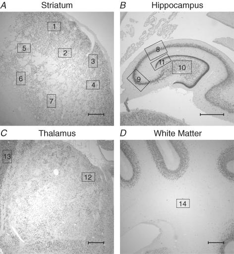
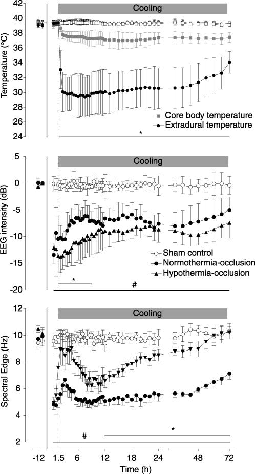
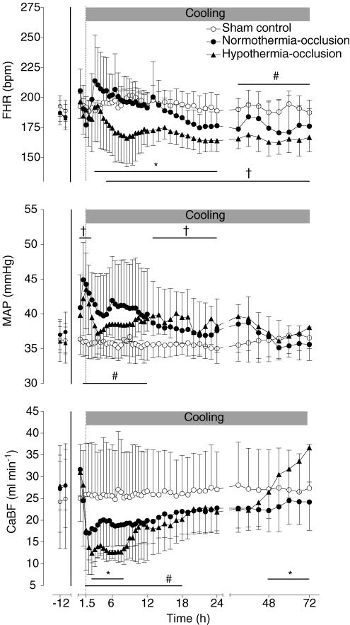
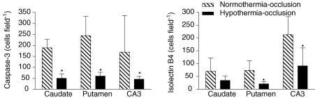
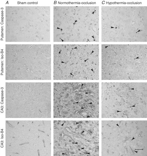
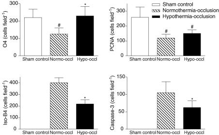
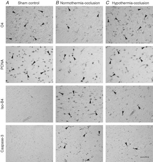
Prolonged, moderate cerebral hypothermia is consistently neuroprotective after experimental hypoxia-ischaemia; however, it has not been tested in the preterm brain. Preterm (0.7 gestation) fetal sheep received complete umbilical cord occlusion for 25 min followed by cerebral hypothermia (fetal extradural temperature reduced from 39.4 +/- 0.3 to 29.5 +/- 2.6 degrees C) from 90 min to 70 h after the end of occlusion or sham cooling. Occlusion led to severe acidosis and profound hypotension, which recovered rapidly after release of occlusion. After 3 days recovery the EEG spectral frequency, but not total intensity, was increased in the hypothermia-occlusion group compared with normothermia-occlusion. Hypothermia was associated with a significant overall reduction in loss of immature oligodendrocytes in the periventricular white matter (P < 0.001), and neuronal loss in the hippocampus and basal ganglia (P < 0.001), with suppression of activated caspase-3 and microglia (isolectin-B4 positive). Proliferation was significantly reduced in periventricular white matter after occlusion (P < 0.05), but not improved after hypothermia. In conclusion, delayed, prolonged head cooling after a profound hypoxic insult in the preterm fetus was associated with a significant reduction in loss of neurons and immature oligodendroglia, with evidence of EEG and haemodynamic improvement after 3 days recovery, but also with a persisting reduction in proliferation of cells in the periventricular region. Further studies are required to evaluate the long-term impact of cooling on brain growth and maturation.


Figures

Figure 1
Figure 1. Photomicrographs showing the fields of view used for histological analysis of brain injury in the striatum (A), the hippocampus (B), the thalamus (C), and the subcortical white matter (D)
Seven fields of view in the striatum (4 caudate, 3 putamen; squares 1–7), one field of view in each of the CA1/2, CA3, CA4 and dentate gyrus (squares 8, 9, 10 and 11, respectively), 2 fields of view in the thalamus (1 medial nucleus, 1 reticular formation; squares 12 and 13, respectively), and one field of view in the periventricular white matter (square 14), in both hemispheres, were used to count numbers of labelled cells. Other regions used for measurement of cortical neuronal survival are not shown. Scale bar, 1 mm.
Figure 2
Figure 2. Time sequence of changes in fetal temperature, electroencephalogram (EEG) intensity (power) and spectral edge (frequency)
The 25 min period of umbilical cord occlusion is shown by the continuous vertical lines, while cooling is shown by the shaded bar. The top panel shows changes in extradural and core body (oesophageal) temperatures in the hypothermia-occlusion and normothermia-occlusion groups; sham control group data are not shown for clarity. Bar plus *P < 0.05 hypothermia-occlusion versus normothermia-occlusion group. Bar plus #P < 0.05 normothermia-occlusion versus sham control group. Data are mean ±s.d.
Figure 3
Figure 3. Time sequence of changes in fetal heart rate (FHR, bpm), mean arterial blood pressure (MAP, mmHg), and total carotid blood flow (CaBF, ml min−1) in the sham control, normothermia-occlusion, and the hypothermia-occlusion groups
The 25 min period of umbilical cord occlusion is shown by the continuous vertical line, while cooling is shown by the shaded bar. Bar plus *P < 0.05 hypothermia-occlusion versus normothermia-occlusion group. Bar plus † P < 0.05 hypothermia-occlusion versus sham control group. Bar plus # P < 0.05 normothermia-occlusion versus sham control group. Data are mean ± s.d.
Figure 4
Figure 4. The effect of hypothermia on neuronal survival assessed with neuronal specific nuclear protein (NeuN) after 3 days recovery from 25 min of umbilical cord occlusion Top
panel, the effect of hypothermia on numbers of NeuN-positive cells in the sham control, normothermia-occlusion, and the hypothermia-occlusion groups. #P < 0.05 versus sham control group; *P < 0.05 versus normothermia-occlusion group. Data are mean ± s.d. Striatum, Th (thalamus), CA (cornu ammonis of the hippocampus), DG (dentate gyrus), thalamic nuclei: Median N (median nucleus), Ret Form (reticular formation) Bottom panel, photomicrographs of the striatum, reticular formation of the thalamus, and CA3 showing NeuN-positive cells (arrowheads) in the sham control group (A), the normothermia-occlusion group (B), and the hypothermia-occlusion group (C). Scale bar, 40 μm.
Figure 5
Figure 5. The effect of hypothermia on numbers of activated caspase-3 (Asp175)- and isolectin-B4-stained cells in subcortical neuronal nuclei after 3 days recovery from 25 min of umbilical cord occlusion
*P < 0.05 versus normothermia-occlusion group. Data are mean ± s.d.
Figure 6
Figure 6. Caspase-3 and isolectin B4 expression in selected neuronal nuclei
Photomicrographs of selected subcortical neuronal nuclei showing activated caspase-3 (Asp175)- and isolectin-B4-stained cells (arrowheads) in the sham control group (A), the normothermia-occlusion group (B), and the hypothermia-occlusion group (C). Scale bar, 40 μm.
Figure 7
Figure 7. The effect of hypothermia on periventricular white matter
The effect of hypothermia on numbers of O4-positive immature oligodendrocytes, activated caspase-3 (Asp175)-, isolectin-B4-, and proliferating cell nuclear antigen (PCNA)-positive cells in the periventricular white matter after 3 days recovery from 25 min of umbilical cord occlusion. #P < 0.05 versus sham control group; *P < 0.05 versus normothermia-occlusion group. Data are mean ± s.d.
Figure 8
Figure 8. Periventricular white matter
Photomicrographs of periventricular white matter showing O4-positive immature oligodendrocytes, activated caspase-3- (Asp175), isolectin-B4-, and proliferating cell nuclear antigen (PCNA)-positive cells (arrowheads) in the sham-control group (A), the normothermia-occlusion group (B), and the hypothermia-occlusion group (C). Scale bar, 40 μm.

References

    1. Argyropoulou MI, Xydis V, Drougia A, Argyropoulou PI, Tzoufi M, Bassounas A, Andronikou S, Efremidis SC. MRI measurements of the pons and cerebellum in children born preterm; associations with the severity of periventricular leukomalacia and perinatal risk factors. Neuroradiology. 2003;45:730–734. - PubMed
    1. Azzopardi D, Robertson NJ, Cowan FM, Rutherford MA, Rampling M, Edwards AD. Pilot study of treatment with whole body hypothermia for neonatal encephalopathy. Pediatrics. 2000;106:684–694. - PubMed
    1. Azzopardi D, Wyatt JS, Cady EB, Delpy DT, Baudin J, Stewart AL, Hope PL, Hamilton PA, Reynolds EO. Prognosis of newborn infants with hypoxic-ischemic brain injury assessed by phosphorus magnetic resonance spectroscopy. Pediatr Res. 1989;25:445–451. - PubMed
    1. Back SA, Han BH, Luo NL, Chricton CA, Xanthoudakis S, Tam J, Arvin KL, Holtzman DM. Selective vulnerability of late oligodendrocyte progenitors to hypoxia-ischemia. J Neurosci. 2002;22:455–463. - PMC - PubMed
    1. Barkovich AJ, Sargent SK. Profound asphyxia in the premature infant: imaging findings. AJNR Am J Neuroradiol. 1995;16:1837–1846. - PMC - PubMed

Publication types

MeSH terms