. 2007 Mar 15;109(6):2461-9.
doi: 10.1182/blood-2006-06-029082. Epub 2006 Nov 9.

Src family kinases mediate neutrophil adhesion to adherent platelets

Affiliations

Src family kinases mediate neutrophil adhesion to adherent platelets

Virgilio Evangelista et al. Blood. .

Abstract

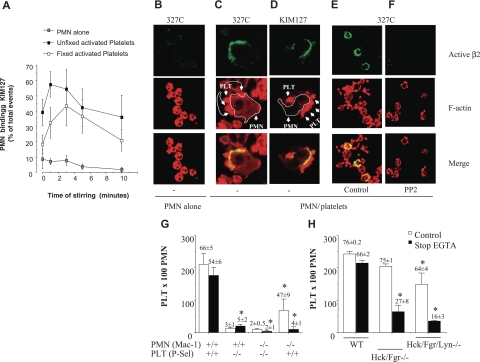
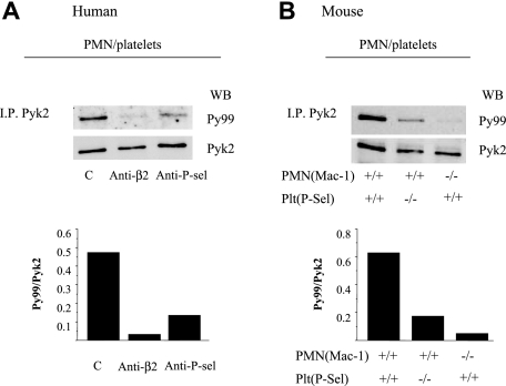
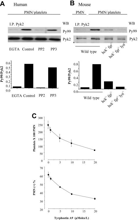
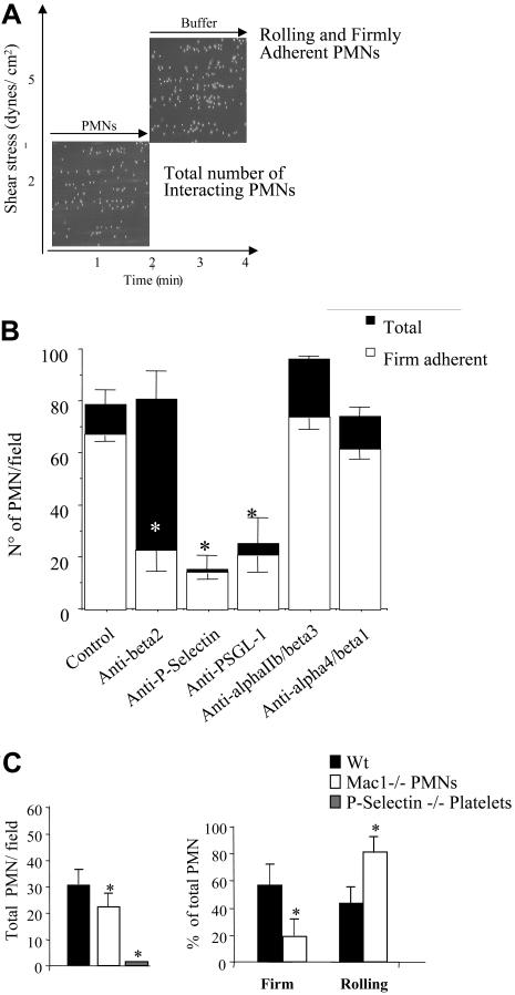
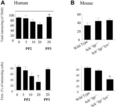
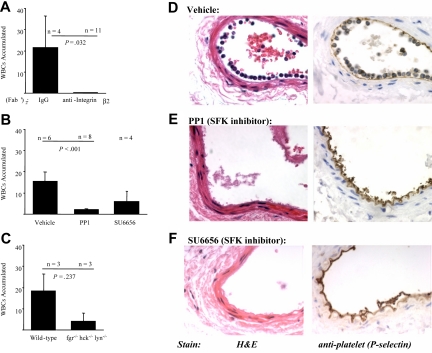
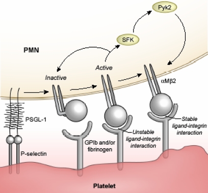
Polymorphonuclear leukocyte (PMN)-platelet interactions at sites of vascular damage contribute to local and systemic inflammation. We sought to determine the role of "outside-in" signaling by Src-family tyrosine kinases (SFKs) in the regulation of alphaMbeta2-integrin-dependent PMN recruitment by activated platelets under (patho)physiologic conditions. Activation-dependent epitopes in beta2 integrin were exposed at the contact sites between PMNs and platelets and were abolished by SFK inhibitors. PMNs from alphaMbeta2(-/-), hck(-/-)fgr(-/-), and hck(-/-)fgr(-/-)lyn(-/-) mice had an impaired capacity to adhere with activated platelets in suspension. Phosphorylation of Pyk2 accompanied PMN adhesion to platelets and was blocked by inhibition as well as by genetic deletion of alphaMbeta2 integrin and SFKs. A Pyk2 inhibitor reduced platelet-PMN adhesion, indicating that Pyk2 may be a downstream effector of SFKs. Analysis of PMN-platelet interactions under flow revealed that SFK signaling was required for alphaMbeta2-mediated shear-resistant adhesion of PMNs to adherent platelets, but was dispensable for P-selectin-PSGL-1-mediated recruitment and rolling. Finally, SFK activity was required to support PMN accumulation along adherent platelets at the site of vascular injury, in vivo. These results definitely establish a role for SFKs in PMN recruitment by activated platelets and suggest novel targets to disrupt the pathophysiologic consequences of platelet-leukocyte interactions in vascular disease.


Figures

Figure 1
Figure 1
SFKs are required for localization of activated β2 integrins at sites of PMN-platelet contacts and for αMβ2-dependent PMN-platelet interactions in suspension. (A) PMNs alone or with PFA-fixed or unfixed thrombin (0.25 U/mL)–activated platelets (1:10 ratio) were incubated for 0 to 10 minutes at 37°C in stirring conditions (1000 rpm). At each time point, stirring was stopped and KIM127 (10 μg/mL) was added to the cells for an additional 5 minutes at 37°C. The cells were then fixed in 2% PFA for 30 minutes, washed, and stained with Alexa-Fluor 488–conjugated goat antimouse antibody at 4°C for 30 minutes. Nonspecific fluorescence was determined by incubation of PMNs with Alexa-Fluor 488–conjugated goat antimouse antibody only. Values are reported as percentages of PMNs expressing KIM127-specific binding. (B) PMNs alone or with thrombin-activated, PFA-fixed platelets (C-D) were stirred at 37°C for 3 minutes, at which time 10 μg/mL mAb 327C (B-C, E-F) or KIM127 (D) was added. Confocal microscopy to detect KIM127 and 327C mAb binding and rhodamine-phalloidin was performed as described in “Materials and methods.” (C-D) The actin staining in these images was captured at a lower amplification than in panels B, E, or F in order to allow clear identification of platelets that contain higher concentration of F-actin than do PMNs. PMNs are identified by a white line around the membrane edge and show intense 327C (C) or KIM127 (D) binding in areas with attached platelets. (E-F) PMNs were pretreated with vehicle (E) or PP2 (F) for 5 minutes at RT before incubation with platelets. PP2 treatment dramatically reduces expression of activated β2 integrins at platelet contact sites. (G) PMNs from wild-type or αM−/− mice were stirred in the absence or in the presence of fixed-activated wild-type or P-selectin–deficient platelets. Samples were immediately fixed with an equal volume of 2% PFA (open bars) or, alternatively, 5 mM EGTA was added to mixed cell suspensions 30 seconds before fixation to disrupt P-selectin–mediated bonds (closed bars designated stop EGTA). The formation of mixed platelet–PMN conjugates was analyzed by flow cytometry, as described in “Materials and methods.” The numbers of platelets recruited by 100 PMNs are graphed and the numbers on the top of the bars are the percentage of PMNs with attached platelets. Average results (mean ± SEM) from 6 different experiments for wild-type cells and 4 experiments for αM−/− or P-selectin–deficient cells are presented. *P < .05 versus WT. (H) PMNs from wild-type or double hck−/−fgr−/− or triple hck−/−fgr−/−lyn−/− mice were stirred in the absence or in the presence of activated, fixed wild-type platelets and treated as described in panel F. The numbers of platelets recruited by 100 PMNs are graphed and the numbers on top of the bars are the percentage of PMNs with attached platelets (mean, n = 4). The figure shows average results (mean ± SEM) from 4 different experiments performed with PMNs isolated from pooled bone marrow obtained from 5 wild-type or 5 knock-out mice. *P < .05 versus WT.
Figure 2
Figure 2
PMN interaction with activated platelets stimulates αMβ2-dependent Pyk2 phosphorylation. (A) Human PMNs were preincubated with the function-blocking anti–β2-integrin mAb IB4 or with an irrelevant mouse monoclonal antibody for 15 minutes in ice. Thrombin-activated fixed platelets were preincubated with control or the anti–P-selectin mAb WAPS for 15 minutes at room temperature. PMNs and platelets were stirred at 1000 rpm at 37°C for 3 minutes. Pyk2 was immunoprecipitated from total cell lysates, and immune complexes were analyzed for phospho-Pyk2 by immunoblotting with PY99, and then the blots were reprobed with anti-Pyk2 antibody to detect total Pyk2. The bars report the ratio between optical density of PY99 andPYK2 (ie, phospho-Pyk2/total Pyk2). The figure is representative of results obtained in 3 different experiments. (B) PMNs from wild-type or αM−/− mice were stirred in the presence of fixed-activated wild-type or P-selectin−/− platelets at 37°C for 3 minutes. The interactions were stopped by the addition of an equal volume of reduced sample buffer, and samples were processed for immunoblot analysis as described for panel A. The bars indicate the ratio between optical density of PY99 and PYK2 (phospho-Pyk2/total Pyk2). The figure is representative of results obtained in 2 different experiments performed with PMNs isolated from pooled bone marrow obtained from 5 wild-type or 5 knock-out mice.
Figure 3
Figure 3
Tyrosine phosphorylation of Pyk2 is mediated by SFKs and is required for shear-resistant PMN attachment to platelets. (A) Untreated PMNs or PMNs pretreated with DMSO, 10 μM PP2, or 10 μM PP3 were stirred for 3 minutes with activated, fixed platelets (ratio 5:1) in the presence or in the absence of 5 μM EGTA. Pyk2 was immunoprecipitated from total cell lysates, and immune complexes were analyzed for phospho-Pyk2 by immunoblotting with PY99 and then reprobed with anti-Pyk2 antibody to detect total Pyk2. The bars report the ratio between optical density of PY99 and PYK2 (ie, phospho-Pyk2/total Pyk2). The figure is representative of results obtained in 3 different experiments. (B) PMNs from wild-type, double hck−/−fgr−/−, or triple hck−/−fgr−/−lyn−/− mice were stirred in the absence or in the presence of activated, fixed wild-type platelets and processed as described in panel A. The bars indicate the ratio between optical density of PY99 and PYK2 (phospho-Pyk2/total Pyk2). The figure is representative of results obtained in 2 different experiments performed with PMNs isolated from pooled bone marrow obtained from 5 wild-type or 5 knock-out mice. (C) Human PMNs pretreated with DMSO or the indicated concentrations of the Pyk2 inhibitor tyrphostin A9 (AG17) were stirred for 3 minutes with activated, fixed platelets (ratio 5:1). Samples were immediately fixed with an equal volume of 2% PFA and the formation of mixed platelet-PMN conjugates was analyzed by flow cytometry, as described in “Materials and methods.” The number of platelets recruited by 100 PMNs and the percentage of PMNs with attached platelets are graphed (mean ± SEM, n = 3).
Figure 4
Figure 4
PMN recruitment by adherent platelets under flow requires P-selectin and αMβ2. (A) Human PMNs (5 × 10⁶/mL) were perfused across a platelet surface at a flow rate of 2 dynes/cm2 for 2 minutes. Then, the chamber was perfused with fresh media for an additional 2 minutes at 5 dynes/cm2, without interruption in the perfusion. The interaction of PMNs with adherent platelets was observed by contrast phase video microscopy with a 20×/0.40 NA objective. The total number of cells interacting at 2 minutes and the fractions of rolling and firmly adhered PMNs in the last 20 seconds of flow (4 minutes) were measured using an ad hoc software for image analysis as described in “Materials and methods.” (B) P-selectin and αIIbβ3 and αvβ3 were blocked by incubating the human platelet–coated slide with the mAbs WAPS12.2 or 7E3, respectively (20 μg/mL) for 15 minutes. β2 or α4/1 integrins on human PMNs were blocked by treating cells with the mAbs IB4 or 2B4 (20 μg/mL) for 15 minutes in ice. Experiments were performed as described for panel A and in “Materials and methods.” The results are presented as means ± SEM for 3 to 8 different experiments performed using cells from different donors. Black bars indicate the total number of PMNs recruited per field, and white bars indicate the number of PMNs that establish firm adhesion. *P < .05 for treatment versus control. (C) PMNs were isolated from wild-type or αM−/− mice bone marrow. Platelets were isolated from wild-type or P-selectin–deficient (P-sel−/−) mice. Experiments were performed as described for panel A and in “Materials and methods.” The results presented are means ± SEM from 4 different experiments each performed using cells collected from 5 wild-type or knock-out mice. *P < .05 for wild-type versus αM−/− PMNs or P-selectin−/− platelets.
Figure 5
Figure 5
SFKs are required for PMN firm adhesion to adherent platelets under conditions of physiologic flow. (A) Human PMNs, pretreated with different concentration of PP2, PP3, or vehicle (DMSO) for 10 minutes, were perfused across a platelet surface at a flow rate of 2 dynes/cm2 for 2 minutes. Then, the chamber was perfused with fresh media for an additional 2 minutes at 5 dynes/cm2, without interruption in the perfusion. The interaction of PMNs with adherent platelets was observed by contrast phase video microscopy with a 20×/0.40 NA objective. The total number of cells interacting at 2 minutes and the fractions of firmly adhered PMNs in the last 20 seconds of flow were measured using an ad hoc software for image analysis as described in “Materials and methods.” The results presented are means ± SEM from 5 different experiments performed using cells from different donors. *P < .05 for treatment versus control. (B) PMNs were isolated from bone marrow of wild-type, hck−/−fgr−/−, or hck−/−fgr−/−lyn−/− mice. Flow experiments were performed exactly as described for human cells. The results presented are mean ± SEM for 6 different experiments each performed in triplicate with cells pooled from 5 wild-type or knock-out mice. *P < .05 for wild-type versus hck−/−fgr−/−lyn−/− cells.
Figure 6
Figure 6
β2 integrins and SFKs mediate PMN recruitment to adherent platelets at the sites of vascular injury in mice. Arterial injury was performed as described in “Materials and methods,” and the vessels were examined one hour after injury. (A) Wild-type mice were pretreated with control F(ab)′2 or anti–β2-integrin F(ab)′2 prior to performing injury. The results presented are the average number of WBCs that accumulated along the vessel (mean ± SEM) in 4 control and 11 anti-integrin–treated animals. (B) Wild-type mice (n = 6) were pretreated with vehicle, PP1 (1.5 mg/kg; n = 8), or SU6656 (0.015 mg; n = 4) prior to injury. The results presented are the average number of WBCs that accumulated along the vessel (mean ± SEM). (C) Arterial injury was performed in wild-type (n = 3) or hck−/−fgr−/−lyn−/− mice (n = 3). The results presented are the average number of WBCs that accumulated along the vessel (mean ± SEM). Representative images of vessel sections taken from a wild-type mouse treated with vehicle (D), PP1 (E), or SU6656 (F) one hour after injury. Sections were stained with hematoxylin and eosin (left panels) or processed for immunohistochemical analysis with a polyclonal antibody to P-selectin (right panels; brown staining).
Figure 7
Figure 7
Hypothetical scheme of a 3-step model of αMβ2 activity during PMN-platelet adhesion. Step 1: platelet-PMN interactions induce initial activation and ligand binding to αMβ2. Step 2: generation of an outside-in, SFK-Pyk2–mediated signal(s). Step 3: stabilization of integrin-ligand binding.

References

    1. Weyrich AS, Prescott SM, Zimmerman GA. Platelets, endothelial cells, inflammatory chemokines, and restenosis: complex signaling in the vascular play book. Circulation. 2002;106:1433–1435. - PubMed
    1. Theilmeier G, Michiels C, Spaepen E, et al. Endothelial von Willebrand factor recruits platelets to atherosclerosis-prone sites in response to hypercholesterolemia. Blood. 2002;99:4486–4493. - PubMed
    1. Massberg S, Brand K, Gruner S, et al. A critical role of platelet adhesion in the initiation of atherosclerotic lesion formation. J Exp Med. 2002;196:887–896. - PMC - PubMed
    1. Huo Y, Schober A, Forlow SB, et al. Circulating activated platelets exacerbate atherosclerosis in mice deficient in apolipoprotein E. Nat Med. 2003;9:61–67. - PubMed
    1. Smyth SS, Reis ED, Zhang W, Fallon JT, Gordon RE, Coller BS. b3-integrin-deficient mice but not P-selectin-deficient mice develop intimal hyperplasia after vascular injury: correlation with leukocyte recruitment to adherent platelets 1 hour after injury. Circulation. 2001;103:2501–2507. - PubMed

Publication types