

Expression of peroxisome proliferator-activated receptor-gamma in macrophage suppresses experimentally induced colitis
- PMID: 17095756
- PMCID: PMC1796914
- DOI: 10.1152/ajpgi.00381.2006
Expression of peroxisome proliferator-activated receptor-gamma in macrophage suppresses experimentally induced colitis
Abstract
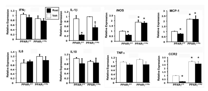
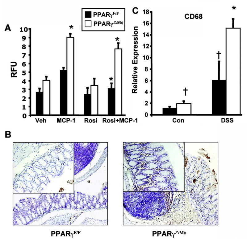
Peroxisome proliferator-activated receptor-gamma (PPAR-gamma) has been shown to be a protective transcription factor in mouse models of inflammatory bowel disease (IBD). PPAR-gamma is expressed in several different cell types, and mice with a targeted disruption of the PPAR-gamma gene in intestinal epithelial cells demonstrated increased susceptibility to dextran sulfate sodium (DSS)-induced IBD. However, the highly selective PPAR-gamma ligand rosiglitazone decreased the severity of DSS-induced colitis and suppressed cytokine production in both PPAR-gamma intestinal specific null mice and wild-type littermates. Therefore the role of PPAR-gamma in different tissues and their contribution to the pathogenesis of IBD still remain unclear. Mice with a targeted disruption of PPAR-gamma in macrophages (PPAR-gamma(DeltaMphi)) and wild-type littermates (PPAR-gamma(F/F)) were administered 2.5% DSS in drinking water to induce IBD. Typical clinical symptoms were evaluated on a daily basis, and proinflammatory cytokine analysis was performed. PPAR-gamma(DeltaMphi) mice displayed an increased susceptibility to DSS-induced colitis compared with wild-type littermates, as defined by body weight loss, diarrhea, rectal bleeding score, colon length, and histology. IL-1beta, CCR2, MCP-1, and inducible nitric oxide synthase mRNA levels in colons of PPAR-gamma(DeltaMphi) mice treated with DSS were higher than in similarly treated PPAR-gamma(F/F) mice. The present study has identified a novel protective role for macrophage PPAR-gamma in the DSS-induced IBD model. The data suggest that PPAR-gamma regulates recruitment of macrophages to inflammatory foci in the colon.
Figures


Similar articles
-
Peroxisome proliferator activated receptor gamma in colonic epithelial cells protects against experimental inflammatory bowel disease.Gut. 2006 Aug;55(8):1104-13. doi: 10.1136/gut.2005.081745. Epub 2006 Mar 17. Gut. 2006. PMID: 16547072 Free PMC article.
-
Effects of peroxisome proliferator-activated receptors-gamma ligands on dextran sodium sulphate-induced colitis in rats.J Physiol Pharmacol. 2011 Jun;62(3):347-56. J Physiol Pharmacol. 2011. PMID: 21893696
-
Oroxyloside prevents dextran sulfate sodium-induced experimental colitis in mice by inhibiting NF-κB pathway through PPARγ activation.Biochem Pharmacol. 2016 Apr 15;106:70-81. doi: 10.1016/j.bcp.2016.02.019. Epub 2016 Mar 4. Biochem Pharmacol. 2016. PMID: 26947454
-
[The peroxisome-proliferator-activated gamma receptor and chronic inflammatory bowel disease (PPARgamma and IBD)].J Soc Biol. 2006;200(2):121-31. doi: 10.1051/jbio:2006015. J Soc Biol. 2006. PMID: 17151549 Review. French.
-
Nuclear Receptors in the Pathogenesis and Management of Inflammatory Bowel Disease.Mediators Inflamm. 2019 Jan 21;2019:2624941. doi: 10.1155/2019/2624941. eCollection 2019. Mediators Inflamm. 2019. PMID: 30804707 Free PMC article. Review.
Cited by
-
Harnessing nature's pharmacy: investigating natural compounds as novel therapeutics for ulcerative colitis.Front Pharmacol. 2024 Aug 14;15:1394124. doi: 10.3389/fphar.2024.1394124. eCollection 2024. Front Pharmacol. 2024. PMID: 39206263 Free PMC article. Review.
-
5-Aminosalicylic Acid Ameliorates Colitis and Checks Dysbiotic Escherichia coli Expansion by Activating PPAR-γ Signaling in the Intestinal Epithelium.mBio. 2021 Jan 19;12(1):e03227-20. doi: 10.1128/mBio.03227-20. mBio. 2021. PMID: 33468700 Free PMC article.
-
NLRP6 function in inflammatory monocytes reduces susceptibility to chemically induced intestinal injury.Mucosal Immunol. 2017 Mar;10(2):434-445. doi: 10.1038/mi.2016.55. Epub 2016 Jun 29. Mucosal Immunol. 2017. PMID: 27353251 Free PMC article.
-
Positive role of CCAAT/enhancer-binding protein homologous protein, a transcription factor involved in the endoplasmic reticulum stress response in the development of colitis.Am J Pathol. 2009 May;174(5):1786-98. doi: 10.2353/ajpath.2009.080864. Epub 2009 Apr 9. Am J Pathol. 2009. PMID: 19359519 Free PMC article.
-
Sumoylation of peroxisome proliferator-activated receptor gamma by apoptotic cells prevents lipopolysaccharide-induced NCoR removal from kappaB binding sites mediating transrepression of proinflammatory cytokines.J Immunol. 2008 Oct 15;181(8):5646-52. doi: 10.4049/jimmunol.181.8.5646. J Immunol. 2008. PMID: 18832723 Free PMC article.
References
-
- Andres PG, Beck PL, Mizoguchi E, et al. Mice with a selective deletion of the CC chemokine receptors 5 or 2 are protected from dextran sodium sulfate-mediated colitis: lack of CC chemokine receptor 5 expression results in a NK1.1+ lymphocyte-associated Th2-type immune response in the intestine. J Immunol. 2000;164:6303–12. - PubMed
-
- Babaev VR, Yancey PG, Ryzhov SV, et al. Conditional knockout of macrophage PPARgamma increases atherosclerosis in C57BL/6 and low-density lipoprotein receptor-deficient mice. Arterioscler Thromb Vasc Biol. 2005;25:1647–53. - PubMed
-
- Bassaganya-Riera J, Reynolds K, Martino-Catt S, et al. Activation of PPAR gamma and delta by conjugated linoleic acid mediates protection from experimental inflammatory bowel disease. Gastroenterology. 2004;127:777–91. - PubMed
Publication types
MeSH terms
Substances
Grants and funding
LinkOut - more resources
Full Text Sources
Molecular Biology Databases
Miscellaneous