. 2007 Jul;56(7):1077-86.
doi: 10.1007/s00262-006-0242-7. Epub 2006 Nov 10.

Oral premalignant lesions induce immune reactivity to both premalignant oral lesions and head and neck squamous cell carcinoma

Affiliations

Oral premalignant lesions induce immune reactivity to both premalignant oral lesions and head and neck squamous cell carcinoma

M Rita I Young et al. Cancer Immunol Immunother. 2007 Jul.

Abstract

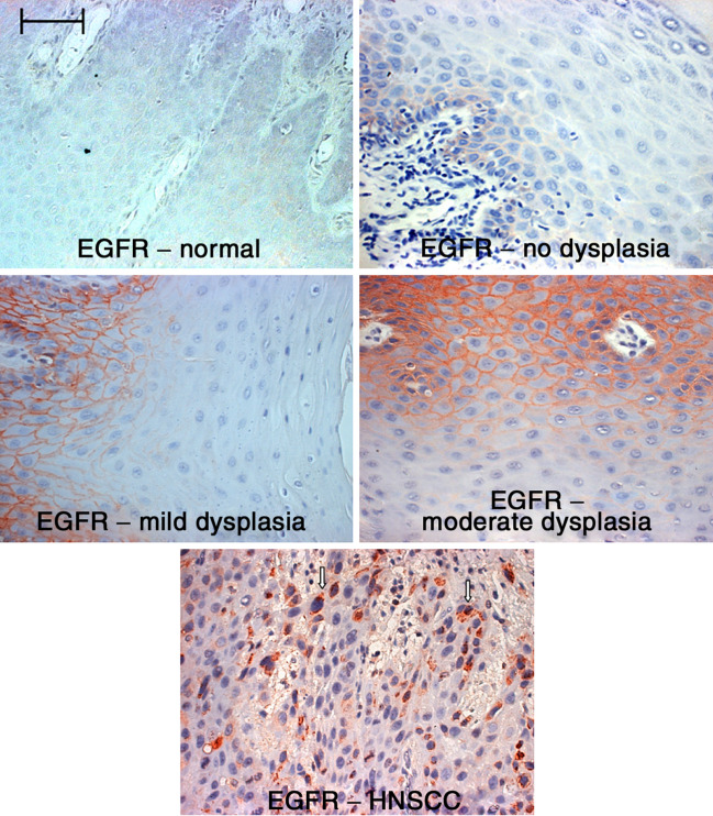
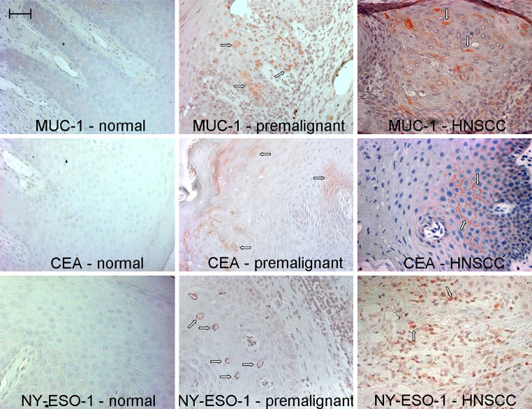
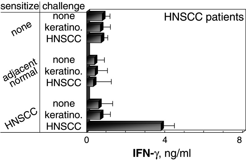
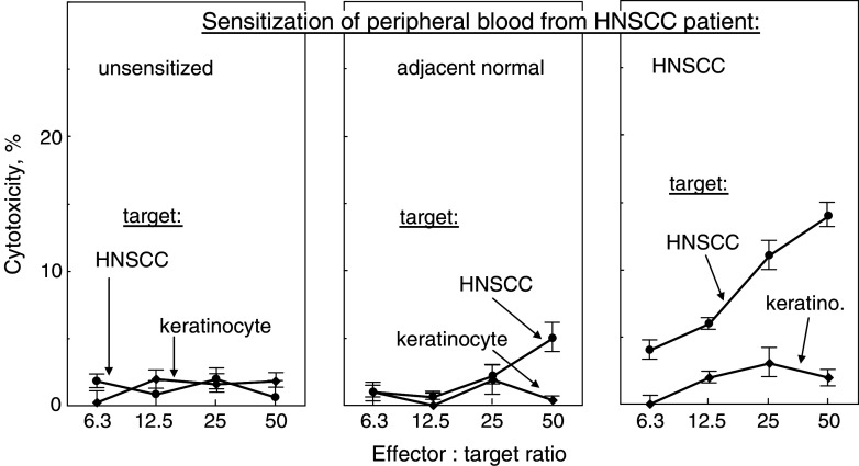
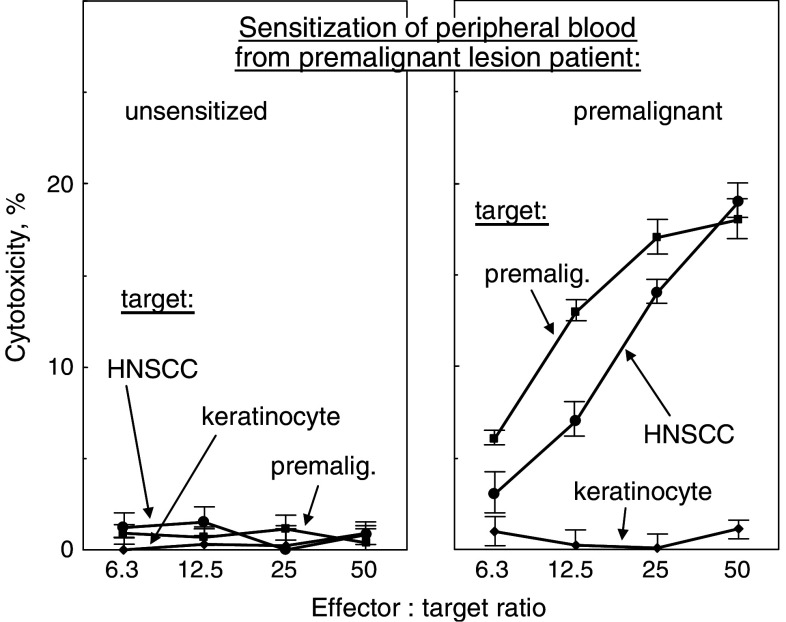
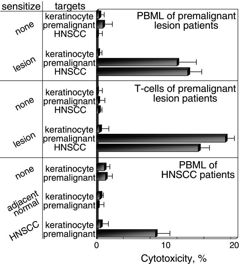
Head and neck squamous cell carcinoma (HNSCC) is an aggressive malignancy, and despite advances in treatments, the 5-year survival has remained at less than 50%. One treatment strategy is to focus on patients with premalignant oral lesions that carry a high-risk for developing recurrent premalignant lesions and HNSCC disease. As an initial attempt to determine if immune therapy has the potential to be protective in these patients, studies determined if premalignant lesions express tumor antigens that have previously been shown to be expressed on HNSCC. Immunohistochemical analyses showed prominent expression of epidermal growth factor receptor in premalignant lesions, even in lesions with mild dysplasia. MUC-1 and carcinoembryonic antigen were expressed in most patient samples, while NY-ESO-1 was less frequently expressed. Each of these antigens was expressed on HNSCC. This provided the rationale for determining if premalignant oral lesions could be used to stimulate autologous peripheral blood mononuclear leukocytes (PBML) to react against heterologous premalignant lesions and HNSCC. Following sensitization with autologous premalignant lesions, PBML responded to a challenge with either heterologous premalignant oral lesion cells or HNSCC by releasing IFN-gamma. In addition, sensitization with autologous premalignant lesion lysates generated cytolytic activity by both PBML and T cells against allogeneic premalignant lesion cells and HNSCC. These studies show the feasibility of using premalignant oral lesions to stimulate immune reactivity against both premalignant oral lesions as well as HNSCC.


Figures

Fig. 1
Fig. 1
Increased expression of EGFR on dysplastic premalignant oral lesions. Tissue sections from premalignant oral lesions with various levels of dysplasia or HNSCC tissue were immunostained for EGFR. Shown are representative microscopic images of immunostained tissue. Scale bar for all panels = 50 μm
Fig. 2
Fig. 2
Immunostaining for tumor antigens on premalignant oral lesions and HNSCC. Sections of pathologically normal tissue adjacent to excised HNSCC, premalignant oral lesions with moderate to severe dysplasia, and HNSCC were immunostained for CEA, MUC-1 and NY-ESO-1. Shown are microscopic images from each category of stained tissue. Scale bar for all panels = 50 μm
Fig. 3
Fig. 3
Comparable levels of expression of HLA-A2 on premalignant lesion cells, HNSCC and normal keratinocytes that were used as challenging cells or targets. Cells were immunostained for HLA-A2 and levels of expression were measure by flow cytometry. Shown are representative histograms of keratinocytes (left panel), premalignant lesion cells (center panel) and HNSCC (right panel) that were used to challenge or as targets for sensitized BPML or T cells
Fig. 4
Fig. 4
Stimulation of IFN-γ release from PBML that were sensitized with autologous HNSCC and challenged with allogeneic HNSCC. PBML of HNSCC patients were cultured with lysates of autologous HNSCC, autologous adjacent normal tissue, or no lysate. After 7 days, they were washed, challenged for 24 h with allogeneic HLA-A2+ HNSCC, normal allogeneic keratinocytes or unchallenged. Levels of IFN-γ released during these 24 h was measured by ELISA. Shown are mean ± SEM from at least five cultures
Fig. 5
Fig. 5
Stimulation of IFN-γ release from PBML that were sensitized with autologous premalignant lesions and challenged with allogeneic premalignant lesion cells or HNSCC. HLA-A2+ PBML were cultured with lysates of autologous premalignant lesions or without lysates. After 7 days, they were washed, challenged with allogeneic HLA-A2+ normal keratinocytes, premalignant lesion cells, HNSCC or left unchallenged for 24 h. The amount of IFN-γ released during these 24 h was measured by ELISA. Shown are mean ± SEM from at least five cultures
Fig. 6
Fig. 6
Generation of cytolytic activity against HNSCC by PBML that were sensitized with autologous HNSCC. HLA-A2+ PBML were cultured with no lysates, or lysates from autologous pathologically normal tissue or autologous HNSCC. After 7 days, they were washed and plated in increasing numbers with 2 × 104 HLA-A2+ allogeneic normal keratinocytes or HNSCC as target cells. The extent of cytotoxicity was measured 4 h later. Shown are mean values from a representative HNSCC patient ± SD from six replicates at each effector to target ratio
Fig. 7
Fig. 7
Generation of cytolytic activity against allogeneic premalignant lesion cells and HNSCC by PBML that were sensitized with autologous premalignant lesions. HLA-A2+ PBML were cultured with no lysates, or lysates from autologous premalignant lesions. After 7 days, they were washed and plated in increasing numbers with 2 × 104 HLA-A2+ allogeneic normal keratinocytes, premalignant lesion cells or HNSCC as target cells. The extent of cytotoxicity was measured 4 h later. Shown are mean values from a representative premalignant lesion patient ± SD from six replicates at each effector to target ratio
Fig. 8
Fig. 8
Generation of cytolytic activity against premalignant lesion cells and HNSCC by PBML and T cells that were sensitized with autologous premalignant lesions. HLA-A2+ PBML were cultured with no lysates, or lysates from autologous premalignant lesions or autologous HNSCC. After 7 days, the sensitized bulk PBML or immunomagnetically isolated T cells were tested for cytolytic activity, using allogeneic HLA-A2+ normal keratinocytes, premalignant lesion cells or HNSCC as targets. Shown are mean values of percent cytotoxicity from 25:1 effector to target ratios. Values are from at least five bulk PBML cultures and two isolated T cell effector cell cultures from premalignant lesion patients or HNSCC patient ± SEM

References

    1. Araujo CS, Graner E, Almeida OP, Sauk JJ, Coletta RD. Histomorphometric characteristics and expression of epidermal growth factor and its receptor by epithelial cells of normal gingiva and hereditary gingival fibromatosis. J Periodontal Res. 2003;38:237–241. doi: 10.1034/j.1600-0765.2003.00013.x. - DOI - PubMed
    1. Banerjee AG, Bhattacharyya I, Vishwanatha JK. Identification of genes and molecular pathways involved in the progression of premalignant oral epithelia. Mol Cancer Ther. 2005;4:865–875. doi: 10.1158/1535-7163.MCT-05-0033. - DOI - PubMed
    1. Bouquot JE, Whitaker SB. Oral leukoplakia—rationale for diagnosis and prognosis of its clinical subtypes or “phases”. Quintessence Int. 1994;25:133–140. - PubMed
    1. Bruzzese F, Di Gennaro E, Avallone A, Pepe S, Arra C, Caraglia M, Tagliaferri P, Budillon A. Synergistic antitumor activity of epidermal growth factor receptor tyrosine kinase inhibitor gefitinib and IFN-α in head and neck cancer cells in vitro and in vivo. Clin Cancer Res. 2006;12:617–625. doi: 10.1158/1078-0432.CCR-05-1671. - DOI - PubMed
    1. Chang SE, Foster S, Betts D, Marnock WE. DOK, a cell line established from human dysplastic oral mucosa, shows a partially transformed non-malignant phenotype. Int J Cancer. 1992;52:869–902. doi: 10.1002/ijc.2910520612. - DOI - PubMed

Publication types

MeSH terms