. 2006 Nov;10(5):425-35.
doi: 10.1016/j.ccr.2006.09.014.

A common signaling cascade may underlie "addiction" to the Src, BCR-ABL, and EGF receptor oncogenes

Affiliations

A common signaling cascade may underlie "addiction" to the Src, BCR-ABL, and EGF receptor oncogenes

Sreenath V Sharma et al. Cancer Cell. 2006 Nov.

Erratum in

  • Cancer Cell. 2009 Nov 6;16(5):448

Abstract

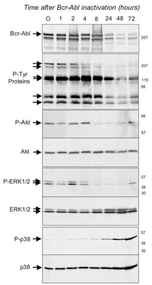
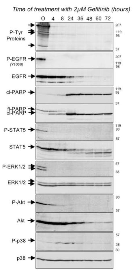
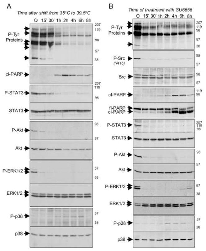
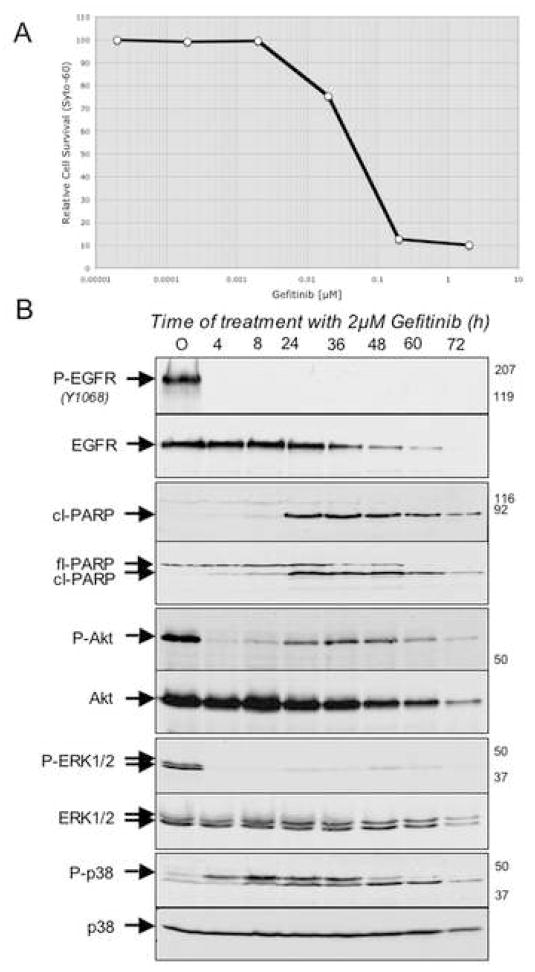
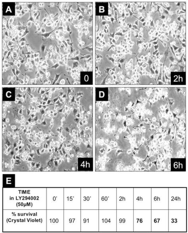
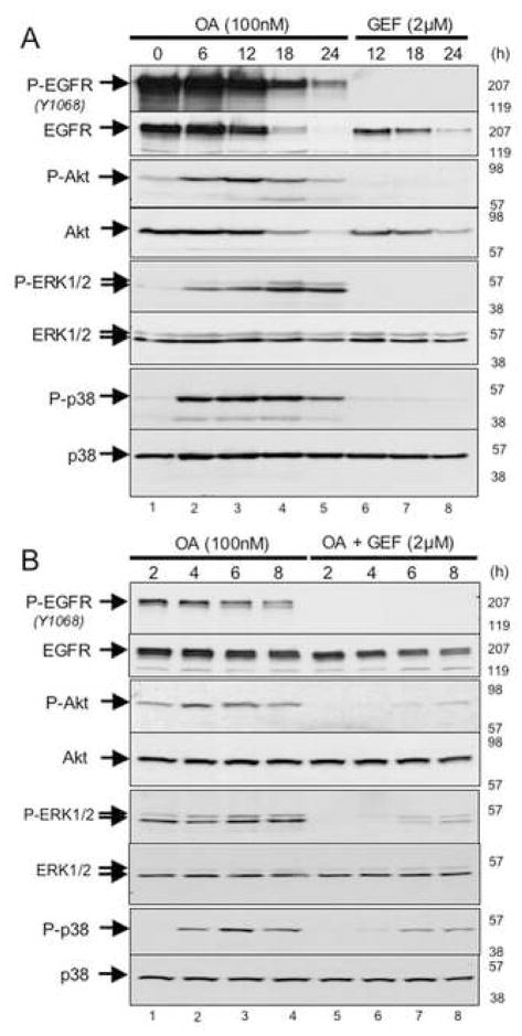
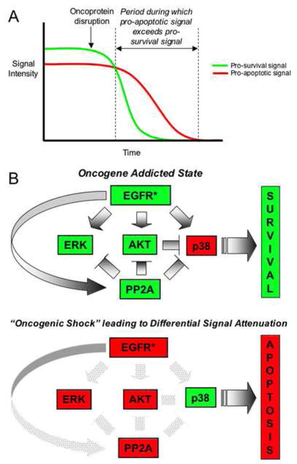
"Oncogene addiction" describes an unexplained dependency of cancer cells on a particular cellular pathway for survival or proliferation. We report that differential attenuation rates of prosurvival and proapoptotic signals in oncogene-dependent cells contribute to cell death following oncogene inactivation. Src-, BCR-ABL-, and EGF receptor-dependent cells exhibit a similar profile of signal attenuation following oncogene inactivation characterized by rapid diminution of phospho-ERK, -Akt, and -STAT3/5, and a delayed accumulation of the proapoptotic effector phospho-p38 MAPK. These findings implicate a transient imbalance in survival and apoptotic oncogenic outputs in the apoptotic response to oncogene inactivation. Moreover, these observations implicate a common profile of signal attenuation for multiple oncogenes and suggest that "addiction" associated with apoptosis reflects an active rather than a passive process.


Figures

Figure 1
Figure 1. Modulation of signaling following acute Bcr-Abl inactivation in TonB210.1 cells
Bcr-Abl was induced in TonB210.1 cells by doxacycline treatment for 18h. Bcr-Abl was then inactivated by the removal of doxacycline and at various times post-inactivation of Bcr-Abl cells were harvested and their proteins were analyzed by SDS-PAGE followed by Western blotting using antibodies directed against Bcr; phospho-tyrosine; phospho-Akt (P-Akt); Akt; phospho-ERK1/2 (P-ERK1/2); ERK1/2; phospho-p38 (P-p38); p38. Times post-inactivation of Bcr-Abl (in hours) are indicated at the top of each lane, the relative migration of the relevant proteins are indicated on the left-hand side of each autoradiogram and to the right of each autoradiogram are indicated the relative migration of molecular weight standards. In the second panel, arrows point to several unidentified proteins whose tyrosine phosphorylation appears to decrease upon Bcr-Abl inactivation.
Figure 2
Figure 2. Modulation of signaling following acute EGFR (L858R) inactivation in BaF3-EGFR(L858R) cells
BaF3-EGFR(L858R) cells constitutively express the L858R activating mutation in EGFR which renders the BaF3 cells independent of IL-3. EGFR (L858R) was inactivated by treating cells with 2μM of Gefitinib and at various times post-treatment cells were harvested and their proteins were analyzed by SDS-PAGE followed by Western blotting using antibodies directed against phospho-tyrosine (P-Tyr proteins); phospho-EGFRY1068 (P-EGFR); EGFR; cleaved PARP (cl-PARP); full-length PARP (fl-PARP); phospho-STAT-5 (P-STAT5); STAT-5; phospho-ERK1/2 (P-ERK1/2); ERK1/2; phospho-Akt (P-Akt); Akt; phospho-p38 (P-p38); p38. Times post-treatment with Gefitinib (in hours) are indicated at the top of each lane, the relative migration of the relevant proteins are indicated on the left-hand side of each autoradiogram and to the right of each autoradiogram are indicated the relative migration of molecular weight standards. In the top panel, arrows point to several unidentified proteins whose tyrosine phosphorylation appears to decrease upon v-src inactivation.
Figure 3
Figure 3. Morphological alterations induced by changes in serum and temperature in Balb-A1 ts-Src cells
A: Balb-A1 ts-Src cells growing in 10% serum containing medium at 35°C display the typical transformed morphology of Src-transformed cells. B: Overnight incubation at 39.5°C in the presence of 10% serum causes the cells to revert to a normal morphology. C: Shown at a slightly higher magnification are Balb-A1 ts Src cells growing over-night in 0.2% serum containing medium at 35°C. D: When cells shown in panel C were shifted to 39.5°C (in 0.2% serum) cellular apoptosis was evident (indicated by jagged arrows) within two hours after the temperature shift. E: Morphology of Balb-A1 ts-Src cells maintained continuously at 39.5°C in the presence of 10% serum, after 15 passages.
Figure 4
Figure 4. Modulation of signaling following acute Src inactivation in Balb-A1 ts-Src cells and Ψ2 v-Src 3T3 cells
A: The ts-Src oncoprotein was inactivated in serum-starved Balb-A1 ts-Src cells by shifting cells to 39.5°C. At various times post-inactivation of Src, cells were harvested and their proteins were analyzed by SDS-PAGE followed by Western blotting using antibodies directed against phospho-tyrosine (P-Tyr proteins); cleaved PARP (cl-PARP); phospho-STAT-3 (P-STAT3); STAT-3; phospho-Akt (P-Akt); Akt; phospho-ERK1/2 (P-ERK1/2); ERK1/2; phospho-p38 (P-p38); p38. Times post-inactivation of Src (in minutes and hours) are indicated at the top of each lane, the relative migration of the relevant proteins are indicated on the left-hand side of each autoradiogram and to the right of each autoradiogram are indicated the relative migration of molecular weight standards. B: V-src oncoprotein was inactivated in serum-starved Ψ2 v-Src 3T3 cells by the addition of 3μM of SU-6656, a src-specific inhibitor. At various times post-inactivation of Src, cells were harvested and their proteins were analyzed by SDS-PAGE followed by Western blotting using antibodies directed against phospho-tyrosine (P-Tyr proteins); activated phospho-src (P-Src); total src; cleaved PARP (cl-PARP); full-length PARP (fl-PARP); phospho-STAT-3 (P-STAT3); STAT-3; phospho-Akt (P-Akt); Akt; phospho-ERK1/2 (P-ERK1/2); ERK1/2; phospho-p38 (P-p38); p38. Times post-inactivation of Src (in minutes and hours) are indicated at the top of each lane, the relative migration of the relevant proteins are indicated on the left-hand side of each autoradiogram and to the right of each autoradiogram are indicated the relative migration of molecular weight standards. In the top panel, arrows point to several unidentified proteins whose tyrosine phosphorylation appears to decrease upon v-src inactivation.
Figure 5
Figure 5. Modulation of signaling following acute EGFR inactivation in PC-9 cells
A: A dose-response curve for cell killing by Gefitinib in sub-confluent PC-9 cells following 72 of treatment with the inhibitor. B: Activated EGFR in PC-9 cells was inactivated by the addition of 2μM of Gefitinib. At various times post-EGFR inactivation, cells were harvested and protein lysates were analyzed by SDS-PAGE followed by Western blotting using antibodies directed against phospho- EGFRY1068 (P-EGFR); total EGFR; cleaved-PARP (cl-PARP); full-length PARP (fl-PARP); phospho-Akt (P-Akt); Akt; phospho-ERK1/2 (P-ERK1/2); ERK1/2; phospho-p38 (P-p38); p38. Times post-inactivation of EGFR (in hours) are indicated at the top of each lane, the relative migration of the relevant proteins are indicated on the left-hand side of each autoradiogram and to the right of each autoradiogram are indicated the relative migration of molecular weight standards.
Figure 6
Figure 6. Effect of LY282004 on Balb-A1 ts-Src cells at 35°C in 0.2% serum
A–D: Serum-starved, Balb-A1 ts-Src cells were treated with 3μM LY294002 and at various times post-treatment (0, 2h, 4h, 6h) cell morphology was documented by photomicrography. E: Cell survival was quantitated by crystal violet staining and tabulated.
Figure 7
Figure 7. Effect of okadaic acid and gefitinib either singly or in combination on modulation of signaling in BaF3-EGFR (l858R) cells
BaF3-EGFR(L858R) cells were treated with either 100nM okadaic acid (OA) alone (lanes 1–5 in panel A; lanes 1–4 in panel B), gefitinib (GEF) alone (lanes 6–8 in panel A) or 100nM okadaic Acid together with 2μM gefitinib (lanes 5–8 in panel B). At various periods of time post-treatment (as indicated at the top of each lane) cells were harvested and their proteins were analyzed by SDS-PAGE followed by Western blotting using antibodies directed against phospho-EGFRY1068 (P-EGFR); EGFR; phospho-Akt (P-Akt); Akt; phospho-ERK1/2 (P-ERK1/2); ERK1/2; phospho-p38 (P-p38); p38. The relative migration of the relevant proteins are indicated on the left-hand side of each autoradiogram and to the right of each autoradiogram are indicated the relative migration of molecular weight standards.
Figure 8
Figure 8. Proposed model of oncogene addiction
A: A graphical depiction of the proposed role of differential signal attenuation in creating a temporal window during which apoptotic signals persist in the absence of pro-survival signals following acute oncogene inactivation to promote cell death. B: This model depicts the proposed role of differential signal attenuation in the cell death response to acute inactivation of oncogenic EGFR (EGFR*) upon which a tumor cell has become dependent. The upper panel illustrates the oncogene “addicted” state in which cells survive, and the lower panel illustrates the shift in the balance of pro-survival and pro-apoptotic signaling shortly following EGFR inactivation. Red=“off”; Green=“on”.

Comment in

Similar articles

Cited by

References

    1. Aoki K, Yoshida T, Matsumoto N, Ide H, Sugimura T, Terada M. Suppression of Ki-ras p21 levels leading to growth inhibition of pancreatic cancer cell lines with Ki-ras mutation but not those without Ki-ras mutation. Mol Carcinog. 1997;20:251–258. - PubMed
    1. Arber N. Janus faces of ras: anti or pro-apoptotic? Apoptosis. 1999;4:383–388. - PubMed
    1. Bandyopadhyay G, Biswas T, Roy KC, Mandal S, Mandal C, Pal BC, Bhattacharya S, Rakshit S, Bhattacharya DK, Chaudhuri U, et al. Chlorogenic acid inhibits Bcr-Abl tyrosine kinase and triggers p38 mitogen-activated protein kinase-dependent apoptosis in chronic myelogenous leukemic cells. Blood. 2004;104:2514–2522. - PubMed
    1. Blake RA, Broome MA, Liu X, Wu J, Gishizky M, Sun L, Courtneidge SA. SU6656, a selective src family kinase inhibitor, used to probe growth factor signaling. Mol Cell Biol. 2000;20:9018–9027. - PMC - PubMed
    1. Brunet CL, Gunby RH, Benson RS, Hickman JA, Watson AJ, Brady G. Commitment to cell death measured by loss of clonogenicity is separable from the appearance of apoptotic markers. Cell Death Differ. 1998;5:107–115. - PubMed

Publication types