. 2006 Dec 29;351(4):958-64.
doi: 10.1016/j.bbrc.2006.10.153. Epub 2006 Nov 7.

Opioid receptor-independent protection of ischemic rat hepatocytes by morphine

Affiliations

Opioid receptor-independent protection of ischemic rat hepatocytes by morphine

Jae-Sung Kim et al. Biochem Biophys Res Commun. .

Abstract

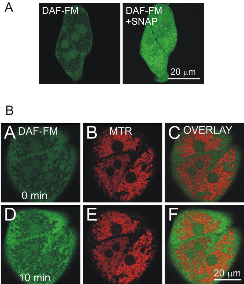
We studied the role of morphine in anoxia/reoxygenation injury to hepatocytes. Overnight cultured rat hepatocytes were incubated in anoxic buffer at pH 6.2 for 4h and reoxygenated at pH 7.4 for 2h to simulate anoxia/reoxygenation. Some hepatocytes were preincubated with 50 microM morphine for 10 min prior to onset of anoxia/reoxygenation. To study the effect of morphine on nitric oxide (NO), hepatocytes were loaded with 4-amino-5-methylamino-2',7'-difluorofluorescein (DAF-FM). Changes in NO concentration were assessed with a multi-well fluorescence reader and confocal microscopy. Morphine substantially improved cell viability after reoxygenation and increased NO generation, which was blocked by ATP-sensitive potassium channel blockers. Confocal images revealed that the increase in NO occurred mainly at the cytosol. However, treatment with opioid receptor antagonists did not reverse cytoprotection by morphine. These results indicate that morphine prevents anoxia/reoxygenation injury to hepatocytes. Protective mechanisms are associated with the potassium channels and NO, but are independent of opioid receptor-mediated signaling.


Figures

Fig. 1
Fig. 1. Protection by morphine against anoxia/reoxygenation injury to hepatocytes
Overnight cultured hepatocytes were incubated in anoxic KRH buffer at pH 6.2 for 4 h, and reoxygenated in aerobic KRH buffer at pH 7.4 at 37ºC for 2 h to simulate ischemia/reperfusion. (A) Some hepatocytes were treated with 50 μM morphine for 10 min prior to anoxia. Cell viability was fluorometrically assessed by measuring propidium iodide uptake. * P < 0.05 vs. Control. (B) Hepatocytes were preincubated for 20 min in KRH containing either 0.1 μM BNTX, a selective opioid δ1 receptor antagonist, or 10 μM naloxone, a nonselective opioid receptor antagonist. The cells were then incubated with morphine for another 10 min. After 4 h of anoxia, cell viability was measured over the time of reoxygenation. Values are mean ± SE from 8 separate cell isolations.
Fig. 2
Fig. 2. Role of NO in morphine-induced cytoprotection
(A) Hepatocytes were preincubated in KRH buffer (pH 7.4) containing 100 μM carboxy-PTIO, an NO chelator, for 20 min prior to the addition of morphine. Following 10 min with morphine, the cells were subjected to 4 h of anoxia and 2 h of reoxygenation. Some hepatocytes were incubated with carboxy-PTIO alone. Cell viability was measured with the time of reoxygenation. Values are mean ± SE from 8 separate cell isolations. * P < 0.05 vs. Control. (B) To measure intracellular NO, hepatocytes were incubated in KRH buffer (pH 7.4) containing 10 μM DAF-FM diacetate at 37ºC for 1 h and further incubated with morphine for 10 min. Some hepatocytes were pretreated with carboxy-PTIO for 20 min. NO was fluorometrically quantified using a multi-well fluorescence scanner. Fluorescence at 10 min after morphine treatment was measured and the background fluorescence was subtracted. Values are mean ± SE from 8 separate cell isolations. * P < 0.05 vs. no addition (Normoxia).
Fig. 3
Fig. 3. Confocal microscopy with DAF-FM
Hepatocytes were loaded with DAF-FM, as described in Materials and methods. The green fluorescence of DAF-FM was imaged by laser scanning confocal microscopy. (A) Representative image at the end of DAF-FM loading. Note that DAF-FM fluorescence was evident in the cytosol and nuclei, leaving the mitochondria as dark voids (left panel). Hepatocytes were further incubated with 100 μM SNAP, an NO donor, for 1–2 min to visualize dye distribution inside the cells (right panel). Note that all compartments became fluorescent with SNAP, indicating that DAF-FM was localized in all compartments. (B) Hepatocytes on glass coverslips were co-loaded with 10 μM DAF-FM and 300 nM Mitotracker Red (MTR). Simultaneous images of green DAF-FM (left panels), red MTR (middle panels), and overlay (right panels) fluorescence were collected by confocal microscopy. Images were collected before (A, B, and C) and after 50 μM morphine addition (D, E, and F). Note that morphine substantially increased cytosolic NO.
Fig. 4
Fig. 4. Effects of the KATP channel blockers on morphine-induced cytoprotection and NO increase
(A) Hepatocytes were preincubated for 20 min in KRH buffer containing 200 μM 5-HD or 50 μM glibenclamide, the KATP channel blockers. The cells were then treated with morphine for 10 min and subjected to simulated anoxia/reoxygenation, as described in Fig. 1. The KATP channel blockers abolished the protection by morphine. Values are mean ± SE. * P < 0.05 vs. morphine. (B) Some hepatocytes were treated with 5-HD or glibenclamide (GC) alone and exposed to simulated anoxia/reoxygenation, showing that the KATP channel blockers alone did not affect cell viability. (C) NO was fluorometrically measured by DAF-FM. *P<0.05 vs. Control.

References

    1. Way EL. Review and overview of four decades of opiate research. Adv Biochem Psychopharmacol. 1979;20:3–27. - PubMed
    1. MacPherson RD. The pharmacological basis of contemporary pain management. Pharmacol Ther. 2000;88:163–185. - PubMed
    1. McPherson BC, Yao Z. Morphine Mimics Preconditioning via Free Radical Signals and Mitochondrial KATP Channels in Myocytes. Circulation. 2001;103:290–295. - PubMed
    1. Schultz JE, Rose E, Yao Z, Gross GJ. Evidence for involvement of opioid receptors in ischemic preconditioning in rat hearts. Am J Physiol. 1995;268:H2157–H2161. - PubMed
    1. Fryer RM, Hsu AK, Gross GJ. Mitochondrial KATP channel opening is important during index ischemia and following myocardial reperfusion in ischemic preconditioned rat hearts. J Mol Cell Cardiol. 2001;33:831–834. - PubMed

LinkOut - more resources