. 2007 Feb;203(2):531-41.
doi: 10.1016/j.expneurol.2006.09.009. Epub 2006 Nov 9.

Parkin is an E3 ubiquitin-ligase for normal and mutant ataxin-2 and prevents ataxin-2-induced cell death

Affiliations

Parkin is an E3 ubiquitin-ligase for normal and mutant ataxin-2 and prevents ataxin-2-induced cell death

Duong P Huynh et al. Exp Neurol. 2007 Feb.

Abstract

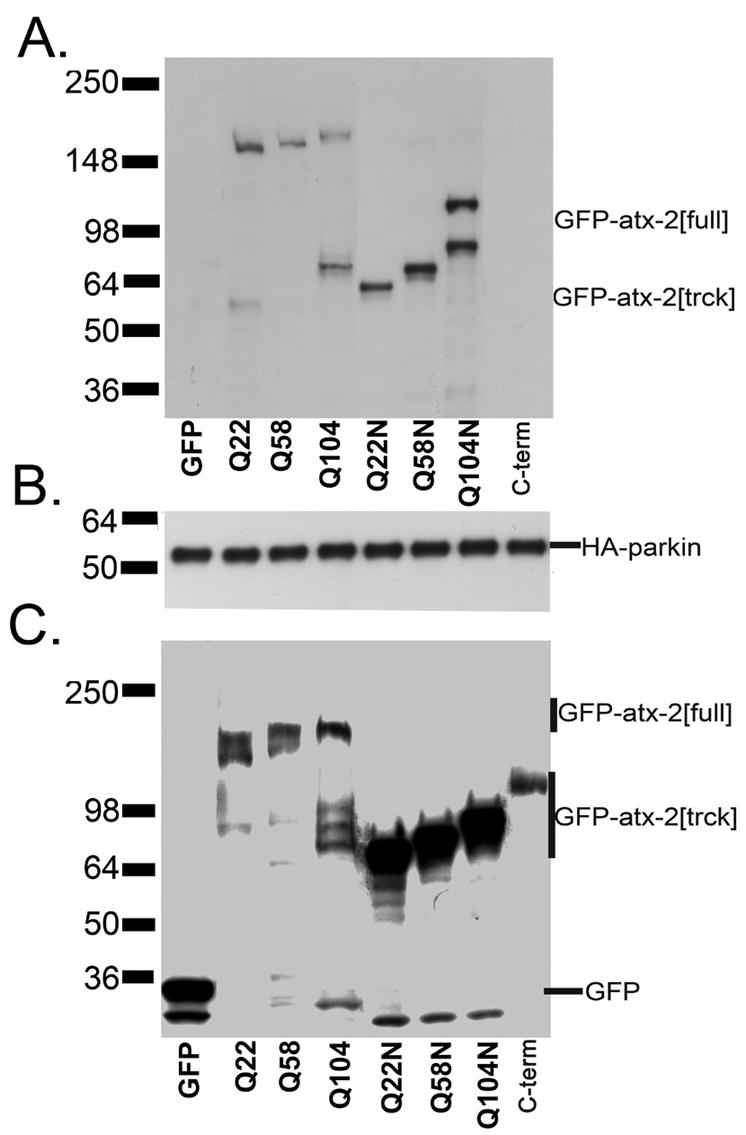
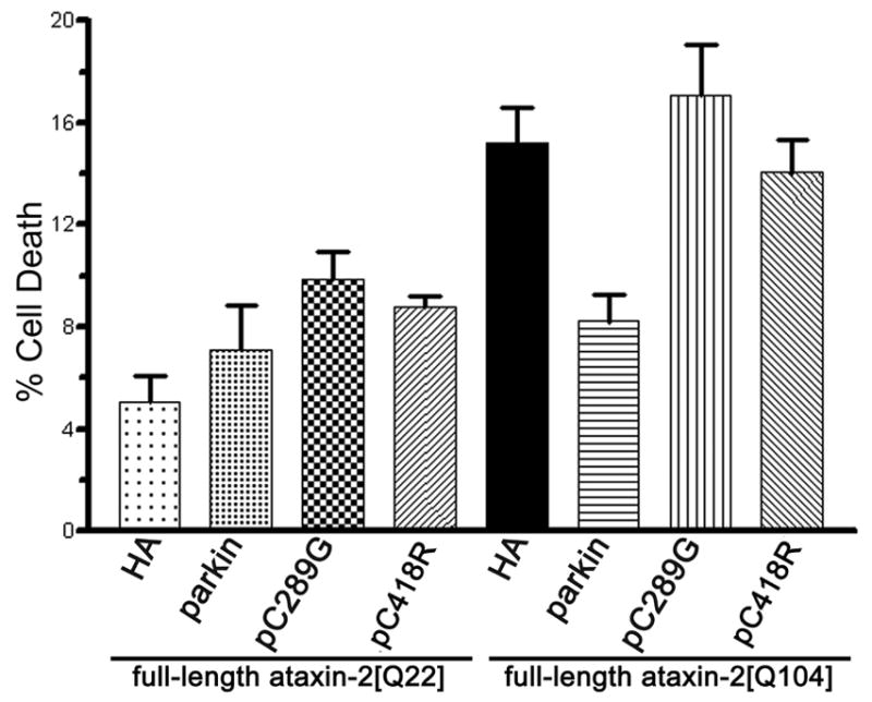
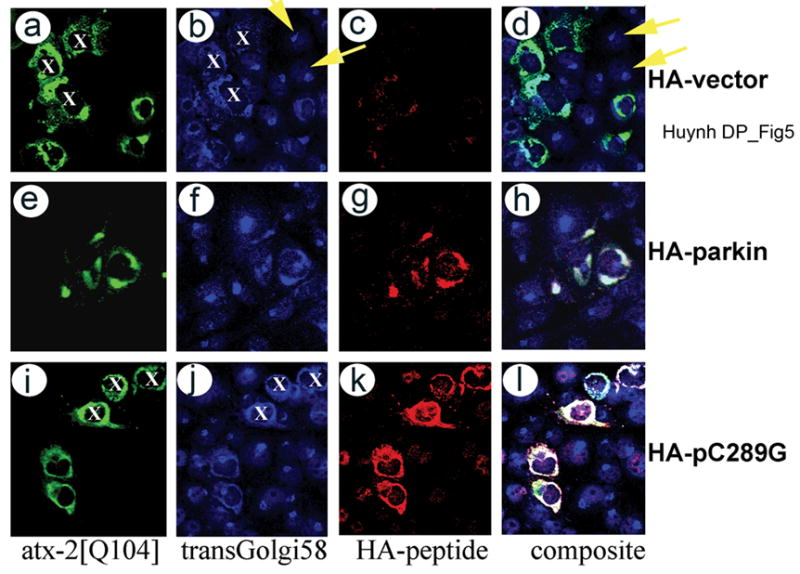
Expansion of the polyQ repeat in ataxin-2 results in degeneration of Purkinje neurons and other neuronal groups including the substantia nigra in patients with spinocerebellar ataxia type 2 (SCA2). In animal and cell models, overexpression of mutant ataxin-2 induces cell dysfunction and death, but little is known about steady-state levels of normal and mutant ataxin-2 and cellular mechanisms regulating their abundance. Based on preliminary findings that ataxin-2 interacted with parkin, an E3 ubiquitin ligase mutated in an autosomal recessive form of Parkinsonism, we sought to determine whether parkin played a role in regulating the steady-state levels of ataxin-2. Parkin interacted with the N-terminal half of normal and mutant ataxin-2, and ubiquitinated the full-length form of both wild-type and mutant ataxin-2. Parkin also regulated the steady-state levels of endogenous ataxin-2 in PC12 cells with regulatable parkin expression. Parkin reduced abnormalities in Golgi morphology induced by mutant ataxin-2 and decreased ataxin-2 induced cytotoxicity. In brains of SCA2 patients, parkin labeled cytoplasmic ataxin-2 aggregates in Purkinje neurons. These studies suggest a role for parkin in regulating the intracellular levels of both wild-type and mutant ataxin-2, and in rescuing cells from ataxin-2-induced cytotoxicity. The role of parkin variants in modifying the SCA2 phenotype and its use as a therapeutic target should be further investigated.


Figures

Figure 1
Figure 1
Both full-length and truncated ataxin-2 interact with parkin (panel A). HEK293 cells were co-transfected with wildtype parkin and GFP-tagged full-length (atx-2[full], N-terminal (N-term), or C-terminal (C-term) fragments of ataxin-2. For the full-length and N-terminal fragments constructs with different polyQ repeats (Q22, Q58, or Q104) were tested. The GFP vector and the GFP-tagged C-terminal domain of ataxin-2 were used as negative controls. Protein extracts were ip with anti-HA antibody conjugated to agarose, and detected with either anti-GFP (A) or anti-HA-peroxidase (B) antibodies. To determine the levels of transfected proteins, western blot of transfected HEK293 cells lysates were detected with anti-GFP (C). Panels D and E show the schematic structure of GFP tagged ataxin-2 (D) and HA-tagged parkin (E) proteins. Panel F demonstrates the colocalization of HA-tagged parkin to GFP tagged full-length and truncated ataxin-2 in COS1 cells. Cells were co-transfected with expression plasmids expressing HA-parkin and GFP-tagged full-length or truncated ataxin-2. Cells were stained with rabbit anti-HA antibody and detected with donkey Rhodamine-RED conjugated anti-rabbit IgG. Images were visualized and acquired by Leica Confocal Laser Microscopy. Bar represents 8 μm.
Figure 1
Figure 1
Both full-length and truncated ataxin-2 interact with parkin (panel A). HEK293 cells were co-transfected with wildtype parkin and GFP-tagged full-length (atx-2[full], N-terminal (N-term), or C-terminal (C-term) fragments of ataxin-2. For the full-length and N-terminal fragments constructs with different polyQ repeats (Q22, Q58, or Q104) were tested. The GFP vector and the GFP-tagged C-terminal domain of ataxin-2 were used as negative controls. Protein extracts were ip with anti-HA antibody conjugated to agarose, and detected with either anti-GFP (A) or anti-HA-peroxidase (B) antibodies. To determine the levels of transfected proteins, western blot of transfected HEK293 cells lysates were detected with anti-GFP (C). Panels D and E show the schematic structure of GFP tagged ataxin-2 (D) and HA-tagged parkin (E) proteins. Panel F demonstrates the colocalization of HA-tagged parkin to GFP tagged full-length and truncated ataxin-2 in COS1 cells. Cells were co-transfected with expression plasmids expressing HA-parkin and GFP-tagged full-length or truncated ataxin-2. Cells were stained with rabbit anti-HA antibody and detected with donkey Rhodamine-RED conjugated anti-rabbit IgG. Images were visualized and acquired by Leica Confocal Laser Microscopy. Bar represents 8 μm.
Figure 2
Figure 2
Parkin ubiquitinates full-length ataxin-2. Ubiquitylation of ataxin-2N-term (Q22N and Q104N) and ataxin-2C-term(C-term) is much less pronounced. HEK293 cells overexpressing HA-parkins or controls with the corresponding myc-ubiquitin and GFP-tagged ataxin-2 were treated with lactacystin for 4 hours, and protein extracts were immunoprecipitated with anti-GFP antibody. IP products of the ubiquitination assays were detected with an antibody to the myc tag (A) and anti-GFP antibody (B). Note the lack of ubiquitinated products in cells expressing HA-parkin and GFP, GFP tagged truncated ataxin-2. Cells expressing parkin mutants and GFP- ataxin-2 produce a lower amount of ubiquitin-conjugated ataxin-2 compared to the wild-type (wt) parkin. The anti-GFP antibody detects near equal amounts of GFP-ataxin-2 monomer (B) in all samples containing GFP-ataxin-2. Controls using GFP and GFP-ataxin-2[Q22] with and without parkin did not show any ubiquitination species. Bottom panel (C) shows western blot analysis of pellets of the protein extracts detected with anti-GFP antibody. Large amounts of high molecular weight species were detected in protein extracts from cells coexpressing both wild parkin and full-length ataxin-2 compared with full-length ataxin-2 alone or full-length ataxin-2 and missense mutated parkin. Although significant amounts of C-terminal truncated ataxin-2 remain in the pellet, no high molecular weight species were detectable.
Figure 3
Figure 3
a) Exogenously expressed wildtype human parkin (hparkin) facilitates the turn over of endogenous wildtype rat ataxin-2. Tet-off hparkin PC12 cells were grown in the presence of different concentrations of Dox for 72 hours. Protein extracts were obtained and proteins were detected with the rabbit anti-ataxin-2 antibody (panel a, top) or the anti-parkin antibody (panel a, bottom). Increased expression of human parkin resulted in decreased abundance of endogenous wildtype ataxin-2. b) Parkin deficiency causes an increase in the level of endogenous ataxin-2 in exon parkin ko mice. Protein extracts from wt and parkin ko mice were detected with anti-ataxin-2 (2 μg/ml SCA2B antibody), anti-parkin (mouse antibody to the C-terminal domain of parkin, 1/500 dil), and antibody to β-actin (1/5000 mouse monoclonal antibody to β-actin). The parkin antibody detects parkin in the wt but not in the ko mice, while ataxin-2 antibody detects a higher level of endogenous ataxin-2 in the ko than the wild type mice. c) Histogram analyses of 3 wt and 3 parkin ko mice using the Image J program. Parkin gene ko causes a 22% increase in endogenous ataxin-2 level (P < 0.007, n =3, unpaired t-test).
Figure 4
Figure 4
Wildtype but not missense mutated parkin inhibits cell death induced by mutant GFP-ataxin-2[Q104]. Cells were co-transfected with wildtype GFP-ataxin-2[Q22] or ataxin-2[Q104] and HA-vector control (vector), HA-parkin (parkin), mutant parkinC289G (pC289G) or parkinC418R(pC418R). Expression of full-length mutant ataxin-2 in HEK293 cells at 48 hours increased the number of cells that were unable to exclude trypan blue. Co-expression with wildtype parkin suppressed toxicity of mutant ataxin-2, while co-expression with mutant parkinC289G and parkinC418R showed no cytoprotective effect. Slight enhancement of cell death was observed for parkinC289G, n=4.
Figure 5
Figure 5
Parkin reduces Golgi dispersion induced by expanded polyQ repeat ataxin-2[Q104]. HEK293 cells were transiently transfected with pGFP-SCA2[Q104] (a, e, i) and labeled with a monoclonal antibody to transGolgi58K (b, f, j), or rabbit anti-HA antibody (c, g, k). Primary antibodies were detected with anti-rabbit Alexa568 (c, g, k) or anti-mouse Alexa660 (b, f, j). As previously described (Huynh et al, 2003b), GFP-ataxin-2[Q104] disrupts the morphology of the Golgi apparatus (panels a–d); co-expression with wildtype parkin (panels e–h) reduces Golgi abnormalities, while missense parkin C289G (panels i–l) shows no effect. Note that co-expression of parkin with mutant ataxin-2 restores the co-localization of ataxin-2 with Golgi markers. Parkin largely colocalizes with the Golgi markers resulting in white color in the composite. Images were acquired using the 40X oil immersion objective of the Leica TCSSP microscope. Yellow arrows indicate untransfected cells with normal Golgi58K labeling. White X labels show transfected cells with Golgi dispersion.
Figure 6
Figure 6
Expression of ataxin-2 and parkin in Purkinje neurons. Cerebella from normal controls (A) and SCA2 patients (B) were labeled with chicken anti-parkin (green, rows a–c of panels A & B) and rabbit anti-ataxin-2 (red) antibodies. Primary antibodies were visualized with FITC-conjugated anti-chick IgY and Rhodamine-RED conjugated anti-rabbit IgG and imaged by confocal laser microscopy. Each image represents an individual 0.5 μm thick section. The composite represents the overlay of individual images corresponding to parkin and ataxin-2 labeling. Light blue arrow in panel A shows parkin and ataxin-2 colocalization in normal Purkinje neurons. Pink and Blue arrows in panel B show cytoplasmic aggregates and a perinuclear aggregate which invaginates into the nucleus. Most ataxin-2 aggregates also label with parkin antibodies. Note the lack of parkin staining in Purkinje cell cytoplasm and Purkinje cell processes, but strong staining in fibers likely representing climbing fibers winding around the Purkinje cell arbor (Yellow arrow). Note also the colocalization of parkin and ataxin-2 in small cells near the Purkinje neuron likely representing Golgi cells (White arrow in panels A and B). Bars represent 40 μm.
Figure 6
Figure 6
Expression of ataxin-2 and parkin in Purkinje neurons. Cerebella from normal controls (A) and SCA2 patients (B) were labeled with chicken anti-parkin (green, rows a–c of panels A & B) and rabbit anti-ataxin-2 (red) antibodies. Primary antibodies were visualized with FITC-conjugated anti-chick IgY and Rhodamine-RED conjugated anti-rabbit IgG and imaged by confocal laser microscopy. Each image represents an individual 0.5 μm thick section. The composite represents the overlay of individual images corresponding to parkin and ataxin-2 labeling. Light blue arrow in panel A shows parkin and ataxin-2 colocalization in normal Purkinje neurons. Pink and Blue arrows in panel B show cytoplasmic aggregates and a perinuclear aggregate which invaginates into the nucleus. Most ataxin-2 aggregates also label with parkin antibodies. Note the lack of parkin staining in Purkinje cell cytoplasm and Purkinje cell processes, but strong staining in fibers likely representing climbing fibers winding around the Purkinje cell arbor (Yellow arrow). Note also the colocalization of parkin and ataxin-2 in small cells near the Purkinje neuron likely representing Golgi cells (White arrow in panels A and B). Bars represent 40 μm.

Similar articles

Cited by

References

    1. Abbas N, Lucking CB, Ricard S, Durr A, Bonifati V, De Michele G, Bouley S, Vaughan JR, Gasser T, Marconi R, Broussolle E, Brefel-Courbon C, Harhangi BS, Oostra BA, Fabrizio E, Bohme GA, Pradier L, Wood NW, Filla A, Meco G, Denefle P, Agid Y, Brice A. A wide variety of mutations in the parkin gene are responsible for autosomal recessive parkinsonism in Europe. French Parkinson's Disease Genetics Study Group and the European Consortium on Genetic Susceptibility in Parkinson's Disease. Hum Mol Genet. 1999;8:567–574. - PubMed
    1. Armstrong J, Bonaventura I, Rojo A, Gonzalez G, Corral J, Nadal N, Volpini V, Ferrer I. Spinocerebellar ataxia type 2 (SCA2) with white matter involvement. Neurosci Lett. 2005;381:247–251. - PubMed
    1. Bingol B, Schuman EM. Activity-dependent dynamics and sequestration of proteasomes in dendritic spines. Nature. 2006;441:1144–1148. - PubMed
    1. Chung KK, Zhang Y, Lim KL, Tanaka Y, Huang H, Gao J, Ross CA, Dawson VL, Dawson TM. Parkin ubiquitinates the alpha-synuclein-interacting protein, synphilin-1: implications for Lewy-body formation in Parkinson disease. Nat Med. 2001;7:1144–1150. - PubMed
    1. Corti O, Hampe C, Koutnikova H, Darios F, Jacquier S, Prigent A, Robinson JC, Pradier L, Ruberg M, Mirande M, Hirsch E, Rooney T, Fournier A, Brice A. The p38 subunit of the aminoacyl-tRNA synthetase complex is a Parkin substrate: linking protein biosynthesis and neurodegeneration. Hum Mol Genet. 2003;12:1427–1437. - PubMed

Publication types