. 2007 Jan 19;365(3):812-24.
doi: 10.1016/j.jmb.2006.10.073. Epub 2006 Oct 26.

Conformation of the HIV-1 Gag protein in solution

Affiliations

Conformation of the HIV-1 Gag protein in solution

Siddhartha A K Datta et al. J Mol Biol. .

Abstract

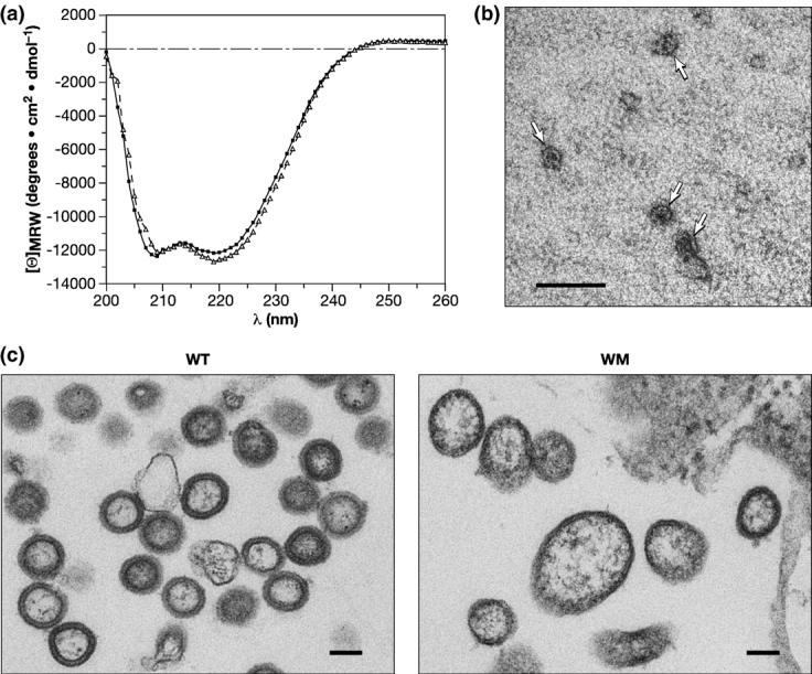
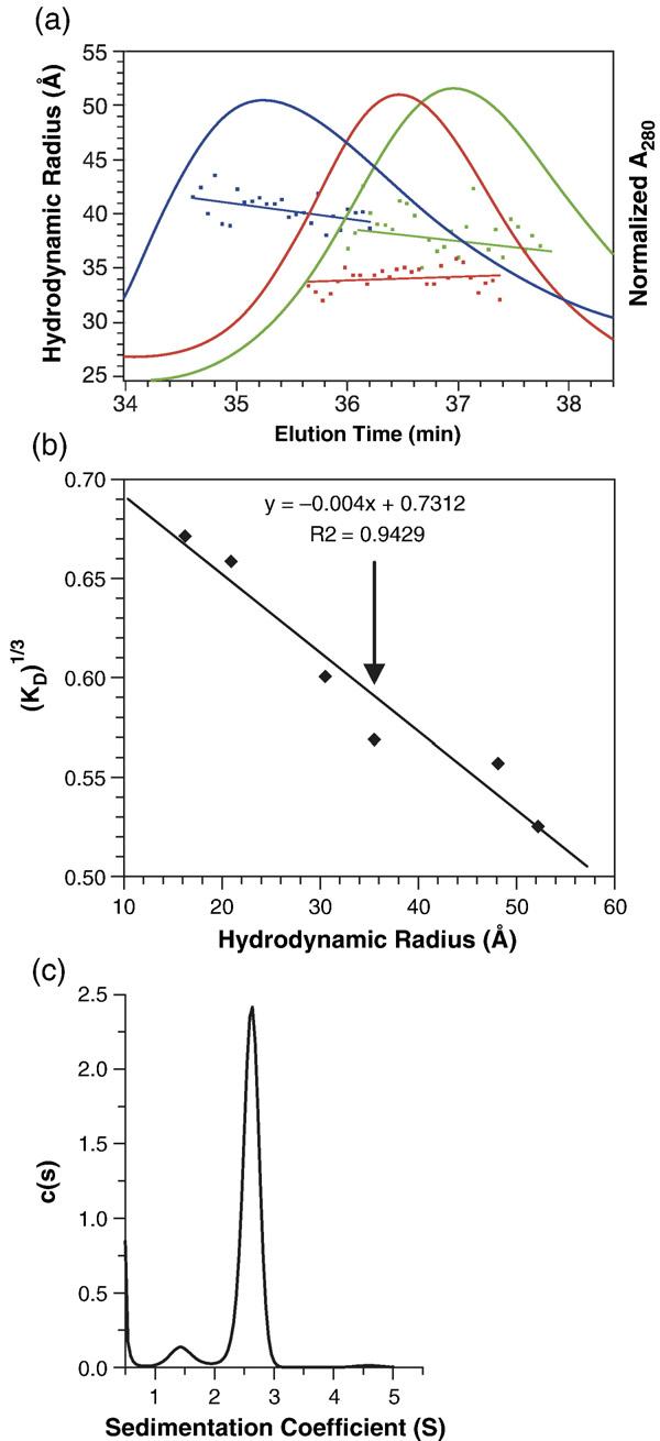
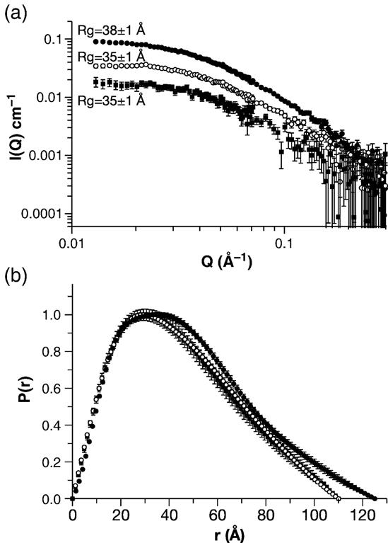
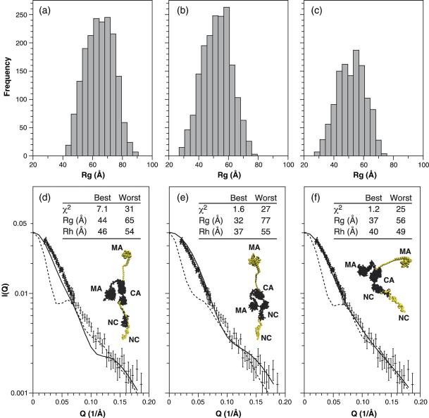
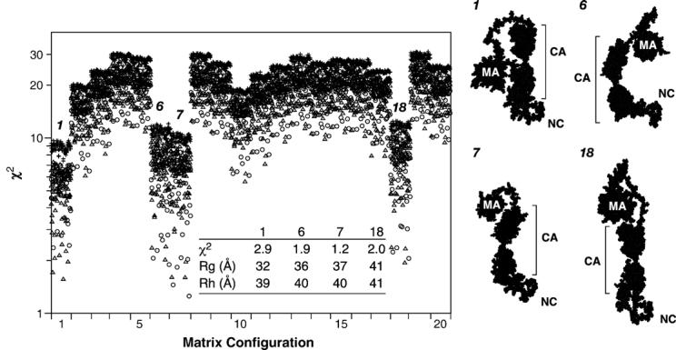
A single multi-domain viral protein, termed Gag, is sufficient for assembly of retrovirus-like particles in mammalian cells. We have purified the human immunodeficiency virus type 1 (HIV-1) Gag protein (lacking myristate at its N terminus and the p6 domain at its C terminus) from bacteria. This protein is capable of assembly into virus-like particles in a defined in vitro system. We have reported that it is in monomer-dimer equilibrium in solution, and have described a mutant Gag protein that remains monomeric at high concentrations in solution. We report that the mutant protein retains several properties of wild-type Gag. This mutant enabled us to analyze solutions of monomeric protein. Hydrodynamic studies on the mutant protein showed that it is highly asymmetric, with a frictional ratio of 1.66. Small-angle neutron scattering (SANS) experiments confirmed its asymmetry and yielded an R(g) value of 34 A. Atomic-level structures of individual domains within Gag have previously been determined, but these domains are connected in Gag by flexible linkers. We constructed a series of models of the mutant Gag protein based on these domain structures, and tested each model computationally for its agreement with the experimental hydrodynamic and SANS data. The only models consistent with the data were those in which Gag was folded over, with its N-terminal matrix domain near its C-terminal nucleocapsid domain in three-dimensional space. Since Gag is a rod-shaped molecule in the assembled immature virion, these findings imply that Gag undergoes a major conformational change upon virus assembly.


Figures

Figure 1
Figure 1
Properties of the WM Gag protein. (a) CD profile of wild-type (circles) and WM Gag (triangles). (b) VLPs assembled in vitro from WM Gag. WM Gag was mixed with tRNA, diluted, and analyzed by negative staining as described in Materials and Methods. VLPs are indicated by arrows. (c) Particles formed by wild-type and WM Gag proteins in mammalian cells. Particles associated with pelleted cells were analyzed by electron microscopy as described in Materials and Methods. The scale bars represent 100 nm.
Figure 2
Figure 2
Hydrodynamic properties of the WM Gag protein. (a) WM Gag (green profile) was injected onto a Superose 12 column at 3 mg/ml, and the eluate was monitored by A280 (green elution profile) and simultaneously analyzed by QELS. The data points under the elution profiles show the Rh values obtained from QELS at each position in the elution and the lines are weighted fits through the points. Also shown are the elution profiles of wild-type Gag, loaded at 3 mg/ml (blue profile), and of BSA (5 mg/ml) (red profile). (b) The Rh value of WM Gag determined from its elution time in (a). The column was calibrated with a series of proteins of known Rh value, as described in Materials and Methods. The graph shows the cube-root of the column distribution coefficient KD plotted against the Rh value of the protein. The vertical arrow indicates the position of WM Gag on the curve. (c) Analysis of WM Gag by SV. WM Gag (0.64 mg/ml) was centrifuged at 58,000 rpm, and the boundary movement was monitored by A280. The graph shows the c(s) versus s plot from the Sednterp software program as described in Materials and Methods.
Figure 3
Figure 3
SANS analysis of the WM Gag protein. WM Gag solutions at 0.5, 1.0, and 2.0 mg/ml were analyzed by SANS as described in Materials and Methods. (a) Guinier plot. (b) Distance distribution function. Filled squares, 0.5 mg/ml; open circles, 1.0 mg/ml; filled circles, 2.0 mg/ml.
Figure 4
Figure 4
Evaluation of model structures for the WM Gag protein. (a)–(c) Rg frequency distributions for model structures with (a) extended random coil p2 structures; (b) helical p2 structures; and (c) partially helical p2 structures. (d)–(f) Detailed evaluation of the structures in each group exhibiting the best and worst fits to the experimental data. The main panel shows the calculated Guinier plot for the best (continuous line) and worst (broken line) in each group, compared with the Guinier plot obtained experimentally at 1 mg/ml. (d) Extended random coil p2 structures. (e) Helical p2 structures. (f) Partially helical p2 structures. Also shown in (d)–(f) are images of the best (black) and worst (colored) structures. The inset table shows the χ2 values for the Guinier plot and the calculated Rg and Rh values for the best and worst structures.
Figure 5
Figure 5
Evaluation of 4800 model structures of the WM Gag protein. For each model structure, the χ2 value for consistency with the SANS data was calculated. The model structures are divided into 20 groups according to the configuration of the MA domain. Structures with extended p2 regions are indicated by plus signs, those with helical p2 by triangles, and those with partially helical p2 by circles. Four groups, arbitrarily numbered 1, 6, 7, and 18, gave the lowest χ2 values. The inset table shows these values, along with the calculated Rg and Rh values, for the best structure in each of these four groups. These structures are depicted on the right side of the Figure. The approximate “vertical” dimensions of these structures are: group 1, 100 Å; groups 2 and 3, 110 Å; group 4, 130 Å.

References

    1. Swanstrom R, Wills JW. Synthesis, assembly, and processing of viral proteins. In: Coffin JM, Hughes SH, Varmus HE, editors. Retro-viruses. Cold Spring Harbor Laboratory Press; Plainview, NY: 1997. pp. 263–334. - PubMed
    1. Campbell S, Vogt VM. Self-assembly in vitro of purified CA-NC proteins from Rous sarcoma virus and human immunodeficiency virus type 1. J. Virol. 1995;69:6487–6497. - PMC - PubMed
    1. Campbell S, Rein A. In vitro assembly properties of human immunodeficiency virus type 1 Gag protein lacking the p6 domain. J. Virol. 1999;73:2270–2279. - PMC - PubMed
    1. Yu F, Joshi SM, Ma YM, Kingston RL, Simon MN, Vogt VM. Characterization of Rous sarcoma virus Gag particles assembled in vitro. J. Virol. 2001;75:2753–2764. - PMC - PubMed
    1. Campbell S, Fisher RJ, Towler EM, Fox S, Issaq HJ, Wolfe T, et al. Modulation of HIV-like particle assembly in vitro by inositol phosphates. Proc. Natl Acad. Sci. USA. 2001;98:10875–10879. - PMC - PubMed

Publication types