

Morphology, localization and accumulation of in vivo microdamage in human cortical bone
- PMID: 17097933
- PMCID: PMC2013741
- DOI: 10.1016/j.bone.2006.09.027
Morphology, localization and accumulation of in vivo microdamage in human cortical bone
Abstract
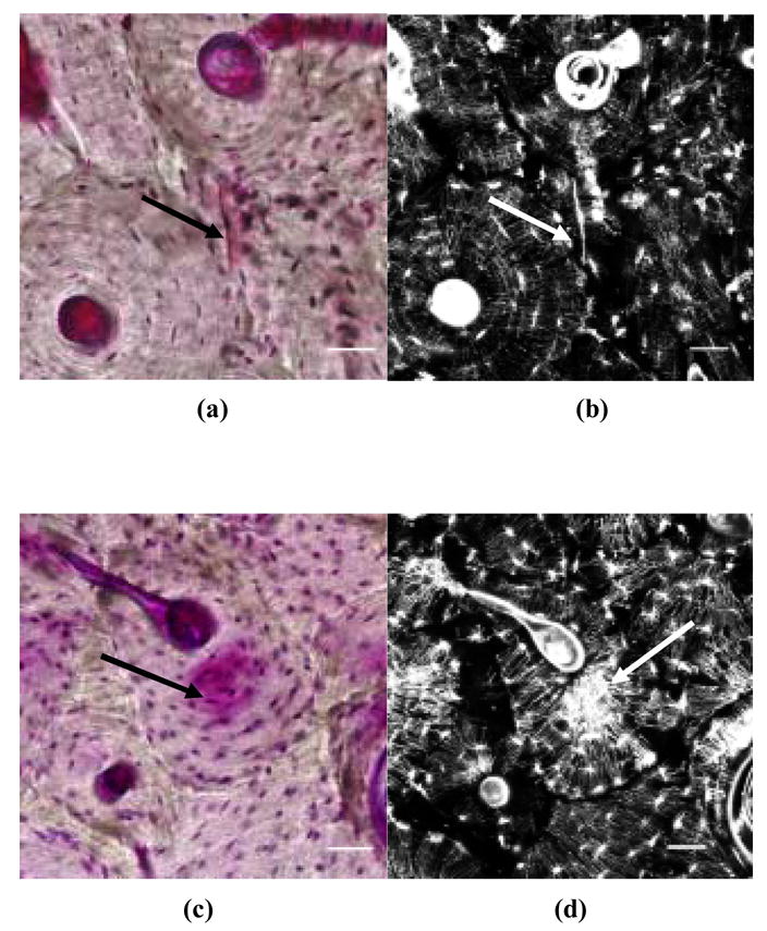
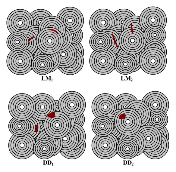
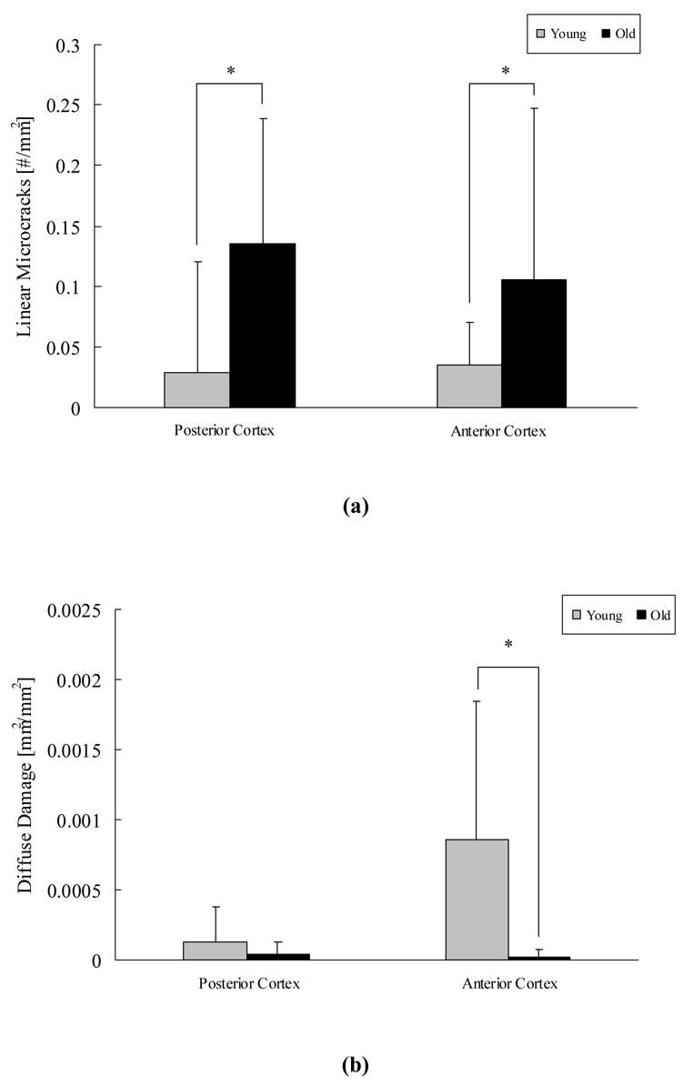
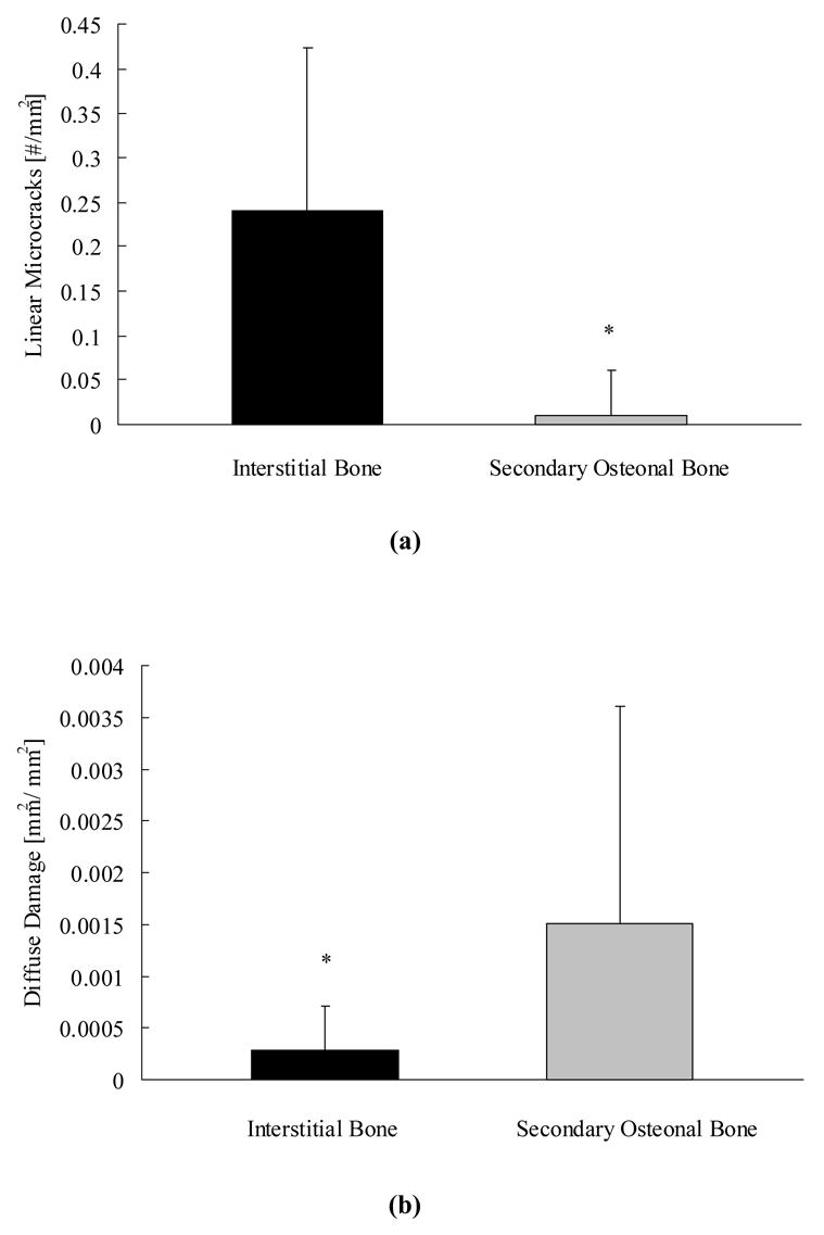
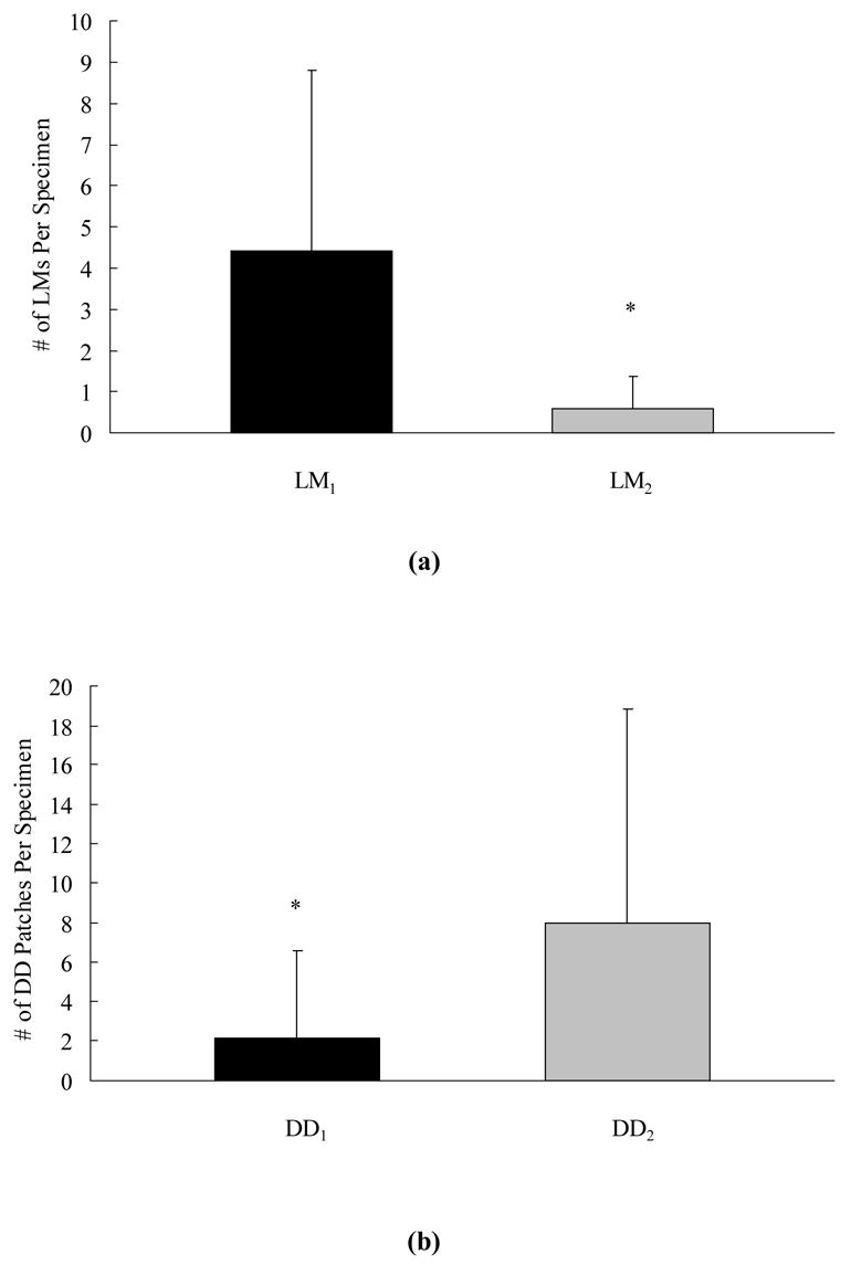
In vivo, microdamage occurs in the form of linear microcracks and diffuse damage. However, it is unknown whether the age-related changes in bone quality predispose bone to form one type of damage morphology over the other during in vivo loading. In this study, histological and histomorphometrical analyses were conducted on transverse cross sections, obtained from the tibiae of aging human bone (age 19 to 89), to investigate the in vivo accumulation and localization of damage morphologies. The results demonstrate that old donor bone (83+/-3 years) contains more linear microcracks than younger donor bone in the cortices predominantly subjected to compressive (p<0.01) and tensile loading (p<0.01). In contrast, young donor bone (40+/-10 years) contains more diffuse damage than older donor bone in the cortex predominantly subjected to tensile loading (p<0.01). The formation of damage morphology showed no correlation with bone geometry parameters and exhibited distinct preferences with bone microstructure. Linear microcracks formed in the interstitial bone (p<0.01) and were either trapped or arrested by the microstructural interfaces (cement line and lamellar interface) (p<0.05). Areas of diffuse damage, however, were preferentially associated with secondary osteonal bone (p<0.01) and had no relationship with the microstructural interfaces (p<0.01). Based upon these findings, we conclude that age-related changes in bone microstructure, but not bone geometry, play a key role in the propensity of old donors to form linear microcrack over diffuse damage under in vivo loading conditions.
Figures
References
-
- Burr DB, Turner CH. Primer on the Metabolic Bone Diseases and Disorders of Mineral Metabolism. J Bone Miner Res. 2005:58–65.
-
- Eswaran SK, Gupta A, Adams MF, Keaveny TM. Cortical and trabecular load sharing in the human vertebral body. J Bone Miner Res. 2006;21:307–314. - PubMed
-
- Burr DB, Forwood MR, Fyhrie DP, Martin RB, Schaffler MB, Turner CH. Bone microdamage and skeletal fragility in osteoporotic and stress fractures. J Bone Miner Res. 1997;12:6–15. - PubMed
-
- Parfitt AM. Bone remodeling and bone loss: understanding the pathophysiology of osteoporosis. Clin Obstet Gynecol. 1987;30:789–811. - PubMed
Publication types
MeSH terms
Grants and funding
LinkOut - more resources
Full Text Sources
Medical
