Comparative Study
. 2006 Dec;36(12):891-903.
doi: 10.1016/j.ibmb.2006.08.010. Epub 2006 Sep 16.

Characterization of hemocytes from the mosquitoes Anopheles gambiae and Aedes aegypti

Affiliations
Comparative Study

Characterization of hemocytes from the mosquitoes Anopheles gambiae and Aedes aegypti

J C Castillo et al. Insect Biochem Mol Biol. 2006 Dec.

Abstract

Hemocytes are an essential component of the mosquito immune system but current knowledge of the types of hemocytes mosquitoes produce, their relative abundance, and their functions is limited. Addressing these issues requires improved methods for collecting and maintaining mosquito hemocytes in vitro, and comparative data that address whether important vector species produce similar or different hemocyte types. Toward this end, we conducted a comparative study with Anopheles gambiae and Aedes aegypti. Collection method greatly affected the number of hemocytes and contaminants obtained from adult females of each species. Using a collection method called high injection/recovery, we concluded that hemolymph from An. gambiae and Ae. aegypti adult females contains three hemocyte types (granulocytes, oenocytoids and prohemocytes) that were distinguished from one another by a combination of morphological and functional markers. Significantly more hemocytes were recovered from An. gambiae females than Ae. aegypti. However, granulocytes were the most abundant cell type in both species while oenocytoids and prohemocytes comprised less than 10% of the total hemocyte population. The same hemocyte types were collected from larvae, pupae and adult males albeit the absolute number and proportion of each hemocyte type differed from adult females. The number of hemocytes recovered from sugar fed females declined with age but blood feeding transiently increased hemocyte abundance. Two antibodies tested as potential hemocyte markers (anti-PP06 and anti-Dox-A2) also exhibited alterations in staining patterns following immune challenge with the bacterium Escherichia coli.


Figures

Fig. 1
Fig. 1
Collection method affects hemocyte number and the level of contaminants recovered from An. gambiae and Ae. aegypti adult females. A. Total hemocyte counts ± SE recovered per mosquito by perfusion, clipping the proboscis, low injection/recovery, and high injection/recovery. A minimum of 10 individuals were bled per treatment (F7, 77= 34.7; p<0.0001). Means with the same letter do not significantly differ from one another (Tukey-Kramer HSD multiple comparison procedure α =0.05). Low magnfication differential interference contrast (DIC) micrographs of samples collected by perfusion (B) or high injection/recovery (C). The perfusion sample contains large amounts of contaminants along with hemocytes while the injection/recovery sample contains primarily hemocytes with low levels of contamination (see text). Representative granulocytes (Gr) and prohemocytes (Pr) are indicated. Scale bar = 40 μm.
Fig. 2
Fig. 2
Differential hemocyte counts for perfusion (A) and high injection/recovery (B) samples from An. gambiae and Ae. aegypti. Counts were made on the same samples used to produce the total hemocyte counts presented in Fig. 1. Hemocytes were classified as granulocytes, oenocytoids, prohemocytes, and adipohemocytes on the basis of morphology and staining with different markers (see text). DIC images of a representitive prohemocyte (C), granulocyte (D) and oenocytoid (E) from an An. gambiae adult female. A prohemocyte (F), granulocyte (G), and oenocytoid (H) from an Ae. aegypti adult female is also shown for comparison. Note the close similarity in morphology and size of each hemocyte type from the two species. Scale bar = 25 μm.
Fig. 3
Fig. 3
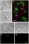
Granulocytes from An. gambiae and Ae. aegypti adult females. (A, B) DIC images of granulocytes from An. gambiae 1 h after placement on a glass slide in Schneider’s medium plus 10% FBS. One cell (A) has spread asymmetrically while the other (B) has spread symmetrically. Granules (G) are visible in the granulocyte presented in B. (C) Confocal image of An. gambiae granulocytes stained with phalloidin that labels F-actin (green) and anti-histone H1 that labels nuclei (red). Filopodia (Fp) and focal adhesions (Fa) associated with F-actin and adhesion to the glass slide are clearly visible. The projected composite image was generated from 6 optical sections that were one half micron in thickness. Scale bar in C = 10 μm with the same magnification in A and B. Low magnification DIC (D) and epifluorescent image (E) of hemocytes from An. gambiae 1 h after incubation with FITC-labeled E. coli. DIC (F) and epifluorescent (G) images of hemocytes from Ae. aegypti 1 h after incubation with FITC-labeled E. coli is also presented. All granulocytes (Gr) from both species phagocytized bacteria (green), whereas oenocytoids (Oe) and prohemocytes (Pr) have not. Scale bar in G= 80 μm with the same magnification in D–F.
Fig. 4
Fig. 4
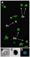
(A) Confocal image of Ae. aegypti hemocytes stained with phalloidin (green) and anti-histone-H1 (red). Spread granulocytes (Gr) are distinguished from round, unspread oenocytoids (Oe) and smaller, unspread prohemocytes (Pr). Also note the visibly higher nuclear to cytoplasmic ratio in the prohemocyte compared to the oenocytoids. Scale bar= 20 μm. The projected composite image was generated from 6 optical sections that were one half micron in thickness. (B) Higher magnification DIC image of an oenocytoid. A DIC image of an oenocytoid following staining for phenoloxidase activity (C), and an epifluorescent image of an oenocytoid following vital staining with monochlorobimane (MCB) (D) are also shown. Staining of An. gambiae oenocytoids for phenoloxidase activity and MCB is virtually identical to the images shown for Ae. aegypti. Scale bar = 15 μm in B with the same magnification in C and D.
Fig. 5
Fig. 5
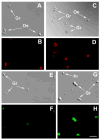
Bacterial challenge alters staining of An. gambiae hemocytes using antibodies to PP06 and PSMD3 (formerly Dox-A2). DIC (A) and confocal (B) micrographs of hemocytes from non-immune challenged mosquitoes stained with anti-PPO6 and a Texas-red conjugated secondary antibody (1:1000). The two oenocytoids in the image are stained strongly whereas the granulocyte is stained weakly. DIC (C) and confocal (D) micrographs of hemocytes collected 3 h post-injection of FITC-conjugated E. coli. Hemocytes were stained with same concentration of PPO6 antiserum and secondary antibody as in A and B. Note that granulocytes are stained more strongly in the challenged sample compared to the non-challenged sample. In contrast, staining intensity of the oenocytoids in the challenged and non-challenged samples are similar. The projected composite images in B and D were generated from 4 optical sections that were one half micron in thickness. Laser intensity, gain, and aperture settings in acquiring the two images were also identical. (E and F) DIC and epifluorescent micrographs of hemocytes from non-immune challenged An. gambiae hemocytes stained with anti-PSMD3 and a FITC-conjugated secondary antibody. All cells in the image are uniformly but weakly labeled. (G and H) DIC and epifluorescent micrographs of hemocytes collected 3 h post-injection of rhodamine-conjugated E. coli. Note the stronger staining of granulocytes (Gr) compared to the non-challenged sample. In contrast, PSMD3 staining of the prohemocyte in the image remains weak. Hemocytes were stained with same concentration of anti-PSMD3 and secondary antibody as in E and F. Camera exposure time and gain settings in acquiring the images in F and H were identical. Scale bar in H= 40 μm with the same magnification in E–G.
Fig. 6
Fig. 6
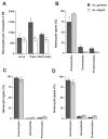
Hemocyte counts from different life stages of An. gambiae and Ae. aegypti larvae. (A) Total hemocyte counts from larvae (third instar), pupae (day 4) and adult males (day 4). (B–D) Differential hemocyte counts from larvae, pupae, and adult males respectively. Hemocytes were collected from each stage using the high injection/recovery method. A minimum of 10 individuals were sampled for each life stage. Hemocytes were placed on glass slides in Schneider’s medium plus 10% FBS for 1 h and then identified by morphology, monochlorobimane, and rhodamine 123 staining. See Fig. 1A and Fig. 2 for total and differential hemocyte counts from adult females.
Fig. 7
Fig. 7
Total hemocyte counts from adult female An. gambiae (A) and Ae. aegypti (B) of increasing age (days). Cohorts of females fed sugar only were sampled daily using the high injection/recovery method. A second cohort of females from the same starting population were blood fed on day 3 and thereafter sampled on days 4, 5 and 6. Asterisks above the bar indicate that the total number of hemocytes collected from blood fed females differed significantly from females of the same age that were fed sugar water only (t-test, α =0.05). A minimum of 10 individuals were bled per time point in each treatment.

References

    1. Abraham EG, Pinto SB, Ghosh A, Vanlangingham DL, Budd A, Higgs S, Kafatos FC, Jacobs-Lorena M, Michel K. An immune-responsive serpin, SRPN6, mediates mosquito defense against malaria parasites. Proceedings of the National Academy of Sciences USA. 2005;102:16327–16332. - PMC - PubMed
    1. Andradis TG, Hall DW. Neoplectana carpocapsae: encapsulation in Aedes aegypti and changes in host hemocytes and hemolymph proteins. Experimental Parasitology. 1976;39:252–261. - PubMed
    1. Ashida M, Ochiae M, Niki T. Immunolocalization of prophenoloxidase among hemocytes of the silkworm Bombyx mori. Tissue and Cell. 1988;20:599–610. - PubMed
    1. Bartholomay LC, Cho WL, Rocheleau TA, Boyle JP, Beck ET, Fuchs JF, Liss P, Rusch M, Butler KM, Wu RCC, Lin SP, Kuo HY, Tsao IY, Huang CY, Liu TT, Hsiao KJ, Tsai SF, Yang UC, Nappi AJ, Perna NT, Chen CC, Christensen BM. Description of the transcriptomes of immune response-activated hemocytes from the mosquito vectors Aedes aegypti and Armigeres subalbatus. Infection and Immunity. 2004;72:4114–4126. - PMC - PubMed
    1. Beck M, Strand MR. Glc1.8 from Microplitis demolitor bracovirus induces a loss of adhesion and phagocytosis by insect High Five and S2 cells. Journal of Virology. 2005;79:1861–1870. - PMC - PubMed

Publication types

LinkOut - more resources