. 2006 Nov;18(11):2929-45.
doi: 10.1105/tpc.106.045617. Epub 2006 Nov 10.

The balance between the MIR164A and CUC2 genes controls leaf margin serration in Arabidopsis

Affiliations

The balance between the MIR164A and CUC2 genes controls leaf margin serration in Arabidopsis

Krisztina Nikovics et al. Plant Cell. 2006 Nov.

Erratum in

Abstract

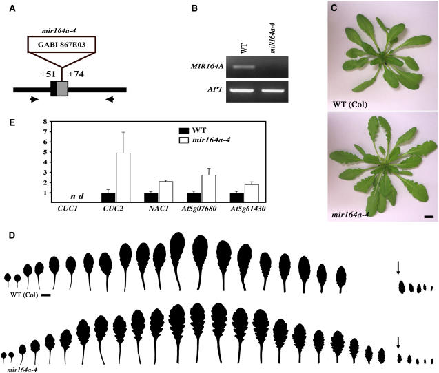
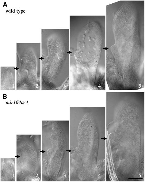
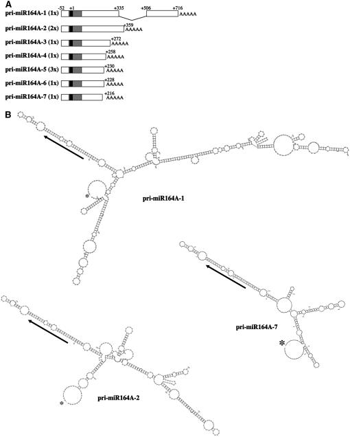
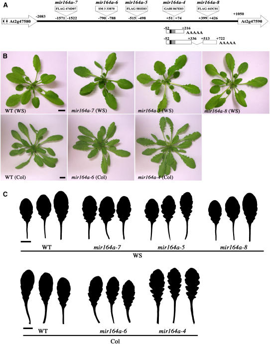
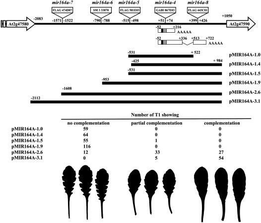
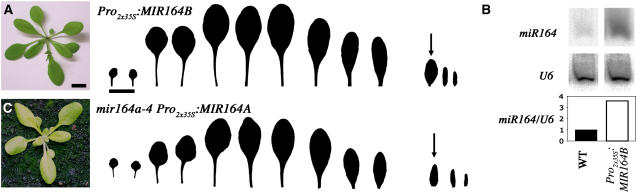
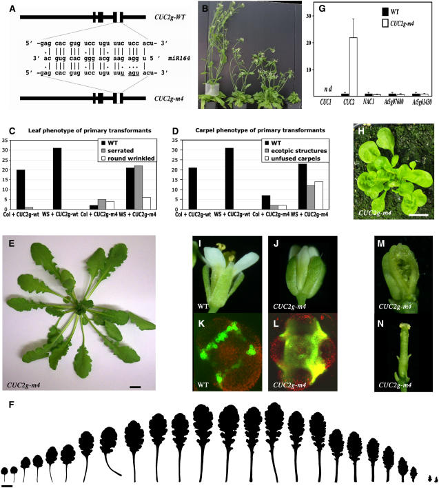
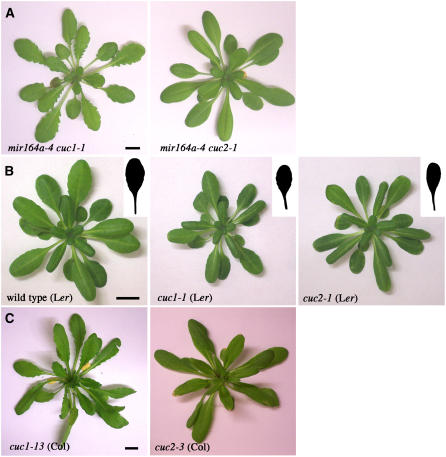
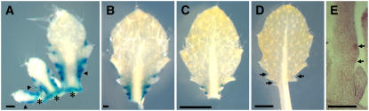
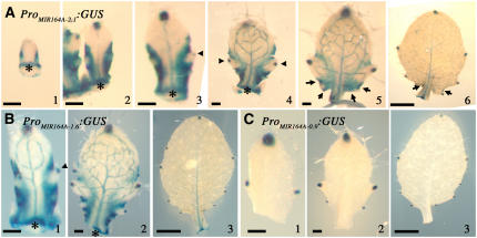
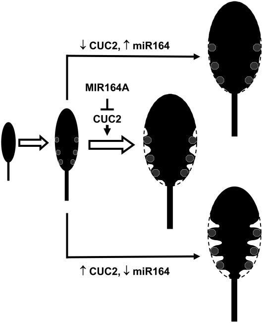
CUP-SHAPED COTYLEDON1 (CUC1), CUC2, and CUC3 define the boundary domain around organs in the Arabidopsis thaliana meristem. CUC1 and CUC2 transcripts are targeted by a microRNA (miRNA), miR164, encoded by MIR164A, B, and C. We show that each MIR164 is transcribed to generate a large population of primary miRNAs of variable size with a locally conserved secondary structure around the pre-miRNA. We identified mutations in the MIR164A gene that deepen serration of the leaf margin. By contrast, leaves of plants overexpressing miR164 have smooth margins. Enhanced leaf serration was observed following the expression of an miR164-resistant CUC2 but not of an miR164-resistant CUC1. Furthermore, CUC2 inactivation abolished serration in mir164a mutants and the wild type, whereas CUC1 inactivation did not. Thus, CUC2 specifically controls leaf margin development. CUC2 and MIR164A are transcribed in overlapping domains at the margins of young leaf primordia, with transcription gradually restricted to the sinus, where the leaf margins become serrated. We suggest that leaf margin development is controlled by a two-step process in Arabidopsis. The pattern of serration is determined first, independently of CUC2 and miR164. The balance between coexpressed CUC2 and MIR164A then determines the extent of serration.


Figures

Figure 1.
Figure 1.
mir164a Mutants Show an Increase in Leaf Margin Serration. (A) Schematic representation of the T-DNA insertion in the mir164a-4 mutant line. The mature miR164 sequence is shown in black, and the predicted pre-miRNA (Reinhart et al., 2002) is shown in gray. Positions are indicated relative to the first nucleotide of the mature miRNA. (B) No pri-miR164As expression is detected in the mir164a-4 mutant line by RT-PCR. The primers used to amplify the pri-miR164As are indicated by arrows in (A). APT is used here as a control. (C) Phenotype at bolting of the wild type and the mir164a-4 mutant. (D) Leaf series of the wild type and the mir164a-4 mutant. The arrows indicate the first cauline leaves. Leaf serration is more pronounced in the mutant but follows the same pattern as in the wild type. (E) Real-time PCR quantification of the expression levels of CUC1, CUC2, NAC1, At5g07680, and At5g61430 in wild-type and mir164a-4 leaves. Expression levels are normalized with respect to Elongation Factor1 expression levels (n = 4; bars = se). Bars = 1 cm in (C) and (D).
Figure 2.
Figure 2.
Early Stages of Leaf Development in the Wild Type and in mir164a-4 Mutants. Four-week-old plants were partly dissected, with leaves 11 to 15 removed. Arrows indicate tooth initiation at the leaf margin. Bar = 100 μm. (A) Developmental sequence of wild-type leaves. (B) Developmental sequence of mir164a-4 mutant leaves.
Figure 3.
Figure 3.
MIR164A Encodes pri-miRNA Transcripts with a Conserved Secondary Structure in the miRNA Region Despite Large Differences in Size. (A) Schematic representation of the pri-miR164A identified by 5′ and 3′ RACE PCR. The number of times each 3′ end was cloned is indicated in parentheses. The mature miR164 sequence is shown in black, and the predicted pre-miRNA (Reinhart et al., 2002) is shown in gray. Positions are indicated relative to the first nucleotide of the mature miRNA (+1). (B) Secondary structure of the pri-miR164A-1, pri-miR164A-2, and pri-miR164A-7 transcripts, as predicted by Mfold (Zuker, 2003). Asterisks indicate the beginning of the transcript, and arrows show the miRNA. The sequences of the pri-miR164B and C and the predicted secondary structures of the other pri-miR164s are available in Supplemental Figure 1 online.
Figure 4.
Figure 4.
Allelic Series of the MIR164A Gene. (A) Insertion positions of an allelic series along the MIR164A locus. The MIR164A locus is represented with the two neighboring genes. The longest and shortest pri-miR164As identified by RACE PCR are indicated. (B) Phenotype at bolting of the mutants. (C) Detail of the three largest rosette leaves (the entire leaf series is available in Supplemental Figure 2 online).
Figure 5.
Figure 5.
Complementation of the mir164a-4 Mutant. The MIR164A locus is represented with the two neighboring genes. The longest and shortest pri-miR164As identified by RACE PCR are indicated. For each genomic construct, the numbers of independent transgenic lines showing no complementation, partial complementation, or full complementation of the mir164a-4 leaf phenotype are indicated. Below are representatives of the largest rosette leaves of noncomplemented, partially complemented, and fully complemented mir164a-4 mutants.
Figure 6.
Figure 6.
Overexpression of miR164 Abolishes Leaf Serration in the Wild Type and mir164a-4 Mutants. (A) Phenotype at bolting and series of leaves of a Pro2x35S:MIR164B transgenic plant forming leaves with a smooth margin. (B) Quantification of miR164 in wild-type and Pro2x35S:MIR164B leaves. (C) Phenotype at bolting and series of leaves from a mir164a-4 mutant expressing the Pro2x35S:MIR164A construct and forming leaves with a smooth margin. The arrows indicate the first cauline leaf. Bars = 1 cm.
Figure 7.
Figure 7.
Expression of an miR164-Resistant CUC2 Gene Phenocopies the mir164a Mutant Leaves. (A) Representation of the wild-type and miR164-resistant CUC2 genes, with detail of the mutations introduced into the miR164 binding site. Thick black lines indicate the exons. (B) General view of the phenotypic changes observed in CUC2g-m4 plants. In CUC2g-m4 lines with a strong phenotype, the inflorescence was not more than a few centimeters long due to severe internode length reduction (plant on the right). CUC2g-m4 lines with weaker phenotypes had shorter inflorescences, with abnormal flower phyllotaxy (two central plants). No decrease in growth rate was observed in any of the CUC2g-wt lines (plant on the left). (C) Numbers of CUC2g-wt and CUC2g-m4 transgenic lines showing normal, serrated (as in [E]), or round and wrinkled leaves (as in [H]). Transgenic plants in the Col and Ws backgrounds were analyzed. (D) Numbers of CUC2g-wt and CUC2g-m4 lines developing normal carpels, carpels with ectopic structures along the replum (as in [N]), or unfused carpels (as in [M]). Transgenic plants in the Col and Ws backgrounds were analyzed. (E) Just after bolting, CUC2g-m4 plants have leaves with exaggerated serration, similar to the leaves of the mir164a-4 mutant. (F) Series of leaves from CUC2g-m4 plants. (G) Real-time PCR quantification of the levels of expression of CUC1, CUC2, NAC1, At5g07680, and At5g61430 in the wild type and in serrated leaves from CUC2g-m4 plants (n = 4; bars = se). (H) CUC2g-m4 plants with a strong phenotype form small rosettes with round, wrinkled leaves. (I) and (J) Wild-type sepals are joined from the base, whereas the sepals of CUC2g-m4 flowers are separated by a gap. (K) and (L) Expression of the boundary marker ProSTM:ALCR-ProalcA:erGFP (Laufs et al., 2004), showing that the boundary domain is larger in CUC2g-m4 flowers than in the wild type. (M) Dissected pistil of a CUC2g-m4 line with a strong phenotype showing carpel fusion defects (N) Dissected pistil of a CUC2g-m4 line with a weak phenotype showing ectopic structures along the replum. Bars = 1 cm.
Figure 8.
Figure 8.
CUC2 Is Required for Leaf Serration in mir164 Mutants and the Wild Type, whereas CUC1 Is Not. (A) Phenotype, at bolting, of mir164a-4 cuc1-1 and mir164a-4 cuc2-1 double mutants. (B) Phenotype of control wild-type Ler, cuc1-1, and cuc2-1 mutants at 8 weeks of short days. Details of the largest leaf are shown. (C) Phenotype, at bolting, of cuc1-13 and cuc2-3 mutants in the Col background. Bars = 1 cm.
Figure 9.
Figure 9.
CUC2 Is Expressed in the Sinus of the Leaves. (A) to (D) ProCUC2:GUS expression in rosette leaves. Arrowheads in (A) indicate regions with weaker GUS staining in which no outgrowth is yet visible, and asterisks indicate the insertion point of the primordium on the apex. Arrows in (D) indicate GUS expression restricted to the sinus. (E) In situ hybridization of CUC2 mRNA in a longitudinal leaf section. Arrows point to cells expressing CUC2. Bars = 100 μm in (A), (B), and (E) and 1 mm in (C) and (D).
Figure 10.
Figure 10.
Expression of MIR164A in the Sinus of the Leaves. (A) Expression of ProMIR164A-2.1:GUS in developing rosette leaves. (B) Expression of ProMIR164A-1.6:GUS in developing rosette leaves. (C) Expression of ProMIR164A-0.9:GUS in developing rosette leaves. Arrowheads indicate GUS expression in the hydathodes, and arrows indicate GUS expression in the sinus. Asterisks indicate the insertion point of the primordium on the apex. Bars = 100 μm in (A1) to (A5), (B1), (B2), (C1), and (C2) and 1 mm in (A6), (B3), and (C3).
Figure 11.
Figure 11.
Model of the Control of Leaf Margin Development by CUC2 and MIR164A. We propose a two-step model of leaf margin development. In the first step, the tips of serration teeth are defined (gray dots). In the second step, CUC2-mediated growth repression controlled by miR164 activity generates the sinus. See text for further details.

References

    1. Aida, M., Ishida, T., Fukaki, H., Fujisawa, H., and Tasaka, M. (1997). Genes involved in organ separation in Arabidopsis: An analysis of the cup-shaped cotyledon mutant. Plant Cell 9 841–857. - PMC - PubMed
    1. Aida, M., Ishida, T., and Tasaka, M. (1999). Shoot apical meristem and cotyledon formation during Arabidopsis embryogenesis: Interaction among the CUP-SHAPED COTYLEDON and SHOOT MERISTEMLESS genes. Development 126 1563–1570. - PubMed
    1. Aloni, R., Schwalm, K., Langhans, M., and Ullrich, C.I. (2003). Gradual shifts in sites of free-auxin production during leaf-primordium development and their role in vascular differentiation and leaf morphogenesis in Arabidopsis. Planta 216 841–853. - PubMed
    1. Alonso, J.M., et al. (2003). Genome-wide insertional mutagenesis of Arabidopsis thaliana. Science 301 653–657. - PubMed
    1. Alvarez, J.P., Pekker, I., Goldshmidt, A., Blum, E., Amsellem, Z., and Eshed, Y. (2006). Endogenous and synthetic microRNAs stimulate simultaneous, efficient, and localized regulation of multiple targets in diverse species. Plant Cell 18 1134–1151. - PMC - PubMed

Publication types