. 2007 Feb;189(4):1288-98.
doi: 10.1128/JB.01168-06. Epub 2006 Nov 10.

Streptococcus pneumoniae DivIVA: localization and interactions in a MinCD-free context

Affiliations

Streptococcus pneumoniae DivIVA: localization and interactions in a MinCD-free context

Daniela Fadda et al. J Bacteriol. 2007 Feb.

Abstract

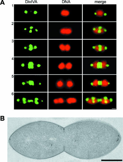
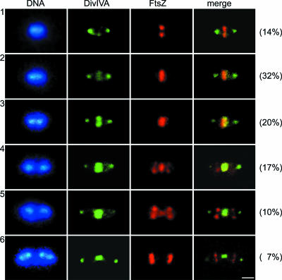
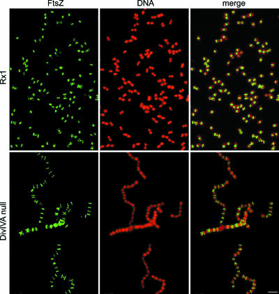
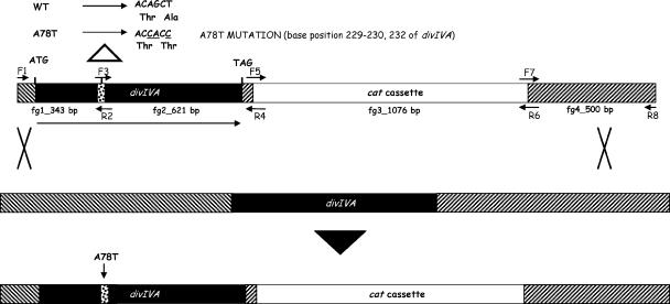
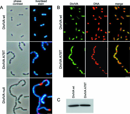
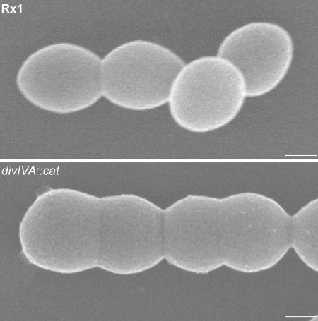
To clarify the function of DivIVA in Streptococcus pneumoniae, we localized this protein in exponentially growing cells by both immunofluorescence microscopy and immunoelectron microscopy and found that S. pneumoniae DivIVA (DivIVA(SPN)) had a unique localization profile: it was present simultaneously both as a ring at the division septum and as dots at the cell poles. Double-immunofluorescence analysis suggested that DivIVA is recruited to the septum at a later stage than FtsZ and is retained at the poles after cell separation. All the other cell division proteins that we tested were localized in the divIVA null mutant, although the percentage of cells having constricted Z rings was significantly reduced. In agreement with its localization profile and consistent with its coiled-coil nature, DivIVA interacted with itself and with a number of known or putative S. pneumoniae cell division proteins. Finally, a missense divIVA mutant, obtained by allelic replacement, allowed us to correlate, at the molecular level, the specific interactions and some of the facets of the divIVA mutant phenotype. Taken together, the results suggest that although the possibility of a direct role in chromosome segregation cannot be ruled out, DivIVA in S. pneumoniae seems to be primarily involved in the formation and maturation of the cell poles. The localization and the interaction properties of DivIVA(SPN) raise the intriguing possibility that a common, MinCD-independent function evolved differently in the various host backgrounds.


Figures

FIG. 1.
FIG. 1.
Localization of the DivIVA protein in S. pneumoniae Rx1 cells. (A) Fluorescence micrographs showing the localization of DivIVA in representative cells at different stages of division, designated 1 to 6. Cells were stained to visualize DivIVA (green) and DNA (red); the merge images show the localization of DivIVA and DNA simultaneously. Scale bar = 0.25 μm. (B) Immunogold labeling of DivIVA. Exponentially growing cells were fixed and freeze-substituted. Thin sections were immunolabeled with specific antibodies and a gold-labeled secondary antibody and analyzed by electron microscopy. Dark grains show the localization of DivIVA. Scale bar = 0.25 μm.
FIG. 2.
FIG. 2.
Fluorescence micrographs showing the double localization of DivIVA and FtsZ in S. pneumoniae Rx1 cells at different stages of division, designated 1 to 6. Cells were stained to visualize DNA (blue), DivIVA (green), and FtsZ (red); the merge images show the localization of DivIVA and FtsZ simultaneously. The fluorescence micrographs are arranged to show the progression through the cell cycle. The percentages of cells in the different stages are indicated on the right. Scale bar = 0.5 μm.
FIG. 3.
FIG. 3.
Fluorescence micrographs showing the localization of the FtsZ cell division protein in wild-type strain Rx1 and in the divIVA null mutant. Cells were stained to visualize FtsZ (green) and DNA (red); the merge images show the localization of DNA and proteins simultaneously. Scale bar = 1.5 μm.
FIG. 4.
FIG. 4.
Immunoprecipitation to determine the specificity of DivIVA interactions. (A) Immunoprecipitation of GFP-DivIVA-cI-Spo0J complex (lane 1), GFP-Spo0J-cI-DivIVA complex (lane 2), GFP-FtsZ-cI-DivIVA complex (lane 3), and GFP-DivIVA-cI-FtsZ complex (lane 4). (B) Immunoprecipitation of cI-DivIVA-GFP-PrfA complex (lane 1). Protein extracts were immunoprecipitated with anti-GFP antibodies and were detected with anti-cI rabbit polyclonal antibodies by Western blotting as described in Materials and Methods. Lane M contained molecular weight markers. In panel B lane 2 contained crude extract of E. coli strain DH5α expressing cI-DivIVA, detected with anti-cI antibodies.
FIG. 5.
FIG. 5.
Schematic diagram of the strategy used for allelic replacement mutagenesis and of the events that occurred after recombination via a double crossover. Four sets of primers were used to generate the final construct by a two-step PCR procedure as described in Materials and Methods. The resulting product was used to transform S. pneumoniae Rx1 competent cells, with selection for chloramphenicol resistance. As a result of the recombination event, in the transformants the cat cassette was inserted 40 bp after the end of divIVA. The correct gene replacement in the transformants was verified by PCR. Similarly, a transformant carrying a wild-type divIVA gene and the cat cassette inserted in same position was constructed as a control. WT, wild type.
FIG. 6.
FIG. 6.
Phenotypic characterization and localization of DivIVA in the wild-type DivIVA strain and the DivIVA A78T mutant. (A) Phase-contrast and live/dead stain fluorescence micrographs. (B) Fluorescence micrographs showing the localization of DivIVA. Cells were stained to visualize DNA (red) and DivIVA (green). The merge images show the localization of DNA and DivIVA simultaneously. Scale bar = 1.5 μm. (C) Western blot analysis of the DivIVA protein. WT, wild type.
FIG. 7.
FIG. 7.
Scanning electron micrographs showing the morphological differences between wild-type Rx1 and Rx1 divIVA::cat cells. Note that the poles of the divIVA null mutant have an oblate rather than prolate shape, which correlated with the approximately 20% difference in cell diameter reported previously (13). Scale bar = 0.25 μm.

Similar articles

Cited by

References

    1. Ben-Yehuda, S., D. Z. Rudner, and R. Losick. 2003. RacA, a bacterial protein that anchors chromosomes to the cell poles. Science 299:532-536. - PubMed
    1. Buddelmeijer, N., and J. Beckwith. 2004. A complex of the Escherichia coli cell division proteins FtsL, FtsB and FtsQ forms independently of its localization to the septal region. Mol. Microbiol. 52:1315-1327. - PubMed
    1. Cha, J. H., and G. C. Stewart. 1997. The divIVA minicell locus of Bacillus subtilis. J. Bacteriol. 179:1671-1683. - PMC - PubMed
    1. Claverys, J. P., M. Prudhomme, I. Mortier-Barriere, and B. Martin. 2000. Adaptation to the environment: Streptococcus pneumoniae, a paradigm for recombination-mediated genetic plasticity? Mol. Microbiol. 35:251-259. - PubMed
    1. Cole, R. M., and J. J. Hahn. 1962. Cell wall replication in Streptococcus pyogenes. Science 135:722-724. - PubMed

Publication types

MeSH terms

LinkOut - more resources