

Mapping the MRI voxel volume in which thermal noise matches physiological noise--implications for fMRI
- PMID: 17101280
- PMCID: PMC1815476
- DOI: 10.1016/j.neuroimage.2006.09.039
Mapping the MRI voxel volume in which thermal noise matches physiological noise--implications for fMRI
Abstract
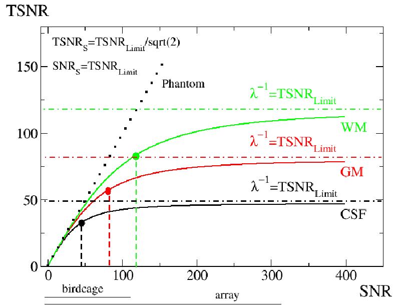
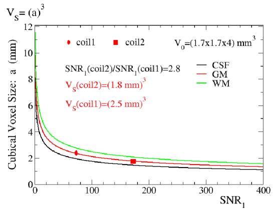
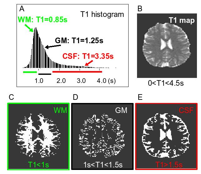
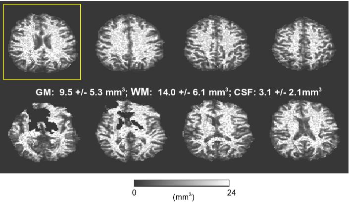
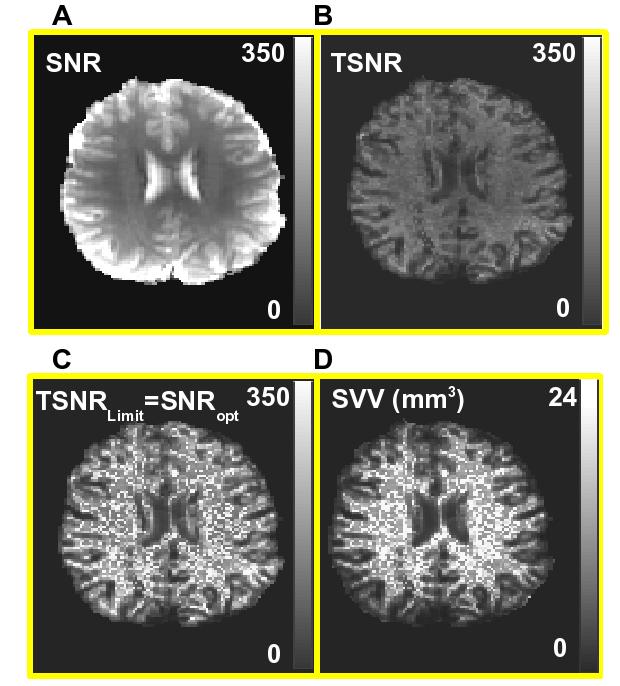
This work addresses the choice of the imaging voxel volume in blood oxygen level dependent (BOLD) functional magnetic resonance imaging (fMRI). Noise of physiological origin that is present in the voxel time course is a prohibitive factor in the detection of small activation-induced BOLD signal changes. If the physiological noise contribution dominates over the temporal fluctuation contribution in the imaging voxel, further increases in the voxel signal-to-noise ratio (SNR) will have diminished corresponding increases in temporal signal-to-noise (TSNR), resulting in reduced corresponding increases in the ability to detect activation induced signal changes. On the other hand, if the thermal and system noise dominate (suggesting a relatively low SNR) further decreases in SNR can prohibit detection of activation-induced signal changes. Here we have proposed and called the "suggested" voxel volume for fMRI the volume where thermal plus system-related and physiological noise variances are equal. Based on this condition we have created maps of fMRI suggested voxel volume from our experimental data at 3T, since this value will spatially vary depending on the contribution of physiologic noise in each voxel. Based on our fast EPI segmentation technique we have found that for gray matter (GM), white matter (WM), and cerebral spinal fluid (CSF) brain compartments the mean suggested cubical voxel volume is: (1.8 mm)3, (2.1 mm)3 and (1.4 mm)3, respectively. Serendipitously, (1.8 mm)3 cubical voxel volume for GM approximately matches the cortical thickness, thus optimizing BOLD contrast by minimizing partial volume averaging. The introduced suggested fMRI voxel volume can be a useful parameter for choice of imaging volume for functional studies.
Figures
References
-
- Beauchamp MS, Argall BD, Bodurka J, Duyn JH, Martin A. Unraveling multisensory integration: patchy organization within human STS multisensory cortex. Nat Neurosci. 2004;7:1190–1192. - PubMed
-
- Bellgowan PSF, Bandettini PA, van Gelderen P, Martin A, Bodurka J. Improved BOLD detection in the medial temporal region using parallel imaging and voxel volume reduction. Neuroimage. 2006;29:1244–1251. - PubMed
-
- Bodurka J, Ledden PJ, van Gelderen P, Chu R, de Zwart JA, Morris D, Duyn JH. Scalable multichannel MRI data acquisition system. Magn. Reson. Med. 2004;51:165–171. - PubMed
-
- Bodurka J, Ye F, Petridou N, Bandettini PA. Determination of the brain voxel specific temporal signal-to-noise ratio limit of 3T BOLD-weighted time course data; Proceedings of the XIII International Society of Magnetic Resonance in Medicine.2005. p. 1554.
-
- Cheng K, Waggoner RA, Tanaka K. Human ocular dominance column as revealed by high-field functional magnetic resonance imaging. Neuron. 2001;32:359–374. - PubMed
Publication types
MeSH terms
Grants and funding
LinkOut - more resources
Full Text Sources
Other Literature Sources
Medical
