. 2007 Feb 4;6(2):190-201.
doi: 10.1016/j.dnarep.2006.09.010. Epub 2006 Nov 13.

The non-homologous end-joining protein Nej1p is a target of the DNA damage checkpoint

Affiliations

The non-homologous end-joining protein Nej1p is a target of the DNA damage checkpoint

Peter Ahnesorg et al. DNA Repair (Amst). .

Abstract

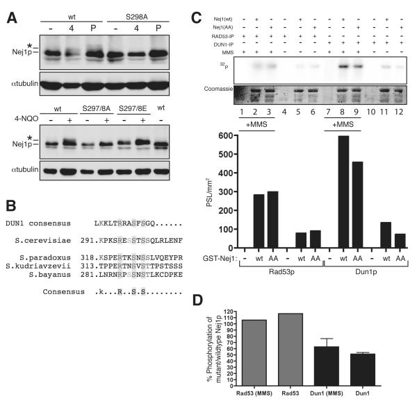
DNA double-strand breaks (DSBs), which are generated by ionizing radiation (IR) and a range of other DNA damaging agents, are repaired by homologous recombination (HR) or non-homologous end-joining (NHEJ). Previous studies have shown that NHEJ in Saccharomyces cerevisiae requires the Yku70p-Yku80p heterodimer and a complex consisting of DNA Ligase IV, Lif1p and Nej1p. Here, we report that Nej1p is phosphorylated in response to DNA damage in a manner that relies on the DNA damage checkpoint kinases Mec1p, Rad53p and Dun1p. By using a mutational approach, we have identified a consensus Dun1p phosphorylation site in Nej1p, and mutation of conserved serine residues within it leads to decreased NHEJ efficiency. These data, together with previous findings that Rad55p--a protein involved in HR--is phosphorylated analogously, point to there being a broad signalling network connecting DNA damage checkpoint responses with the regulation of DNA DSB repair activities.


Figures

Figure 1
Figure 1. Nej1p is rapidly phosphorylated in response to DNA damage
(A,B,D-F); Extracts prepared from the indicated strains were run on 10% SDS-gels and analyzed by Western blotting with a mouse anti-myc monoclonal antibody to detect Nej1p. The asterisk represents the phosphorylated, slower migrating form of Nej1p generated in response to DNA damage, while “Loading” represents an unspecific band (~50 kDa) recognized by the mouse anti-Myc antibody in the absence or presence of 13-myc-Nej1p. A nej1Δ cells were transformed with pNej1 or empty vector (pRS415) and treated with phleomycin or 4-NQO for 2 h. B Native protein extracts (80 μg) prepared from untreated cells or cells treated with 4-NQO (top) or phleomycin (bottom), were treated with or without 800 U of λ-phosphatase (NEB) and with or without 50 mM EDTA as indicated. C Asynchronously growing cells transformed with pNej1 untreated or treated with 0.075% of MMS, 90 min; 100 mM of HU, 90 min; 1μg/ml of 4-NQO, 90 min; 25 μg/ml of phleomycin , 90 min; 230 Gy of IR; 100 J/m2 of UV. Extracts were prepared and resolved on a 8% SDS-PAGE before immunoblotting with mouse anti-myc antibody. D G1 arrested cells were released into the cell cycle. Samples were taken and phosphorylation was analyzed by Western blotting (upper panel). As a control, cycling cells were treated with 4-NQO. Progression through the cell cycle was monitored by light microscopy (lower panel). E Nej1-13myc cells were grown asynchronously and extracts prepared at indicated time intervals after drug addition. F nej1Δ cells were transformed with pNej1 and either grown asynchronously, arrested at G2/M with 15 μg/ml of nocodazole, or arrested in G1 with 5 μg/ml of α-factor. Cycling and arrested cultures were treated with 4-NQO for 90 min before extract preparation.
Figure 2
Figure 2. Genetic dependencies of Nej1p phosphorylation
(A-E); Extracts prepared from the indicated strains were run on 10% SDS-gels and analyzed by Western blotting with anti-Myc antibody to detect Nej1p. The asterisk represents the phosphorylated, slower migrating form of Nej1p generated in response to DNA damage. Nej1p phosphorylation depends on the DNA damage checkpoint kinases Mec1p, Rad53p and Dun1p. A W303a, mec1Δ::trp1, tel1Δ::his3 or chk1Δ::trp1 strains were transformed with pNej1. Cultures were treated with drug for 2 h as indicated. Loading represents an unspecific band (~50 kDa) recognized by the anti-Myc antibody in the absence or presence of 13-myc-Nej1p. B W303a, rad53Δ::his3 and rad53Δ::rad53-1 and rad53Δ::his3 + pRad53 strains were transformed with pNej1. Cultures were treated with drug for 2 h as indicated. C dun1Δ::his3 nej1Δ::kanMX cells were transformed with pNej1 and pDun1 as indicated. Nej1-13myc cells were used as control. Cultures were treated with drug for 90 min as indicated. D Nej1p phosphorylation is independent of the NHEJ machinery. nej11Δ::kanMX, lif11Δ::ura3 and yku80Δ::kanMX cells were transformed with pNej1. Cultures were treated with drug for 90 min as indicated. Extracts were prepared, resolved by 10% SDS-PAGE and analyzed by western blotting with a mouse anti-Myc antibody to detect Nej1p. E Nej1p phosphorylation is enhanced in the absence of function HR. nej1Δ::kanMX (W303) or rad52Δ::trp1 strains were transformed with pNej1. Cells were grown as indicated in the absence or presence of 4-NQO. Extracts were run on SDS-gels and analyzed as described above. Loading represents an unspecific band (~50 kDa) recognized by the anti-Myc antibody in the absence or presence of 13-myc-Nej1p.
Figure 3
Figure 3. Nej1p serines 297 and 298 are involved the DNA damage dependent phosphorylation of Nej1p
A Site-directed mutagenesis of potential phosphorylation sites in Nej1p. nej11Δ::kanMX cells were complemented with pNej1(wt, W303) or pNej1 plasmids harbouring the amino acid substitutions Ser-298 to Ala, Ser-297/Ser-298 to Ala, or Ser-297/Ser-298 to Glu. Transformed strains were treated with drug for 90 min with phleomycin or 4-NQO and extracts were resolved by 10% SDS-PAGE and western blotting with anti-Myc antibody to detect Nej1p. Tubulin was detected with a rat anti-tubulin monoclonal antibody. The asterisk represents the phosphorylated, slower migrating form of Nej1p generated in response to DNA damage. B Alignment of Nej1p homologues of different yeast species to the Dun1p consensus peptide [31]. C In vitro phosphorylation reactions with wild-type Nej1p (wt) and Nej1p mutated on Ser residues 297 and 298 (AA) expressed as GST fusion proteins. Phosphorylation of Nej1p was monitored by autoradiography (upper panel 32P; Coomassie shows the loading of GST-Nej1p) and quantified with a phosphorimager (histogram in lower panel). D Substrate specificity of Rad53p and Dun1p. Ratio (in %) of phosphorylated Nej1p mutated at Ser residues 297 and 298 to wild-type Nej1p obtained with quantification data from in vitro phosphorylation reactions. Error bars indicate the mean and standard deviation of two independent experiments.
Figure 4
Figure 4. Nej1p serines 297 and 298 are required for efficient NHEJ and survival following acute phleomycin treatment
A Plasmid repair assay. nej1::kanMX (Δnej1) cells complemented with full-length Nej1p (pNej1), empty vector (pRS415), Nej1p point mutants (S298A, S298E, S297/8A, R294A, S280G, S58G/T60G/S63G/S68G STSS), Nej1p deleted for amino acids 243-323, or dun1::HIS3 cells were transformed with 0.5 μg of pBTM116. Plasmid substrate was linearized with EcoRI and equal amounts of circular plasmid were transformed in parallel to correct for differences in transformation efficiency. Error bars indicate the mean and standard deviation of three independent experiments. B End joining of different DSB end substrates. nej1::kanMX (Δnej1) cells complemented with full-length Nej1p (pNej1), empty vector (pRS415) or Nej1p S298A point mutants were transformed with 0.5 μg pRS416 digested with either EcoRI (5′-overhang, MCS), SmaI (blunt end, MCS) or NcoI (5′-overhang, outside MCS). Equal amounts of circular plasmid were transformed in parallel to correct for differences in transformation efficiency. The NHEJ rate in cells complemented with wild-type Nej1p was set to 100% as a reference. Error bars indicate mean and standard deviation of three independent experiments. C Phleomycin survival assay. Nej11Δ::kanMX cells were complemented with pNej1 or pNej1 plasmids harbouring the amino acid substitutions Ser-298 to Ala, Ser-297/Ser-298 to Ala, Ser-298 to Glu or Ser-297/Ser-298 to Glu. Early log phase cells were grown in the presence of 5 μg/ml, 10 μg/ml or 20 μg/ml phleomycin for 4 h and plated. Surviving colonies were counted after three days. Data represent % surviving colonies after treatment compared to untreated cells.
Figure 5
Figure 5. Lif1p nuclear localization and chromatin binding are not abolished in the absence of Nej1p
A Localization of EGFP-Lif1p. nej1Δ::kanMX lif1Δ::ura3 cells (Δnej1 Δlif1) were transformed with both pNej1 and pLif1-EGFP in parallel or separately. The GFP signal was monitored by fluorescence microscopy. B Δnej1 Δlif1 cells were either transformed with pNej1 and pLif1 in parallel or separately. Cultures were grown and treated with 0.0125% of MMS as described above. As a control, whole cell extract (WCE) is shown. Cells were fractionated to generate a soluble pool that contains the combined soluble nuclear and cytoplasmic proteins (S), a soluble chromatin pool that contains weakly chromatin-bound proteins (SC), and an insoluble chromatin fraction (ISC) containing tightly chromatin-bound proteins. Nej1p was detected with an anti-Myc antibody, EGFP-Lif1p was detected with an anti-GFP antibody; tubulin as a control for the soluble fraction was detected with an anti-tubulin antibody. Orc2p as a control for the chromatin fractions, was detected with an anti-Orc2p antibody. Phosphorylation of Nej1p observed in these experiments in the absence of DNA damage was found to be due to background level activation of the Rad53p kinase cascade by oxalyticase treatment.

Similar articles

Cited by

References

    1. Kolodner RD, Putnam CD, Myung K. Maintenance of genome stability in Saccharomyces cerevisiae. Science. 2002;297:552–557. - PubMed
    1. Rouse J, Jackson SP. Interfaces between the detection, signaling, and repair of DNA damage. Science. 2002;297:547–551. - PubMed
    1. Sancar A, Lindsey-Boltz LA, Unsal-Kaccmaz K, Linn S. Molecular Mechanisms of Mammalian DNA Repair and the DNA Damage Checkpoints. Annu Rev Biochem. 2004;73:39–85. - PubMed
    1. Longhese MP, Foiani M, Muzi-Falconi M, Lucchini G, Plevani P. DNA damage checkpoint in budding yeast. EMBO Journal. 1998;17:5525–5528. - PMC - PubMed
    1. Melo J, Toczyski D. A unified view of the DNA-damage checkpoint. Current Opinion in Cell Biology. 2002;14:237–245. - PubMed

Publication types

MeSH terms