. 2006 Nov 27;203(12):2715-25.
doi: 10.1084/jem.20061401. Epub 2006 Nov 13.

Interleukin-17 is a negative regulator of established allergic asthma

Affiliations

Interleukin-17 is a negative regulator of established allergic asthma

Silvia Schnyder-Candrian et al. J Exp Med. .

Abstract

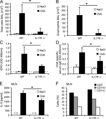
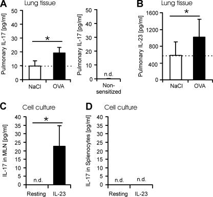
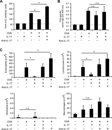
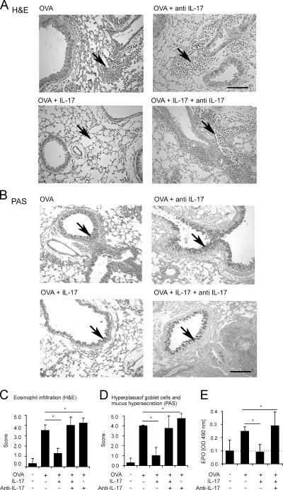
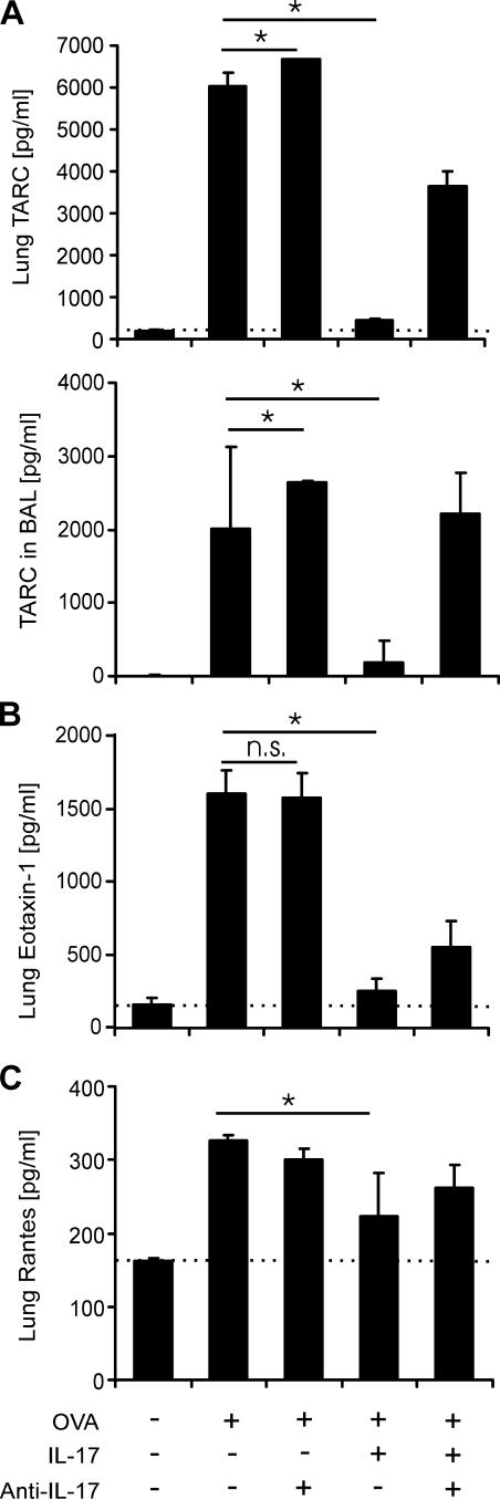
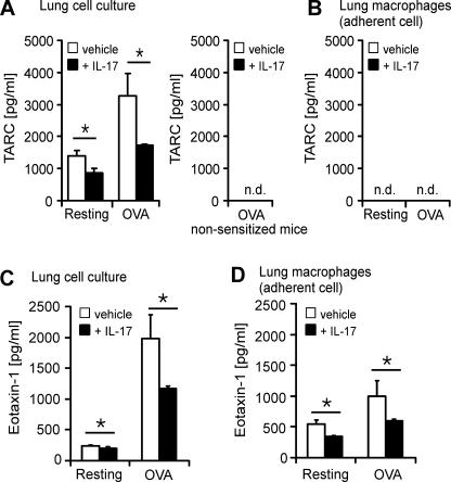
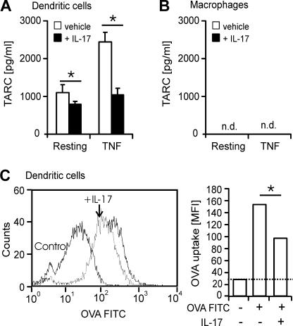
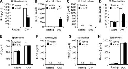
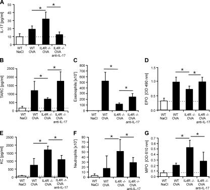
T helper (Th)17 cells producing interleukin (IL)-17 play a role in autoimmune and allergic inflammation. Here, we show that IL-23 induces IL-17 in the lung and IL-17 is required during antigen sensitization to develop allergic asthma, as shown in IL-17R-deficient mice. Since IL-17 expression increased further upon antigen challenge, we addressed its function in the effector phase. Most strikingly, neutralization of IL-17 augmented the allergic response in sensitized mice. Conversely, exogenous IL-17 reduced pulmonary eosinophil recruitment and bronchial hyperreactivity, demonstrating a novel regulatory role of IL-17. Mechanistically, IL-17 down modulated eosinophil-chemokine eotaxin (CCL11) and thymus- and activation-regulated chemokine/CCL17 (TARC) in lungs in vivo and ex vivo upon antigen restimulation. In vitro, IL-17 reduced TARC production in dendritic cells (DCs)-the major source of TARC-and antigen uptake by DCs and IL-5 and IL-13 production in regional lymph nodes. Furthermore, IL-17 is regulated in an IL-4-dependent manner since mice deficient for IL-4Ralpha signaling showed a marked increase in IL-17 concentration with inhibited eosinophil recruitment. Therefore, endogenous IL-17 is controlled by IL-4 and has a dual role. Although it is essential during antigen sensitization to establish allergic asthma, in sensitized mice IL-17 attenuates the allergic response by inhibiting DCs and chemokine synthesis.


Figures

Figure 1.
Figure 1.
Role of IL-17 in development of allergic inflammation, as assessed in receptor IL-17R KO mice. OVA/alum-sensitized C57BL/6 mice (WT) and IL-17R KO (IL-17R−/−) mice were challenged three times intranasally with OVA. 48 h after the third challenge, the BAL cells and eosinophils were counted (A and B), and lung tissue activities of EPO (C) and OVA-specific serum IgE concentrations (D) were determined. Mediastinal lymph nodes (MLNs) were removed from antigen-sensitized and challenged mice, restimulated in vitro with 50 μg/ml OVA, and analyzed for IL-5 production (E). In F, the cell composition of freshly excised MLNs was determined by FACS analysis. The percentages of CD3-, CD11b- and CD11c-positive cells are shown. The bars represent the mean ± SD (n = 8 animals per group). *, P ≤ 0.05; dotted line indicates basal levels.
Figure 2.
Figure 2.
IL-17 is induced in the lung upon allergen challenge. Mice were sensitized and challenged with OVA, and IL-17 and IL-23 were determined by ELISA in the lung homogenate (A and B). In naive, nonsensitized mice, no IL-17 was detected (A). In C and D, cells from mediastinal lymph nodes (MLNs) or splenocytes of sensitized and challenged mice were restimulated in vitro with 300 ng/ml IL-23. The bars represent the mean ± SD (n = 8 animals per group). n.d., not detected. *, P ≤ 0.05; dotted line indicates basal levels.
Figure 3.
Figure 3.
Exogenous IL-17 inhibits methacholine response upon allergen challenge. OVA-sensitized C57BL/6 mice were challenged intranasally with either saline, OVA alone, IL-17 alone, OVA with IL-17, or OVA with IL-17 plus neutralizing IL-17 antibodies. 24 h after the challenges, the methacholine response was measured using whole-body plethysmography. The intensity is measured in Penh arbitrary units, and the calculated area under the Penh time-curves (AUC) is shown. 48 h after the third challenge, OVA-specific serum IgE concentrations were determined, given in absorbance (OD 405 nm) values (B), and the BAL cells were counted. Eosinophil, lymphocyte, macrophage, and neutrophil counts (C) are presented. The same effect of IL-17 was found in mice sensitized with OVA/alum (unpublished data). The results represent the mean ± SD of n = 8 animals per group. *, P ≤ 0.05; dotted line indicates basal levels.
Figure 4.
Figure 4.
IL-17 inhibits cell recruitment to the lungs and mucus hypersecretion. Lung sections of OVA-sensitized C57BL/6 mice killed 2 d after the third challenge with either saline, OVA, OVA with IL-17 antibodies, OVA with IL-17, or OVA with IL-17 plus IL-17 antibodies are shown. The formalin-fixed tissue sections were stained with hematoxylin and eosin to visualize cell recruitment (A) and with periodic acid Schiff reagent (PAS) to visualize mucus (B), as shown by the arrows. Bars, 200 μm. In C and D, the histology sections were quantitated for eosinophil recruitment and mucus hypersecretion, respectively. A score scale from 0 to 5 is given on the y axis. The EPO in the lung tissue homogenates was determined 48 h after the last challenge, given in OD values (OD 490 nm) (E). The results represent the mean ± SD of n = 8 animals per group. n.d., not detected. *, P ≤ 0.05; dotted line indicates basal levels.
Figure 5.
Figure 5.
IL-17 inhibits pulmonary contents of the mononuclear cell attracting chemokines TARC, eotaxin and Rantes. OVA-sensitized C57BL/6 mice were challenged three times intranasally with either saline, OVA alone, OVA plus IL-17–neutralizing antibodies, OVA with IL-17, or OVA with IL-17 plus IL-17 antibodies. 48 h after the third challenge, BAL was performed and the lung tissues were excised. The chemokine contents in the liquid phase of the lung homogenate and in the BAL were measured by ELISA specific for TARC (A), eotaxin (B), and Rantes (C). The results represent the mean ± SD (n = 8 animals per group). *, P ≤ 0.05; dotted line indicates basal levels.
Figure 6.
Figure 6.
IL-17 inhibits TARC and eotaxin production ex vivo in lung explants. Lung explants (A and C) and lung adherent cells (B and D) were prepared from OVA-sensitized and challenged animals. Cells were treated with saline, 50 μg/ml OVA alone, IL-17 alone, or OVA plus 30 ng/ml IL-17 for 24 h. The concentrations of TARC (A and B) and eotaxin (C and D) were measured by ELISA. In lung cell cultures of naive, nonsensitized mice no TARC was detected (A). The results represent the mean ± SD of 8 animals per group. n.d., not detected. *, P ≤ 0.05.
Figure 7.
Figure 7.
IL-17 inhibits TARC production and OVA uptake in DCs. DCs (A and C) and macrophages (B) were differentiated in vitro from naive bone marrow–derived cells. Cells were treated with saline, IL-17 alone, 3 ng/ml TNF, or TNF with 30 ng/ml IL-17 for 24 h. The concentrations of TARC (A and B) were measured by ELISA. The results represent the mean ± SD of 8 animals per group. In C, uptake of 100 μg/ml OVA-FITC by DCs in the absence and presence of IL-17 (+IL-17) was analyzed by FACS. A histogram is shown in the left panel. The mean fluorescence intensity (MFI) thereof is given in the right hand panel. n.d., not detected. *, P ≤ 0.05.
Figure 8.
Figure 8.
IL-17 inhibits IL-5, IL-13, and Rantes production in mediastinal lymph nodes (MLNs). MLN (A–D) and splenocyte (E–H) cultures were prepared from OVA-sensitized and challenged animals. Cells were treated with saline, 50 μg/ml OVA alone, IL-17 alone, or OVA plus 30 ng/ml IL-17 for 24 h. The concentrations of IL-5, IL-13, TARC, and Rantes were measured by ELISA. The results represent the mean ± SD of 8 animals per group. n.d., not detected. *, P ≤ 0.05; dotted line indicates basal levels.
Figure 9.
Figure 9.
IL-4 signaling suppresses pulmonary IL-17 production. OVA/alum-sensitized BALB/c mice (WT) and IL-4Rα KO (IL4R−/−) mice were challenged twice intranasally with either saline or OVA alone. In parallel, the IL-4Rα KO mice were challenged with OVA plus neutralizing anti–IL-17 antibodies. 48 h after the last challenge, IL-17, TARC, and KC concentrations in the lung homogenates were measured by ELISAs (A, B, and E), and the cells infiltrating the BAL were counted (C and F). Lung tissue activities of EPO and myeloperoxidase (D and G). The results represent the mean ± SD of 8 animals per group.

References

    1. Wills-Karp, M. 1999. Immunologic basis of antigen-induced airway hyperresponsiveness. Annu. Rev. Immunol. 17:255–281. - PubMed
    1. D'Ambrosio, D., A. Iellem, R. Bonecchi, D. Mazzeo, S. Sozzani, A. Mantovani, and F. Sinigaglia. 1998. Selective up-regulation of chemokine receptors CCR4 and CCR8 upon activation of polarized human type 2 Th cells. J. Immunol. 161:5111–5115. - PubMed
    1. Herrick, C.A., and K. Bottomly. 2003. To respond or not to respond: T cells in allergic asthma. Nat. Rev. Immunol. 3:405–412. - PubMed
    1. Fossiez, F., J. Banchereau, R. Murray, C. Van Kooten, P. Garrone, and S. Lebecque. 1998. Interleukin-17. Int. Rev. Immunol. 16:541–551. - PubMed
    1. Yao, Z., S.L. Painter, W.C. Fanslow, D. Ulrich, B.M. Macduff, M.K. Spriggs, and R.J. Armitage. 1995. Human IL-17: a novel cytokine derived from T cells. J. Immunol. 155:5483–5486. - PubMed

Publication types

MeSH terms