. 2007 Jan;27(2):633-50.
doi: 10.1128/MCB.00461-06. Epub 2006 Nov 13.

The yeast PH domain proteins Slm1 and Slm2 are targets of sphingolipid signaling during the response to heat stress

Affiliations

The yeast PH domain proteins Slm1 and Slm2 are targets of sphingolipid signaling during the response to heat stress

Alexes Daquinag et al. Mol Cell Biol. 2007 Jan.

Abstract

The PH domain-containing proteins Slm1 and Slm2 were previously identified as effectors of the phosphatidylinositol-4,5-bisphosphate (PI4,5P(2)) and TORC2 signaling pathways. Here, we demonstrate that Slm1 and Slm2 are also targets of sphingolipid signaling during the heat shock response. We show that upon depletion of cellular sphingolipid levels, Slm1 function becomes essential for survival under heat stress. We further demonstrate that Slm proteins are regulated by a phosphorylation/dephosphorylation cycle involving the sphingolipid-activated protein kinases Pkh1 and Pkh2 and the calcium/calmodulin-dependent protein phosphatase calcineurin. By using a combination of mass spectrometry and mutational analysis, we identified serine residue 659 in Slm1 as a site of phosphorylation. Characterization of Slm1 mutants that mimic dephosphorylated and phosphorylated states demonstrated that phosphorylation at serine 659 is vital for survival under heat stress and promotes the proper polarization of the actin cytoskeleton. Finally, we present evidence that Slm proteins are also required for the trafficking of the raft-associated arginine permease Can1 to the plasma membrane, a process that requires sphingolipid synthesis and actin polymerization. Together with previous work, our findings suggest that Slm proteins are subject to regulation by multiple signals, including PI4,5P(2), TORC2, and sphingolipids, and may thus integrate inputs from different signaling pathways to temporally and spatially control actin polarization.


Figures

FIG. 1.
FIG. 1.
Pathways involved in sphingolipid biosynthesis in S. cerevisiae. A schematic overview of the sphingolipid biosynthetic and degradation pathways is shown. Steps in sphingolipid biosynthesis blocked by chemical inhibitors myriocin and aureobasidin A are indicated. Deletion mutants used to test for genetic interactions with the slm1Δ mutant are boxed.
FIG. 2.
FIG. 2.
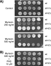
Deletion of SLM1 confers supersensitivity to myriocin. (A) Isogenic wild-type, slm1Δ, and slm2Δ mutant strains were grown to stationary phase in YPD medium, and serial dilutions of cultures were spotted on YPD plates containing either drug vehicle alone or the indicated concentrations of myriocin. (B) Addition of exogenous PHS (10 μM) to YPD plates containing 0.5 μg/ml myriocin rescues growth of the slm1Δ mutant. All plates were incubated at 30°C and photographed after 3 days.
FIG. 3.
FIG. 3.
Synthetic genetic interactions between slm1Δ mutants and mutants with mutations in the sphingolipid biosynthesis pathway. (A) Serial dilutions of isogenic wild-type, slm1Δ, csg2Δ, fen1Δ, and lcb4Δ single-mutant, and slm1Δ csg2Δ, slm1Δ fen1Δ, and slm1Δ lcb4Δ double-mutant yeast cultures were spotted on YPD plates. Plates were incubated at 26°C and 38°C and photographed after 3 days. (B) Exponentially growing wild-type, slm1Δ, csg2Δ, fen1Δ, and lcb4Δ single-mutant and slm1Δ csg2Δ, slm1Δ fen1Δ, and slm1Δ lcb4Δ double-mutant yeast cultures grown at 26°C were shifted to 38°C for 2 h. Cells were fixed and stained with Alexa594-phalloidin to visualize the actin cytoskeleton. (C) Small- to medium-budded cells from panel B were scored for their actin polarization state. Cells were classified as having an actin cytoskeleton that was polarized (containing cables and polarized actin patches), partially polarized (containing cables and partially polarized patches), or depolarized (containing no cables and depolarized patches). One hundred cells per sample were counted.
FIG. 4.
FIG. 4.
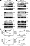
Slm1 and Slm2 phosphorylation in response to heat stress is dependent on sphingolipid synthesis. Western blot analysis of phosphorylated Slm1 (A) and Slm2 (B) proteins is shown. Cells expressing Slm1-TAP and Slm2-TAP were grown to mid-logarithmic phase in YPD medium at 26°C; aliquots of cells were then treated with vehicle alone (dimethyl sulfoxide), myriocin (2 μg/ml), or exogenous PHS (10 μM) for 30 min at RT and heat shock-shifted to 38°C for the indicated times. Cell extracts were prepared and subjected to TAP purification using IgG-Sepharose beads. Bound proteins were eluted with SDS-PAGE buffer, separated by SDS-PAGE, and immunoblotted with antibodies directed against phosphoserine (Q5) and phosphothreonine (Q7). Note that Slm2 Western blots were exposed to ECL reagents eight times longer than Slm1. (C) Quantitation of kinetics of Slm1 and Slm2 phosphorylation in response to heat stress in the presence or absence of drugs. Densitometric measurements of Western blots in panels A and B were made with the Image Gauge 4.0 program. Q5 and Q7 signals were normalized to the TAP signal for each time point. Results from two independent experiments are shown.
FIG. 5.
FIG. 5.
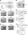
The Ca2+/calmodulin-dependent protein phosphatase calcineurin dephosphorylates Slm1. (A) Sequence alignment of Slm1 and Slm2 with known calcineurin binding sites. The consensus calcineurin-binding motif is given below the sequence alignment. (B) In vitro-transcribed and -translated 35S-labeled Cna1 was incubated with beads alone or with beads containing bound recombinant His6-Slm1 or His6-Slm2 proteins or Slm mutant variants lacking the calcineurin-binding motif. Bound 35S-labeled proteins that remained after washing were analyzed by SDS-PAGE and autoradiography. The molecular masses of protein markers are indicated on the left. (C) Cells expressing Slm1-TAP were grown to mid-logarithmic phase in YPD medium at 26°C, divided into aliquots, and treated for 30 min at RT with vehicle alone (dimethyl sulfoxide), 100 mM CaCl2, 10 μM BAPTA, or FK506 (2 μg/ml). Cells were shifted to 38°C for the indicated times, and cell extracts were prepared and subjected to TAP purification using IgG-Sepharose beads. Bound proteins were separated by SDS-PAGE and immunoblotted with antibodies directed against phosphoserine (Q5) and phosphothreonine (Q7). (D) Densitometric analysis of phosphorylation levels of Slm1-TAP from panel C. Western blots were digitized with the Image Gauge 4.0 program, and the levels of phosphorylation were normalized to the TAP signal at each time point. (E) Wild-type cells expressing Slm1-TAP or the Slm1ΔCN-TAP mutant from a low-copy-number vector under the control of the Gal1 promoter were grown at 26°C in the presence of galactose for 2.5 h to induce expression of the TAP fusion protein. Glucose was then added to shut off expression, and incubation was continued for 30 min at 26°C. Cells were then heat shocked at 38°C for the indicated times. Extracts were prepared and analyzed as described for panel C. (F) Densitometric analysis of phosphorylation levels of Slm1-TAP from panel E. Results from two independent experiments are shown.
FIG. 6.
FIG. 6.
FK506 and exogenous PHS rescue the lethality of slm1-ts slm2Δ mutant cells. Serial dilutions of isogenic wild type, and slm1-ts slm2Δ mutant yeast cultures were spotted on YPD plates either containing drug vehicle (Tween 20-ethanol [Control]) or supplemented with FK506 (2 μg/ml), PHS (10 μM), or both. Plates were incubated at 37°C and photographed after 3 days.
FIG. 7.
FIG. 7.
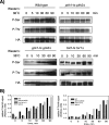
Slm1 phosphorylation is dependent on the sphingolipid-activated kinases Pkh1/2 and Pkh2 but is independent of Ypk1/2 and Tor2. (A) Exponentially growing wild-type or temperature-sensitive pkh1-ts pkh2Δ, ypk1-ts ypk2Δ (strain YPT40), or tor1Δ tor2-ts (strain SH121) mutant cells that contain HA-Slm1 under the control of a galactose-inducible promoter were grown at 26°C in the presence of galactose for 2.5 h to induce expression of HA-Slm1. Glucose was then added to shut off expression, and incubation was continued for 30 min at 26°C. Cells were then heat shocked at 38°C for the indicated times. Extracts were prepared and subjected to affinity purification on anti-HA-Sepharose beads. Bound proteins were separated by SDS-PAGE and immunoblotted with antibodies directed against phosphoserine (Q5), phosphothreonine (Q7), and HA. (B) Western blot densitometric analysis of phosphorylation levels of HA-Slm1. Blots were digitized with the Image Gauge 4.0 program, and the levels of phosphorylation were normalized to the HA signal.
FIG. 8.
FIG. 8.
Mutation of Ser659 abrogates Slm1 phosphorylation and Slm1-dependent survival under heat stress conditions. (A) Cells expressing HA-tagged Slm1S659A under control of the GAL1 promoter were grown at 26°C in the presence of galactose for 2.5 h to induce expression of HA-Slm1. Glucose was then added to shut off expression, and incubation was continued for 30 min at 26°C. Cells were then heat shocked at 38°C for the indicated times. Extracts were prepared and subjected to affinity purification on anti-HA-Sepharose beads. Bound proteins were separated by SDS-PAGE and immunoblotted with antibodies directed against phosphoserine (Q5), phosphothreonine (Q7), and HA. (B) Densitometric analysis of phosphorylation levels of HA-Slm1S659A from panel A. (C) Growth of strain JK520 transformed with LEU2 plasmids containing either wild-type SLM1 or the SLM1S659A and SLM1ΔC mutant variants at 30°C and 38°C on medium containing 5-FOA, which counterselects the SLM1 URA3 plasmid.
FIG. 9.
FIG. 9.
SLM1 overexpression partially corrects growth and actin polarization defects of pkh1-ts pkh2Δ mutant cells. (A) Vector alone or plasmids containing PKH1, SLM1, SLM1S659D, or SLM1ΔCN, under the control of the galactose-inducible GAL1 promoter, were transformed into the temperature-sensitive pkh1-ts pkh2Δ mutant strain. Growth of transformants was tested on synthetic complete medium containing galactose as the carbon source and supplemented with 1 M sorbitol (SG, Sorb). Plates were incubated at the indicated temperatures and photographed after 3 to 4 days. (B) pkh1-ts pkh2Δ cells containing the indicated plasmids were grown at 26°C in medium supplemented with 1 M sorbitol and galactose. Cells were then shifted to 38°C for 2 h and fixed, and the actin cytoskeleton was visualized with Alexa594-phalloidin. (C) Small- to medium-budded pkh1-ts pkh2Δ mutants cells transformed with the indicated plasmids from panel B were scored for their actin polarization state. Cells were classified as having an actin cytoskeleton that was polarized (containing cables and polarized actin patches), partially polarized (containing cables and partially polarized patches), or depolarized (containing no cables and depolarized patches). One hundred fifty cells per sample were counted.
FIG. 10.
FIG. 10.
Loss of Slm function results in defective delivery of the arginine transporter Can1 to the plasma membrane. (A) A plasmid encoding Can1p-GFP was expressed in wt (W303), slm1-ts slm2Δ, pkh1-ts pkh2Δ, erg24Δ, and lcb1-100 strains and Can1-GFP was visualized in living cells grown at the permissive (26°C) or nonpermissive (38°C) temperature for 1 h. Representative examples of GFP localization are shown. GFP signal is shown in the top panels; DIC images of the same cells are shown in the bottom panels. (B) Can1-GFP localization in slm1-ts slm2Δ mutant cells (strain JK515) containing empty vector (left and right panels) or SLM1 expressed from a high-copy-number vector under the control of the GPD promoter (2μm, TRP1; middle panel). Cells were either left untreated or incubated in the presence of PHS (7.5 μM) and FK506 (2 μg/ml) for 1 h at 26°C before being shifted to 38°C for 2 h, and GFP fluorescence was visualized. (C) Can1-GFP localization is shown in pkh1-ts pkh2Δ mutant cells containing empty vector (left panel) or SLM1 expressed from a high-copy-number vector under the control of the GPD promoter (2μm, TRP1; right panel). Vector-transformed cells were left untreated, whereas cells expressing SLM1 were incubated in the presence of FK506 (2 μg/ml) for 1 h at 26°C before being shifted to 38°C for 2 h, and GFP fluorescence was visualized. (D) Serial dilutions of wild-type (W303), lcb1-100, and slm1-ts slm2Δ (JK515) strains transformed with empty vector or pCAN1-GFP were plated on media lacking Arg to verify plating (left panel) and on medium lacking Arg and containing the arginine analog canavanine (0.5 μg/ml). Plates were incubated at 30°C for 3 days. (E) Wild-type cells (W303) containing a plasmid encoding for Can1p-GFP were treated with latrunculin A (5 μM) at 30°C, and GFP fluorescence in living cells was recorded at the indicated times.

Similar articles

Cited by

References

    1. Audhya, A., and S. D. Emr. 2002. Stt4 PI 4-kinase localizes to the plasma membrane and functions in the Pkc1-mediated MAP kinase cascade. Dev. Cell 2:593-605. - PubMed
    1. Audhya, A., M. Foti, and S. D. Emr. 2000. Distinct roles for the yeast phosphatidylinositol 4-kinases, Stt4p and Pik1p, in secretion, cell growth, and organelle membrane dynamics. Mol. Biol. Cell 11:2673-2689. - PMC - PubMed
    1. Audhya, A., R. Loewith, A. B. Parsons, L. Gao, M. Tabuchi, H. Zhou, C. Boone, M. N. Hall, and S. D. Emr. 2004. Genome-wide lethality screen identifies new PI4,5P2 effectors that regulate the actin cytoskeleton. EMBO J. 23:3747-3757. - PMC - PubMed
    1. Bagnat, M., S. Keranen, A. Shevchenko, and K. Simons. 2000. Lipid rafts function in biosynthetic delivery of proteins to the cell surface in yeast. Proc. Natl. Acad. Sci. USA 97:3254-3259. - PMC - PubMed
    1. Beeler, T., D. Bacikova, K. Gable, L. Hopkins, C. Johnson, H. Slife, and T. Dunn. 1998. The Saccharomyces cerevisiae TSC10/YBR265w gene encoding 3-ketosphinganine reductase is identified in a screen for temperature-sensitive suppressors of the Ca2+-sensitive csg2Delta mutant. J. Biol. Chem. 273:30688-30694. - PubMed

Publication types

MeSH terms