. 2007 Jan;27(2):579-94.
doi: 10.1128/MCB.01192-06. Epub 2006 Nov 13.

Distinct promoters mediate the regulation of Ebf1 gene expression by interleukin-7 and Pax5

Affiliations

Distinct promoters mediate the regulation of Ebf1 gene expression by interleukin-7 and Pax5

Stephanie Roessler et al. Mol Cell Biol. 2007 Jan.

Abstract

Early differentiation of B lymphocytes requires the function of multiple transcription factors that regulate the specification and commitment of the lineage. Loss- and gain-of-function experiments have provided important insight into the transcriptional control of B lymphopoiesis, whereby E2A was suggested to act upstream of EBF1 and Pax5 downstream of EBF1. However, this simple hierarchy cannot account for all observations, and our understanding of a presumed regulatory network, in which transcription factors and signaling pathways operate, is limited. Here, we show that the expression of the Ebf1 gene involves two promoters that are differentially regulated and generate distinct protein isoforms. We find that interleukin-7 signaling, E2A, and EBF1 activate the distal Ebf1 promoter, whereas Pax5, together with Ets1 and Pu.1, regulates the stronger proximal promoter. In the absence of Pax5, the function of the proximal Ebf1 promoter and accumulation of EBF1 protein are impaired and the replication timing and subcellular localization of the Ebf1 locus are altered. Taken together, these data suggest that the regulation of Ebf1 via distinct promoters allows for the generation of several feedback loops and the coordination of multiple determinants of B lymphopoiesis in a regulatory network.


Figures
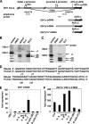
FIG. 1.
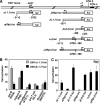
FIG. 1.
The Ebf1 gene is regulated by two promoters, the distal α and proximal β promoters. (A) Schematic overview of the distal Ebf1α and proximal Ebf1β promoters. Two alternative first exons, exon 1a and exon 1b, are indicated. The proximal Ebf1β promoter is located within the first intron of Ebf1α mRNA and 879 nucleotides upstream of the ATG-β, which is spliced out in Ebf1α mRNA. Ebf1α mRNA and Ebf1β mRNA can be distinguished by RT-PCR with primer pairs specific for exon 1a or exon 1b sequences. Indicated are the positions of the PCR amplicons and of the primer used for primer extensions and S1 nuclease protection experiments. (B) Primer extension experiments show multiple bands corresponding to potential 5′ ends of Ebf1β mRNA located 879 nucleotides (nt) upstream of the ATG-β. Potential transcriptional start sites are visible only in 70/Z3 pre-B cells and in spleen, not in Ebf1-negative EL4 T cells. (C) S1 nuclease protection experiments show a similar pattern of protected Ebf1β 5′ ends as in the primer extension experiments. nt, nucleotides. (D) Alignment of the human and mouse Ebf1β promoter transcriptional start sites. (E) Quantitative RT-PCR analysis of EL4 T cells and sorted lymphoid cells shows the upregulation of Ebf1 mRNA during early B-cell development. HSC (Lin IL-7R+ c-kithigh Sca-1high), CLP (Lin IL-7R+ c-kitlow Sca-1low), pro-B cell Fr. A (B220+ AA4.1+ c-kit+ CD19) and Fr. B and Fr. C according to the work of Hardy and Hayakawa (18) (Fr. B, B220+ CD43+ HSA+ BP-1; Fr. C, B220+ CD43+ HSA+ BP-1+), and marginal zone (MZ; B220+ CD21high CD23low) and follicular (FO; B220+ CD21low CD23high) B cells have been sorted by FACS to more than 98% purity. Ebf1 mRNA levels were normalized to β-actin, and the expression level of HSC was set to 1. (F) Quantitative RT-PCR analysis of endogenous levels of Ebf1α and Ebf1β mRNA. The ratio of Ebf1β to Ebf1α mRNA is displayed. Ebf1β mRNA is predominantly expressed in all analyzed cell types, and the ratio increases during pro-B-cell development, with the highest ratio being in fraction C pre-B cells.
FIG. 2.
FIG. 2.
Cloning of the proximal Ebf1β promoter and comparison with the distal Ebf1α promoter. (A) Schematic overview of the Ebf1α and Ebf1β promoter luciferase reporter constructs. (B) Comparison of the promoter activities of the two Ebf1 promoters in transient-transfection assays of B- and non-B-cell lines. Three micrograms of α(−1.1)-luc, β(−1.7)-luc, or pGl3-luc was transfected together with 1 μg cytomegalovirus β-galactosidase reporter into PD36 pre-B cells, Raji mature B cells, 3T3L1 preadipocytes, EL4 pre-T cells, or NIH 3T3 fibroblasts. The activation (n-fold) compared to that of pGl3-luc is depicted. (C) Transient transfection of Ebf1β promoter constructs into Raji B cells. The empty-vector control pGl3-luc was compared to the Ebf1 promoter constructs α(−1.1)-luc, α(flip)-luc, β(−1.7)-luc, β(flip)-luc, β(−0.5)-luc, α-β-luc, and α(flip)-β-luc. Three micrograms of each luciferase construct was transfected together with 1 μg cytomegalovirus β-galactosidase reporter into Raji B cells. The levels of luciferase activity, normalized to the activity of the cotransfected β-galactosidase reporter, are expressed as activation (n-fold) relative to the luciferase levels of cells transfected with pGl3-luc. All transfections were performed at least three times, and results from representative experiments with standard deviations are shown.
FIG. 3.
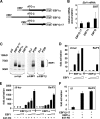
FIG. 3.
The two isoforms of EBF1 protein, EBF1α and EBF1β, differ in the first 14 amino acids but have the same transactivation potential. (A) Schematic overview of EBF1 protein expression vectors. EBF1α and EBF1β comprise the full 5′ UTR of the Ebf1α and Ebf1β mRNA, respectively, whereas EBF1β-17 differs from EBF1β in its 5′ UTR, which comprises only 45 nucleotides. (B) Real-time RT-PCR of 293 cells transfected with 2 μg of EBF1β-17, EBF1α, or EBF1β together with LEF1 expression vector. Forty-eight hours after transfection Ebf1 and Lef1 mRNA levels were analyzed by RT-PCR and Ebf1 mRNA levels were normalized to Lef1 mRNA. (C) Immunoblot analysis of EBF1 isoforms was performed by transfecting 2 μg of EBF1β-17, EBF1α, EBF1β, or empty vector into 293 cells. After 48 h cells were harvested and total cell extracts were subjected to anti-myc immunoblotting. Cell extracts of EL4 T cells and PD36 B cells were analyzed by anti-EBF1 and anti-EBF1β immunoblotting. (D) Ba/F3 cells were transiently transfected with 2 μg of λ5 luciferase reporter construct, containing nucleotides −299 to +131 of the λ5 gene, together with 1 μg cytomegalovirus β-galactosidase reporter and 0, 0.3, 1, or 3 μg of EBF1β or EBF1α expression plasmids as indicated. (E) Ba/F3 cells were transiently transfected with 2 μg of λ5 luciferase reporter construct together with 1 μg cytomegalovirus β-galactosidase reporter; 3 μg of E47-FD; and 0.3, 1, or 3 μg of EBF1β-17 or EBF1α expression plasmids alone or in pairwise combinations as indicated. (F) Transient transfection of Ba/F3 cells with empty vector only or 3 μg E47-FD in combination with 3 μg EBF1β-17 or EBF1α followed by quantitative RT-PCR of endogenous λ5 mRNA. λ5 mRNA levels were normalized to β-actin mRNA, and the expression level of the empty-vector control was set to 1. Error bars represent the standard deviations of the means of three experiments.
FIG. 4.
FIG. 4.
Activation of the Ebf1α promoter by STAT5, EBF1, and E47. (A) Transient transfection of Ba/F3 cells with 10 μg STAT5-CA expression plasmid or empty vector. Ebf1 mRNA levels were determined by quantitative RT-PCR analysis and normalized to β-actin mRNA. The expression level of the empty-vector control was set to 1. (B) Transfection of Ba/F3 cells with 3 μg luciferase reporter constructs containing either multimerized STAT-binding sites of the cyclin D1 promoter (D1), Ebf1α(−1.1) promoter sequences (Ebf1α), or the Ebf1β(−1.7) promoter (Ebf1β), together with 1 μg Rous sarcoma virus β-galactosidase reporter as a transfection control. The cells were incubated with or without IL-3 for 12 h prior to the luciferase and β-galactosidase assays. (C) Ba/F3 cells were transiently transfected with 3× D1-SIE1-luc, Ebf1α(−1.1)-luc, or Ebf1β(−1.7)-luc, together with 1 μg Rous sarcoma virus β-galactosidase reporter and increasing amounts of STAT5-CA (0, 3, or 10 μg). IL-3 was withdrawn 12 h prior to the luciferase and β-galactosidase assays. (D) Transient transfection of Ba/F3 cells with empty vector or with 3 μg E47-FD and 3 μg EBF1β-17, followed by quantitative RT-PCR to detect endogenous Ebf1α or Ebf1β mRNA. mRNA levels were normalized to β-actin, and the expression level of the empty-vector control was set to 1. Error bars represent the standard deviations of the means of three experiments. (E) Real-time RT-PCR analysis of fraction B pro-B cells of Ebf1+/ mice show a reduction of Ebf1α, Ebf1β, and total Ebf1 mRNA, relative to wild-type (wt) fraction B pro-B cells (Fr. B; B220+ CD43+ HSA+ BP-1). Error bars represent the standard deviations of the means of three experiments. (F) Schematic overview of the Ebf1β-luc and Ebf1β(Pu.1mut)-luc promoter luciferase reporter constructs. (G) Transient transfection of HeLa cells with 300 ng of the Ebf1β-luc promoter construct together with 0, 0.3, 1, or 3 μg of Pu.1 and 200 ng of cytomegalovirus β-galactosidase reporter.
FIG. 5.
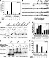
FIG. 5.
Pax5 and Ets1 collaborate to activate the Ebf1β but not Ebf1α promoter. (A) Ba/F3 cells were transiently transfected with 1 μg cytomegalovirus β-galactosidase reporter and 2 μg of Ebf1α or Ebf1β luciferase reporter construct, together with 3 μg of Pax5 and 3 μg of Ets1 expression plasmids alone or in pairwise combinations as indicated. (B) Overview of the Ebf1β promoter luciferase reporter constructs. Indicated are the Pax5-binding sites at positions −318, −25, +222, and +257 and the Ets1-binding sites at −75 and +303. (C) Electrophoretic mobility shift assays show binding of Pax5 to binding sites at −318, +222, and +257 in the Ebf1β promoter. Recombinant Pax5 protein (0, 50, or 200 ng) was incubated with labeled wild-type or mutant oligonucleotide probes. (D) Mutation of all four Pax5-binding sites in the Ebf1β promoter leads to a twofold reduction of transactivation by Pax5. Ba/F3 cells were transiently transfected with 1 μg cytomegalovirus β-galactosidase reporter and 2 μg of Ebf1β(0.5)-luc or Ebf1β(Pax5mut)-luc reporter construct, together with increasing amounts of Pax5 expression plasmids (0.3, 1, and 3 μg). (E) Electrophoretic mobility shift assays show binding of Ets1 to binding sites at −75 and +303 in the Ebf1β promoter. The indicated amount of recombinant Ets1 protein (0, 100, or 300 ng) was incubated with labeled oligonucleotide probes and assayed in the presence of a 200-fold excess of specific (mb1) or unspecific competitor. (F) Mutation of the Ets1-binding sites at positions −75 and +303 reduces fourfold the potential transactivation by Ets1 and Pax5 on the Ebf1β promoter. Ba/F3 cells were transiently transfected with 1 μg cytomegalovirus β-galactosidase reporter and 2 μg of Ebf1β(0.2)-luc or Ebf1β(Ets1mut)-luc reporter construct, together with 3 μg of Pax5 and 3 μg of Ets1 expression plasmids as indicated.
FIG. 6.
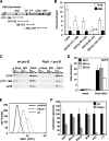
FIG. 6.
Binding of Pax5 to Ebf1β regulatory sequences in vivo. (A) Schematic representation of the Ebf1β promoter and regions analyzed by ChIP. (B) ChIP was performed in the indicated cell lines with anti-Pax5 and the control IgG antibody. Enrichment (n-fold) comparing immunoprecipitate with the specific anti-Pax5 antibody and the control IgG was determined by real-time PCR; n.d., not detected. (C) Semiquantitative PCR was performed with serial dilutions of template DNA from cultured bone marrow pro-B cells of wild-type (wt) and Pax5−/− mice. Binding can be detected with anti-Pax5 antibodies but not with nonspecific IgG on both the Ebf1β and CD19 promoters. The anti-Pax5 antibodies do not bind to the JunB promoter. (D) Transient transfection of Ba/F3 cells with empty vector only or with 3 μg Pax5 and 3 μg Ets1 followed by quantitative RT-PCR of endogenous Ebf1α, Ebf1β, or total Ebf1 mRNA. mRNA levels were normalized to β-actin, and the expression level of the empty-vector control was set to 1. Error bars represent the standard deviations of the means of three experiments. (E) IL-7-cultured pro-B cells of Pax5-deficient mice have reduced EBF1 protein levels. Intracellular staining of IL-7-cultured pro-B cells was performed with monoclonal anti-EBF1 antibody and secondary anti-rat-FITC antibody. For the control staining only secondary antibody was used. (F) Fraction B pro-B cells of mice deficient for Pax5 show a reduction of Ebf1β mRNA and total Ebf1 mRNA. Real-time RT-PCR analysis was performed with sorted fraction B cells of wild-type (wt) and Pax5−/− mouse fetal liver (B220+ CD43+ HSA+ BP-1). Ebf1α, Ebf1β, and total Ebf1 mRNAs have been amplified by oligonucleotides specific for exon 1a, exon 1b, or both isoforms. As a control, RT-PCR of B29, mb1, and λ5 was performed. mRNA levels were normalized to β-actin, and the expression level of the wild type was set to 100%. Error bars represent the standard deviations of the means of three experiments.
FIG. 7.
FIG. 7.
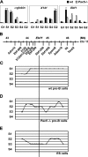
Replication timing of the Ebf1 locus is shifted to later replication in Pax5-deficient cells. (A) Replication timing of the early-replicating α-globin gene locus, the late-replicating X141 locus, and the Ebf1 locus in wild-type (wt) and Pax5-deficient pro-B cells is depicted. (B) Schematic overview of the Ebf1 locus. The locations of the primer pairs used for analysis of the replication timing are indicated. (C) In wild-type pro-B cells, Ebf1 replication peaked at the first quarter of S phase (S1). (D) Pax5-negative pro-B cells display later replication (S2) of the Ebf1 locus. (E) An even later replication was observed in ES cells, where Ebf1 is silent. A domain of later replication “centered” at Ebf1 was observed in Pax5-negative and ES cells, which extended to an area of more than 1 Mb, spanning the 37324131I11Rik and EpsinR genes that are active in both pro-B and ES cells.
FIG. 8.
FIG. 8.
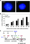
Fluorescence in situ hybridization shows differential Ebf1 locus positioning in Pax5-deficient compared to wild-type (wt) pro-B cells. (A) Representative images of 2D-FISH analysis of Pax5-deficient and wild-type pro-B cells. (B) Analysis of the nuclear position of the Ebf1 locus shows that the Ebf1 locus is more often detected at the periphery of the nucleus in Pax5-deficient than in wild-type pro-B cells. (C) A complex regulatory network regulates the expression of the two Ebf1 promoters.

Similar articles

Cited by

References

    1. Arney, K. L., and A. G. Fisher. 2004. Epigenetic aspects of differentiation. J. Cell Sci. 117:4355-4363. - PubMed
    1. Azuara, V., K. E. Brown, R. R. Williams, N. Webb, N. Dillon, R. Festenstein, V. Buckle, M. Merkenschlager, and A. G. Fisher. 2003. Heritable gene silencing in lymphocytes delays chromatid resolution without affecting the timing of DNA replication. Nat. Cell Biol. 5:668-674. - PubMed
    1. Bain, G., E. C. Maandag, D. J. Izon, D. Amsen, A. M. Kruisbeek, B. C. Weintraub, I. Krop, M. S. Schlissel, A. J. Feeney, M. van Roon, et al. 1994. E2A proteins are required for proper B cell development and initiation of immunoglobulin gene rearrangements. Cell 79:885-892. - PubMed
    1. Bertolino, E., K. Reddy, K. L. Medina, E. Parganas, J. Ihle, and H. Singh. 2005. Regulation of interleukin 7-dependent immunoglobulin heavy-chain variable gene rearrangements by transcription factor STAT5. Nat. Immunol. 6:836-843. - PubMed
    1. Chambeyron, S., N. R. Da Silva, K. A. Lawson, and W. A. Bickmore. 2005. Nuclear re-organisation of the Hoxb complex during mouse embryonic development. Development 132:2215-2223. - PubMed

Publication types

MeSH terms

LinkOut - more resources