. 2007 Jan;27(2):758-67.
doi: 10.1128/MCB.01470-06. Epub 2006 Nov 13.

Variable and tissue-specific subunit composition of mitochondrial m-AAA protease complexes linked to hereditary spastic paraplegia

Affiliations

Variable and tissue-specific subunit composition of mitochondrial m-AAA protease complexes linked to hereditary spastic paraplegia

Mirko Koppen et al. Mol Cell Biol. 2007 Jan.

Abstract

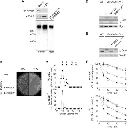
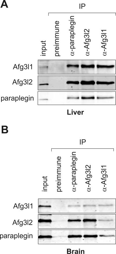
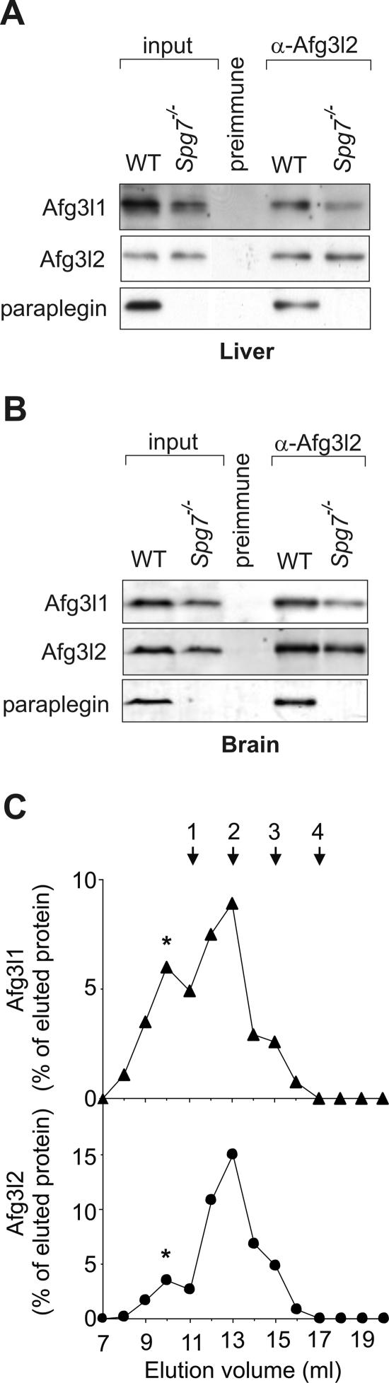
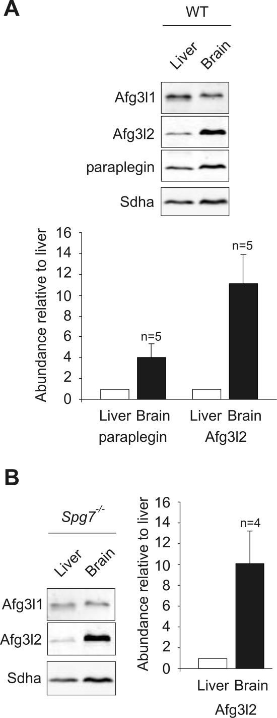
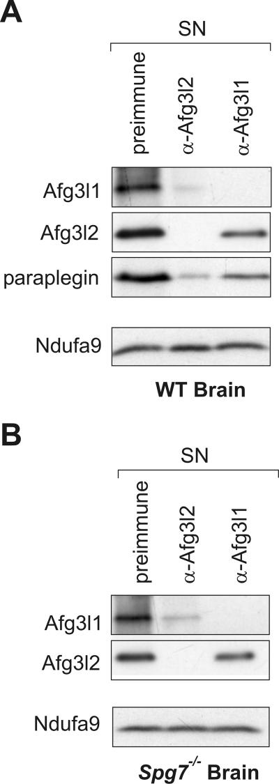
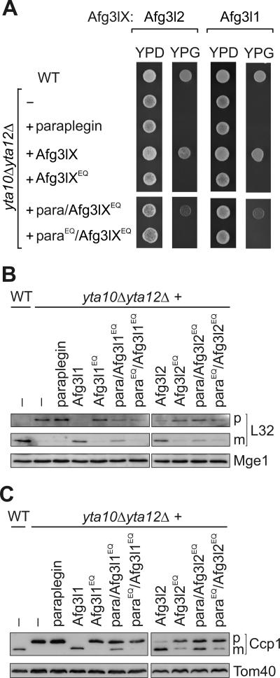
The m-AAA protease, an ATP-dependent proteolytic complex in the mitochondrial inner membrane, controls protein quality and regulates ribosome assembly, thus exerting essential housekeeping functions within mitochondria. Mutations in the m-AAA protease subunit paraplegin cause axonal degeneration in hereditary spastic paraplegia (HSP), but the basis for the unexpected tissue specificity is not understood. Paraplegin assembles with homologous Afg3l2 subunits into hetero-oligomeric complexes which can substitute for yeast m-AAA proteases, demonstrating functional conservation. The function of a third paralogue, Afg3l1 expressed in mouse, is unknown. Here, we analyze the assembly of paraplegin into m-AAA complexes and monitor consequences of paraplegin deficiency in HSP fibroblasts and in a mouse model for HSP. Our findings reveal variability in the assembly of m-AAA proteases in mitochondria in different tissues. Homo-oligomeric Afg3l1 and Afg3l2 complexes and hetero-oligomeric assemblies of both proteins with paraplegin can be formed. Yeast complementation studies demonstrate the proteolytic activity of these assemblies. Paraplegin deficiency in HSP does not result in the loss of m-AAA protease activity in brain mitochondria. Rather, homo-oligomeric Afg3l2 complexes accumulate, and these complexes can substitute for housekeeping functions of paraplegin-containing m-AAA complexes. We therefore propose that the formation of m-AAA proteases with altered substrate specificities leads to axonal degeneration in HSP.


Figures

FIG. 1.
FIG. 1.
hAFG3L2 forms a homo-oligomeric m-AAA protease complex with proteolytic activity. (A) A homo-oligomeric hAFG3L2 complex in mitochondria isolated from HSP fibroblasts. Mitochondria were solubilized in digitonin and analyzed by SDS-PAGE (50 μg mitochondrial protein; upper panel) or BN-PAGE (150 μg mitochondrial protein; lower panel), followed by immunoblotting using paraplegin-specific (upper panel) and hAFG3L2-specific (upper and lower panel) antisera. For the control, yeast yta10Δ yta12Δ mitochondria containing hAFG3L2 were fractionated by BN-PAGE in parallel. Thyroglobulin (669 kDa) and apoferritin (443 kDa) were used for calibration. (B) Maintenance of respiratory growth of yta10Δ yta12Δ yeast cells by hAFG3L2. Wild-type (WT) cells, yta10Δ yta12Δ cells, and yta10Δ yta12Δ cells expressing hAFG3L2 or the proteolytically inactive variant hAFG3L2E575Q (hAFG3L2EQ), were grown on fermentable (yeast extract-peptone-dextrose [YPD]) and nonfermentable (glycerol-containing media [YPG]) carbon sources at 30°C. (C) Homo-oligomeric hAFG3L2 complexes in yta10Δ yta12Δ mitochondria. hAFG3L2 or hAFG3L2E575Q (hAFG3L2EQ) were expressed in yta10Δ yta12Δ cells, and mitochondria were isolated. After solubilization in digitonin-containing buffer, mitochondrial extracts (1 mg mitochondrial protein) were fractionated by Superose 6 sizing chromatography. Eluate fractions were TCA precipitated and analyzed by SDS-PAGE and immunoblotting using hAFG3L2-specific antibodies. hAFG3L2 (filled squares) or hAFG3L2E575Q (filled circles) present in eluate fractions was quantified by laser densitometry, and results are given as a percent of the respective protein in the total eluate. A smaller hAFG3L2-containing complex, marked with an asterisk, most likely results from partial dissociation of the large complex. The following marker proteins were used for calibration: 1, Hsp60 (840 kDa); 2, apoferritin (443 kDa); 3, alcohol dehydrogenase (150 kDa); 4, bovine serum albumine (66 kDa). (D and E) Processing of yeast MrpL32 and Ccp1 by hAFG3L2. Protein processing was analyzed in wild-type (WT) cells, yta10Δ yta12Δ cells, and yta10Δ yta12Δ cells expressing either hAFG3L2 or hAFG3L2E575Q (hAFG3L2EQ) by SDS-PAGE. (D) Maturation of MrpL32 (L32) was monitored in isolated mitochondria (30 μg mitochondrial protein) by immunoblotting using polyclonal antisera directed against the mature and the precursor forms of MrpL32 and, as a loading control, against matrix-localized Mge1. (E) Ccp1 processing was examined in cell extracts using antisera directed against Ccp1 and, for control, the outer membrane protein Tom40. p, precursor; m, mature form. (F) Degradation of nonassembled membrane proteins by hAFG3L2 expressed in yeast. Radiolabeled Yme2ΔC (upper panel) or Atp7 (lower panel) was imported for 10 min at 25°C into mitochondria isolated from the yeast strains used for panels D and E. The stability of newly imported proteins at 37°C was determined by SDS-PAGE and autoradiography. The average of three independent experiments (± standard error of the mean) is shown. Filled squares, wild type; filled triangles, yta10Δ yta12Δ; open triangles, yta10Δ yta12Δ+hAFG3L2; open squares, yta10Δ yta12Δ+hAFG3L2E575Q.
FIG. 2.
FIG. 2.
Assembly of paraplegin, Afg3l1, and Afg3l2 in murine mitochondria. Mitochondria (400 μg mitochondrial protein) were isolated from (A) liver and (B) brain and solubilized with digitonin. Ten (liver) or 20% (brain) of the sample was removed for control (input). Coimmunoprecipitations were carried out using affinity-purified paraplegin-, Afg3l1-, and Afg3l2-specific polyclonal antibodies. Preimmune antiserum was used as a negative control. Immunoprecipitates (IP) were analyzed by SDS-PAGE and immunoblotting.
FIG. 3.
FIG. 3.
A hetero-oligomeric Afg3l1/Afg3l2 complex in mitochondria of Spg7−/− mice. Digitonin extracts of mitochondria (400 μg mitochondrial protein) from (A) liver and (B) brain of wild-type (WT) and paraplegin-deficient Spg7−/− mice were subjected to coimmunoprecipitations using affinity-purified Afg3l2-specific polyclonal antibodies as described in the legend for Fig. 2. Preimmune serum was used as a negative control. Precipitates were analyzed by SDS-PAGE and immunoblotting using paraplegin-, Afg3l1-, and Afg3l2-specific polyclonal antibodies. The amounts of Afg3l1 in the precipitates derived from wild-type and Spg7−/− mitochondria varied slightly in different experiments. (C) A high-molecular-mass complex containing Afg3l1 and Afg3l2 in Spg7−/− mitochondria. Extracts (1 mg mitochondrial protein) of liver mitochondria isolated from Spg7−/− mice were fractionated by Superose 6 sizing chromatography. Eluate fractions were TCA precipitated and analyzed by SDS-PAGE and immunoblotting using Afg3l1- and Afg3l2-specific polyclonal antibodies. Afg3l1 (filled triangles) and Afg3l2 (filled circles) in the eluate were quantified by laser densitometry and are given as a percent of the respective protein in the total eluate. An asterisk marks a high-molecular-mass complex of Afg3l1 and Afg3l2 which coelutes with the prohibitins Phb1 the prohibitins Phb1 and Phb2, suggesting an interaction of the m-AAA protease complex with prohibitins, as observed in yeast mitochondria (31). The following marker proteins were used for calibration: 1, dimeric ATP synthase (1.5 MDa); 2, monomeric ATP synthase (750 kDa); 3, apoferritin (443 kDa); 4, alcohol dehydrogenase (150 kDa).
FIG. 4.
FIG. 4.
Relative abundance of paraplegin, Afg3l1 and Afg3l2 in liver and brain mitochondria. Mitochondria were isolated from (A) wild-type or (B) Spg7−/− murine liver and brain. Extracts containing equal amounts of Afg3l1 were analyzed by SDS-PAGE and immunoblotting using paraplegin-, Afg3l1-, and Afg3l2-specific antibodies as well as a monoclonal antibody directed against the 70-kDa subunit of succinate dehydrogenase (Sdha). Paraplegin and Afg3l2 present in liver and brain mitochondria were quantified by fluorescence-based imaging using Afg3l1 as a loading control. The ratio of signals detected for Afg3l2 and paraplegin in brain versus liver mitochondria was calculated to monitor the relative abundance of m-AAA protease subunits in both tissues. The average of n independent experiments (± standard error of the mean) using tissues isolated from different, randomly chosen animals is shown.
FIG. 5.
FIG. 5.
Coexistence of m-AAA proteases built up of different subunits in brain mitochondria. Mitochondria (150 μg mitochondrial protein) from (A) wild-type (WT) and (B) Spg7−/− brain were lysed in digitonin and incubated with saturating amounts of preimmune serum or affinity-purified Afg3l2- or Afg3l1C-specific antibodies. After the removal of the precipitate, supernatant fractions (SN) were analyzed by SDS-PAGE and examined for the presence of paraplegin, Afg3l1, and Afg3l2 by immunoblotting. A monoclonal antibody directed against the 39-kDa subunit of complex I (Ndufa9) was used to control for equal gel loading. Whereas Afg3l1 and Afg3l2 could be completely depleted from the supernatant fraction by using affinity-purified Afg3l1- or Afg3l2-specific antibodies, respectively, extracts could not be depleted of paraplegin using available paraplegin-specific antibodies (data not shown).
FIG. 6.
FIG. 6.
Proteolytic activity of homo-oligomeric and hetero-oligomeric m-AAA protease complexes in yeast. (A) Respiratory growth of yta10Δ yta12Δ cells expressing murine m-AAA protease subunits. Wild-type (WT) cells, yta10Δ yta12Δ cells, and yta10Δ yta12Δ cells expressing either paraplegin, Afg3l1 (Afg3lX), Afg3l2 (Afg3lX), or their mutant variants Afg3l1E567Q or Afg3l2E574Q (Afg3lXEQ) were grown at 30°C on glucose-containing (YPD) or glycerol-containing (YPG) media to examine the respiratory competence of the cells. To assess the activity of hetero-oligomeric complexes, the mutant variants Afg3l1E567Q or Afg3l2E574Q were coexpressed with paraplegin (para/Afg3lXEQ) or with parapleginE575Q (paraEQ/Afg3lXEQ) in yta10Δ yta12Δ cells and cell growth was analyzed as above. (B and C) Processing of yeast MrpL32 and Ccp1 by murine m-AAA proteases. Protein processing was analyzed in yta10Δ yta12Δ cells harboring murine m-AAA protease subunits by SDS-PAGE as described for panel A and immunoblotting as described in the legend for Fig. 1 by using MrpL32 (L32)-specific antisera (B) or Ccp1-specific antisera (C). p, precursor forms; m, mature forms. Mge1 and Tom40 were used to control for equal gel loading.

References

    1. Alexander, C., M. Votruba, U. E. Pesch, D. L. Thiselton, S. Mayer, A. Moore, M. Rodriguez, U. Kellner, B. Leo-Kottler, G. Auburger, S. S. Bhattacharya, and B. Wissinger. 2000. OPA1, encoding a dynamin-related GTPase, is mutated in autosomal dominant optic atrophy linked to chromosome 3q28. Nat. Genet. 26:211-215. - PubMed
    1. Antonicka, H., F. Sasarman, N. G. Kennaway, and E. A. Shoubridge. 2006. The molecular basis for tissue specificity of the oxidative phosphorylation deficiencies in patients with mutations in the mitochondrial translation factor EFG1. Hum. Mol. Genet. 15:1835-1846. - PubMed
    1. Arlt, H., G. Steglich, R. Perryman, B. Guiard, W. Neupert, and T. Langer. 1998. The formation of respiratory chain complexes in mitochondria is under the proteolytic control of the m-AAA protease. EMBO J. 17:4837-4847. - PMC - PubMed
    1. Arlt, H., R. Tauer, H. Feldmann, W. Neupert, and T. Langer. 1996. The YTA10-12-complex, an AAA protease with chaperone-like activity in the inner membrane of mitochondria. Cell 85:875-885. - PubMed
    1. Atorino, L., L. Silvestri, M. Koppen, L. Cassina, A. Ballabio, R. Marconi, T. Langer, and G. Casari. 2003. Loss of m-AAA protease in mitochondria causes complex I deficiency and increased sensitivity to oxidative stress in hereditary spastic paraplegia. J. Cell Biol. 163:777-787. - PMC - PubMed

Publication types

MeSH terms