. 2006 Nov 28;114(22):2342-50.
doi: 10.1161/CIRCULATIONAHA.105.598524. Epub 2006 Nov 13.

X-ray fused with magnetic resonance imaging (XFM) to target endomyocardial injections: validation in a swine model of myocardial infarction

Affiliations

X-ray fused with magnetic resonance imaging (XFM) to target endomyocardial injections: validation in a swine model of myocardial infarction

Ranil de Silva et al. Circulation. .

Abstract

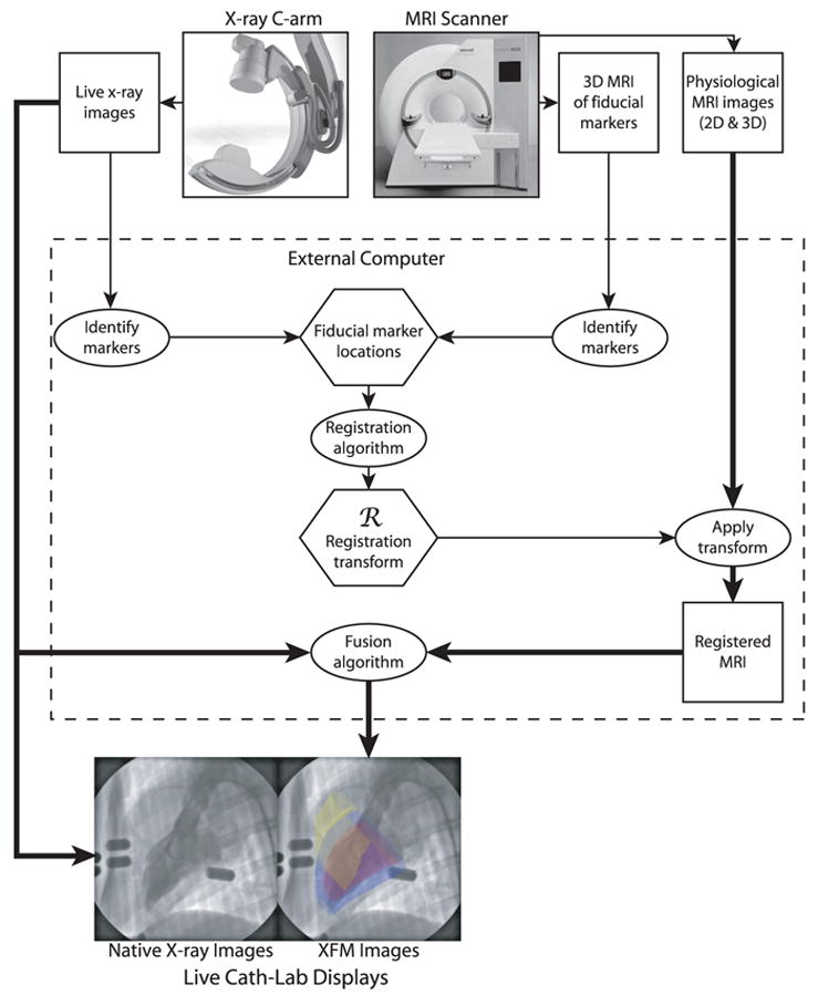
Background: Magnetic resonance imaging (MRI) permits 3-dimensional (3D) cardiac imaging with high soft tissue contrast. X-ray fluoroscopy provides high-resolution, 2-dimensional (2D) projection imaging. We have developed real-time x-ray fused with MRI (XFM) to guide invasive procedures that combines the best features of both imaging modalities. We tested the accuracy of XFM using external fiducial markers to guide endomyocardial cell injections in infarcted swine hearts.

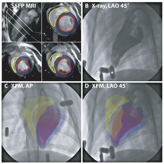
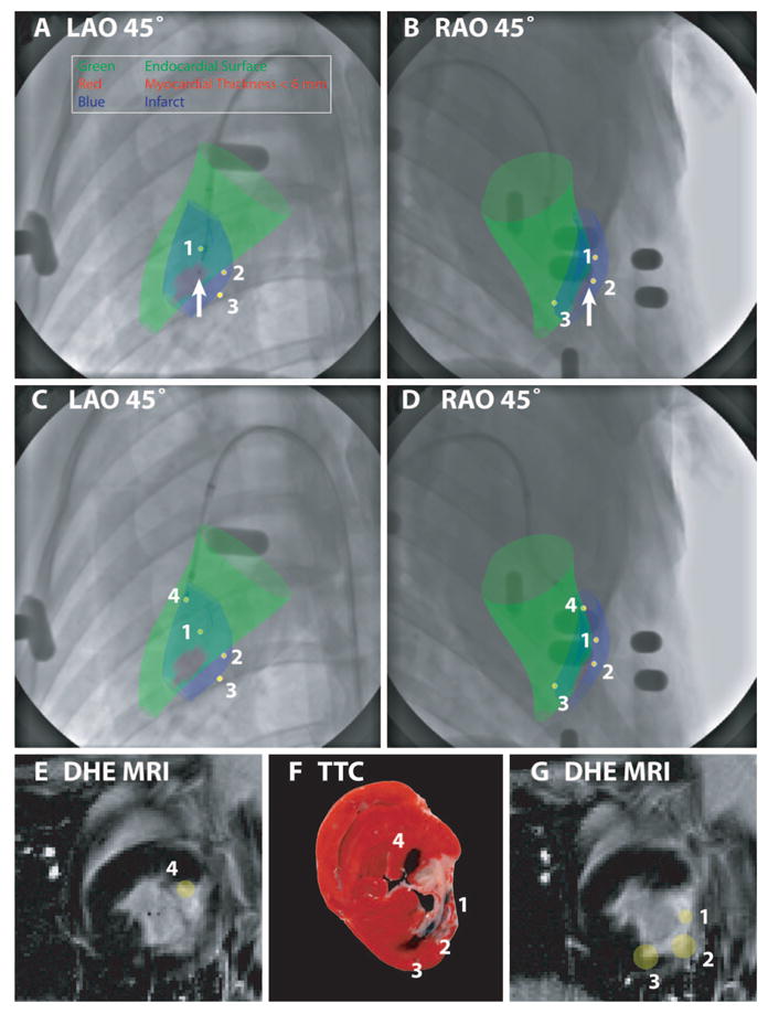
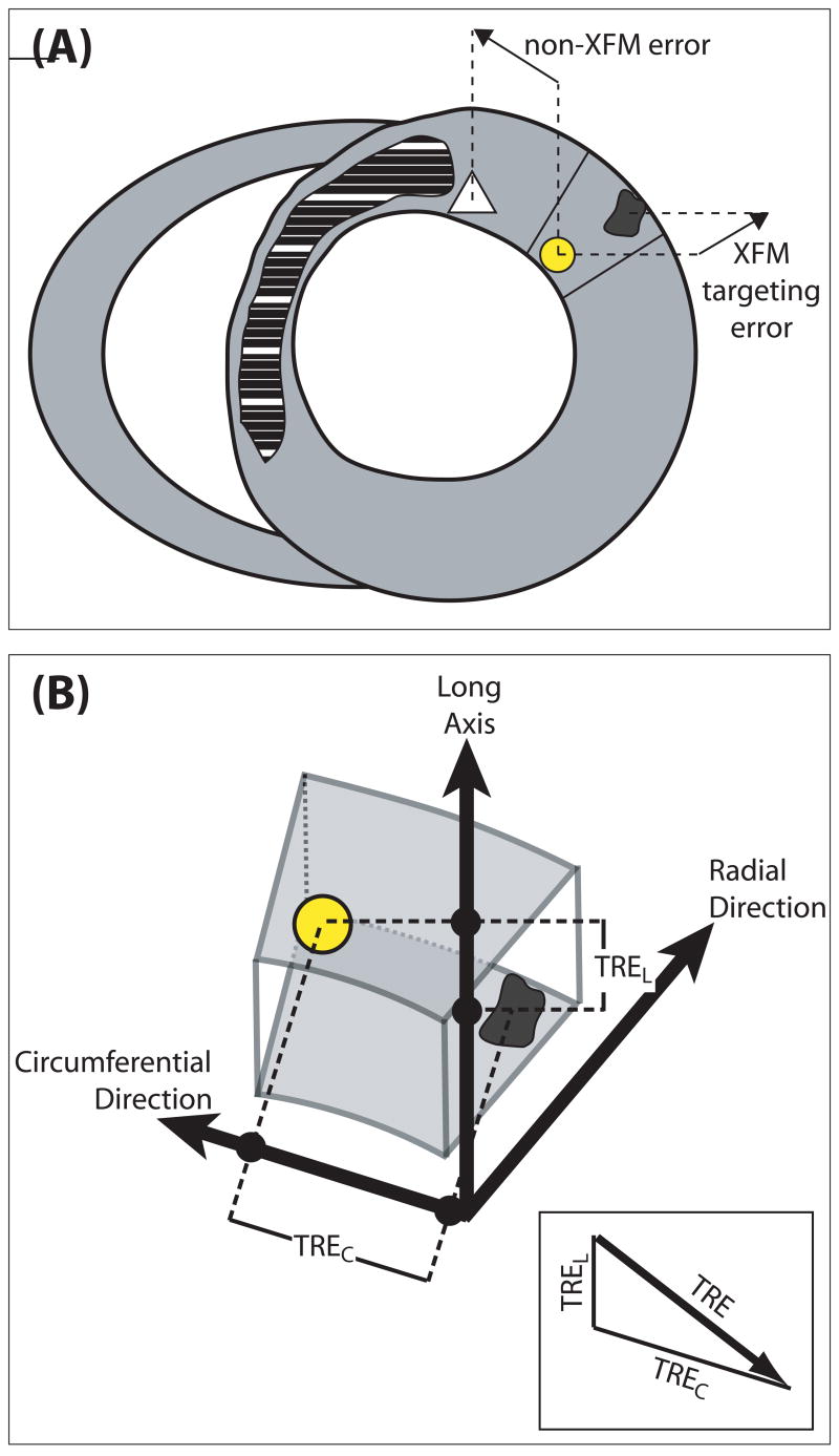
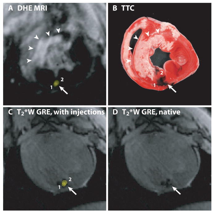
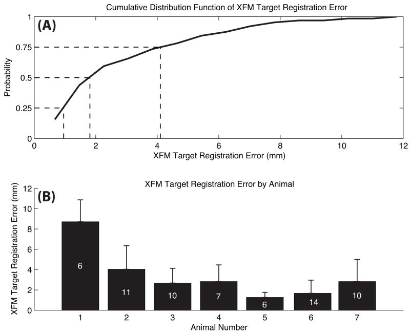
Methods and results: Endomyocardial injections of iron-labeled mesenchymal stromal cells admixed with tissue dye were performed in previously infarcted hearts of 12 Yucatan miniswine (weight, 33 to 67 kg). Features from cardiac MRI were displayed combined with x-ray in real time to guide injections. During 130 injections, operators were provided with 3D surfaces of endocardium, epicardium, myocardial wall thickness (range, 2.6 to 17.7 mm), and infarct registered with live x-ray images to facilitate device navigation and choice of injection location. XFM-guided injections were compared with postinjection MRI and with necropsy specimens obtained 24 hours later. Visual inspection of the pattern of dye staining on 2,3,5-triphenyltetrazolium chloride-stained heart slices agreed (kappa=0.69) with XFM-derived injection locations mapped onto delayed hyperenhancement MRI and the susceptibility artifacts seen on the postinjection T2*-weighted gradient echo MRI. The distance between the predicted and actual injection locations in vivo was 3.2+/-2.6 mm (n=64), and 75% of injections were within 4.1 mm of the predicted location.

Conclusions: Three-dimensional to two-dimensional registration of x-ray and MR images with the use of external fiducial markers accurately targets endomyocardial injection in a swine model of myocardial infarction.


Figures

Figure 1
Figure 1
Schematic illustration of the components of XFM using external fiducial markers. In the example XFM image, the left ventricular endocardium is depicted in red, left ventricular epicardium in blue, and right ventricular endocardium in yellow.
Figure 2
Figure 2
Fusion of 3D MRI-derived surfaces with radiocontrast ventriculograms. End-diastolic short-axis SSFP images from apex to base are segmented to define left ventricular endocardial (red), left ventricular epicardial (blue), and right ventricular endocardial (yellow) contours (A). Three-dimensional surfaces are generated from these contours as described in the text and overlaid on the live x-ray display during ventriculography (B) (The asterisk denotes the external fiducial marker) acquired in anteroposterior (AP) (C) and left anterior oblique (LAO) 45° (D) projections.
Figure 3
Figure 3
XFM targeting of endomyocardial injections according to infarct location (blue surface) and regional myocardial wall thickness (colored green for wall thickness >6 mm and red for wall thickness ≤6 mm) in an animal with a chronic LCX infarct. These surfaces were displayed over x-ray in orthogonal projections (A and B). The catheter is positioned where the wall thickness is ≤6 mm (arrow); this location was therefore rejected. C and D demonstrate relocation of the injection catheter to a “safe” peri-infarct location with wall thickness >6 mm. After deployment of the needle, orthogonal x-ray views allow reconstruction of the injection location in 3D (yellow spot, numbered 4). Previous injection locations (yellow spots, numbered 1 to 3) are also displayed to help avoid overlapping injections. The 3D injection locations are also displayed superimposed on the prior DHE MRI (E and G). A postmortem TTC-stained heart slice (F), located between the MRI slices displayed in E and G, shows tissue dye–staining patterns that correlate well with the XFM-derived injection locations.
Figure 4
Figure 4
A, Schematic short-axis slice, with normal myocardium in gray and the infarct in cross-hatch. The yellow spot represents an XFM-predicted injection location. The dark spot represents the susceptibility artifact on T2*W-GRE imaging due to injected Fe-MSC. The white triangle denotes an ideal target. The distance between the white triangle and the yellow dot represents errors due to device and operator factors, whereas the distance between the yellow dot and dark spot represents intrinsic targeting errors of our registration method. B, Calculation of the target registration error (TRE). A 3D vector relates the end-diastolic XFM-derived injection location (yellow) and the centroid of the corresponding iron injection (black) on the postinjection end-diastolic T2*W-GRE MRI images. The target registration error is the length of the circumferential (TREC) and long-axis (TREL) components of this vector. The radial component of the vector, which is not controlled by the operator, is ignored (see text).
Figure 5
Figure 5
The locations of 2 XFM-guided injections of Fe-MSC mixed with tissue dye are shown relative to areas of infarction demonstrated in vivo by DHE MRI (A) and ex vivo by TTC staining (B). The XFM-guided injection locations (yellow spots, numbered 1 and 2) are superimposed on the preinjection DHE-MR to target injections (A) and on the postinjection T2*W GRE image (C) to validate in vivo accuracy of XFM-guided injection locations. The susceptibility artifact on this image resulting from injected Fe-MSC is better appreciated in D. The distributions of tissue dye stains on the TTC-stained heart slice (B) and susceptibility artifacts (D) are very similar.
Figure 6
Figure 6
In vivo validation of XFM-guided targeting accuracy. A, Cumulative distribution function of target registration errors of all 64 injections performed in 7 animals with evaluable data. B, Mean±SD target registration error for each animal independently. The number within bars indicates the number of injections in each individual animal.

Similar articles

Cited by

References

    1. Dohmann HF, Perin EC, Takiya CM, Silva GV, Silva SA, Sousa AL, Mesquita CT, Rossi MI, Pascarelli BM, Assis IM, Dutra HS, Assad JA, Castello-Branco RV, Drummond C, Dohmann HJ, Willerson JT, Borojevic R. Transendocardial autologous bone marrow mononuclear cell injection in ischemic heart failure: postmortem anatomicopathologic and immunohistochemical findings. Circulation. 2005;112:521–526. - PubMed
    1. Dick AJ, Guttman MA, Raman VK, Peters DC, Pessanha BS, Hill JM, Smith S, Scott G, McVeigh ER, Lederman RJ. Magnetic resonance fluoroscopy allows targeted delivery of mesenchymal stem cells to infarct borders in swine. Circulation. 2003;108:2899–2904. - PMC - PubMed
    1. Kawamoto A, Tkebuchava T, Yamaguchi J, Nishimura H, Yoon YS, Milliken C, Uchida S, Masuo O, Iwaguro H, Ma H, Hanley A, Silver M, Kearney M, Losordo DW, Isner JM, Asahara T. Intramyocardial transplantation of autologous endothelial progenitor cells for therapeutic neo-vascularization of myocardial ischemia. Circulation. 2003;107:461–468. - PubMed
    1. Perin EC, Dohmann HF, Borojevic R, Silva SA, Sousa AL, Mesquita CT, Rossi MI, Carvalho AC, Dutra HS, Dohmann HJ, Silva GV, Belem L, Vivacqua R, Rangel FO, Esporcatte R, Geng YJ, Vaughn WK, Assad JA, Mesquita ET, Willerson JT. Transendocardial, autologous bone marrow cell transplantation for severe, chronic ischemic heart failure. Circulation. 2003;107:2294–2302. - PubMed
    1. Chazaud B, Hittinger L, Sonnet C, Champagne S, Le Corvoisier P, Benhaiem-Sigaux N, Unterseeh T, Su J, Merlet P, Rahmouni A, Garot J, Gherardi R, Teiger E. Endoventricular porcine autologous myoblast transplantation can be successfully achieved with minor mechanical cell damage. Cardiovasc Res. 2003;58:444–450. - PubMed

Publication types