. 2006 Nov 28;103(48):18344-9.
doi: 10.1073/pnas.0604073103. Epub 2006 Nov 13.

Short-chain ubiquitination mediates the regulated endocytosis of the aquaporin-2 water channel

Affiliations

Short-chain ubiquitination mediates the regulated endocytosis of the aquaporin-2 water channel

Erik-Jan Kamsteeg et al. Proc Natl Acad Sci U S A. .

Abstract

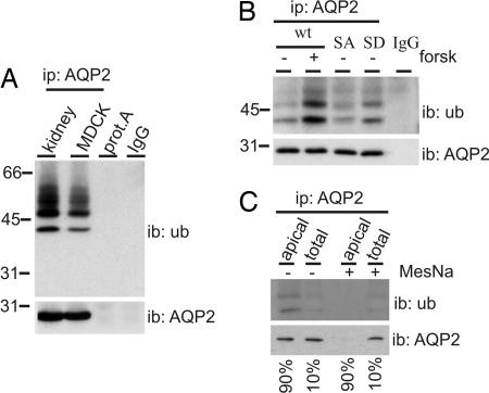
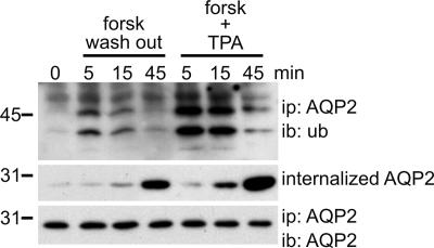
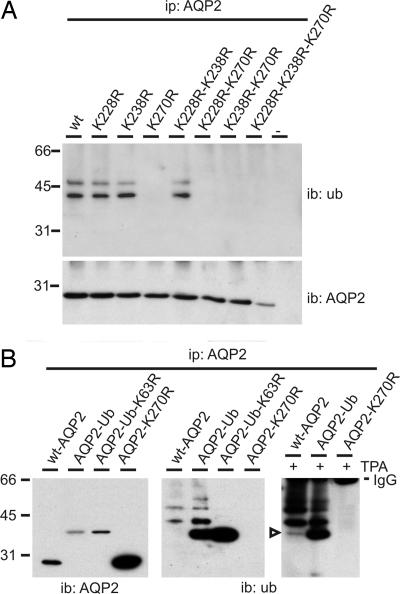
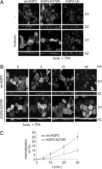
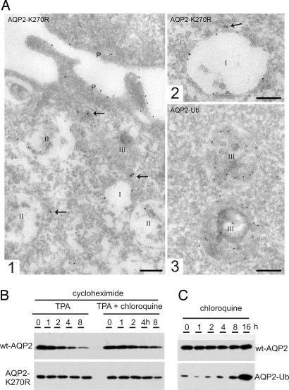
To regulate mammalian water homeostasis, arginine-vasopressin (AVP) induces phosphorylation and thereby redistribution of renal aquaporin-2 (AQP2) water channels from vesicles to the apical membrane. Vice versa, AVP (or forskolin) removal and hormones activating PKC cause AQP2 internalization, but the mechanism is unknown. Here, we show that a fraction of AQP2 is modified with two to three ubiquitin moieties in vitro and in vivo. Mutagenesis revealed that AQP2 is ubiquitinated with one K63-linked chain at K270 only. In Madin-Darby canine kidney cells, AQP2 ubiquitination occurs preferentially when present in the apical membrane, is transiently increased with forskolin removal or PKC activation, and precedes its internalization. Internalization kinetics assays with wild type (wt) and ubiquitination-deficient (K270R) AQP2 revealed that ubiquitination enhances AQP2 endocytosis. Electron microscopy showed that a translational fusion of AQP2 with ubiquitin (AQP2-Ub) localized particularly to internal vesicles of multivesicular bodies (MVBs), whereas AQP2-K270R largely localized to the apical membrane, early endosomes, and the limiting membrane of MVBs. Consistent with this distribution pattern, lysosomal degradation was extensive for AQP2-Ub, low for AQP2-K270R, and intermediate for wt-AQP2. Our data show that short-chain ubiquitination is involved in the regulated endocytosis, MVB sorting, and degradation of AQP2 and may be the mechanism used by AVP removal and PKC-activating hormones to reduce renal water reabsorption. Moreover, because several other channels are also (short-chain) ubiquitinated, our data suggest that ubiquitination may be a general mediator for the regulated endocytosis and degradation of channels in higher eukaryotes.


Conflict of interest statement

The authors declare no conflict of interest.

Figures

Fig. 1.
Fig. 1.
Ubiquitination of AQP2 occurs at the apical plasma membrane. (A) AQP2 was immunoprecipitated (ip: AQP2) from solubilized goat kidney inner medulla membranes (kidney) or MDCK-AQP2 cells (MDCK). Solubilized kidney membranes were also subjected to precipitation with protein A-coupled beads without antibodies (prot.A). Throughout this study, nonequivalents (5% and 95%) of the (immuno)precipitates were immunoblotted (ib) for AQP2 or ubiquitin (ub), respectively. AQP2 antibodies (IgG) serve as negative control. (B) MDCK cells expressing wt-AQP2 (wt), AQP2-S256A (SA) or AQP2-S256D (SD) were left untreated (−) or pretreated (±) with forskolin (forsk) for 45 min. Subsequently, AQP2 was immunoprecipitated and immunoblotted for ubiquitin or AQP2. (C) MDCK cells expressing wt-AQP2 were treated with forskolin for 45 min, surface-biotinylated, treated with MesNa to strip surface biotin where indicated (+), and lysed. AQP2 was immunoprecipitated, released from the beads, and a part (10%) of the sample was used for direct immunoblotting (total), whereas the remainder (90%) was first subjected to streptavidin pulldowns to recover biotinylated AQP2 (apical) and subsequently immunoblotted.
Fig. 2.
Fig. 2.
Ubiquitination of AQP2 occurs before its internalization. MDCK cells expressing wt-AQP2 were pretreated with forskolin for 45 min (0). Subsequently, forskolin was washed out (forsk wash out) or the phorbol ester TPA was added (forsk + TPA) for the indicated times. Cells were lysed, and AQP2 was immunoprecipitated and immunoblotted for ubiquitin or AQP2. In parallel, forskolin-treated MDCK-AQP2 cells were surface-biotinylated in the cold, followed by an incubation at 37°C without forskolin (forsk wash out) or with forskolin and TPA (forsk + TPA) for the indicated times. Then, surface-bound biotin was removed with MesNa, and the cells were lysed. Biotinylated proteins were recovered by using immobilized streptavidin and immunoblotted for AQP2 (internalized AQP2).
Fig. 3.
Fig. 3.
AQP2 is ubiquitinated at K270 with one short K63 linked chain. (A) Lysates of nontransfected MDCK cells (−) or of those expressing wt-AQP2 (wt) or its indicated mutants were subjected to AQP2 immunoprecipitation. Immunoprecipitates were immunoblotted for ubiquitin or AQP2. (B) MDCK cells stably expressing wt-AQP2 or its mutants were not treated or treated (+) with TPA. AQP2 immunoprecipitates from these cells were immunoblotted for ubiquitin or AQP2. The open arrowhead indicates AQP2 modified with one ubiquitin molecule. Signals from immunoglobulins (IgG) used in the immunoprecipitations are indicated.
Fig. 4.
Fig. 4.
Ubiquitination of AQP2 increases its endocytosis. (A) MDCK cells expressing wt-AQP2, AQP2-K270R or AQP2-Ub were left untreated (−) or treated with forskolin, fixed, and subjected to immunocytochemistry using AQP2 antibodies. Confocal images were obtained in the XY and XZ planes. (B) MDCK cells expressing wt-AQP2 or AQP2-K270R were treated with forskolin, followed by TPA addition (forsk. + TPA) for the indicated times. Then, cells were fixed and analyzed as under A. (C) Internalization of wt-AQP2 or AQP2-K270R after forskolin treatment followed by TPA addition (forsk. + TPA) was determined as in Fig. 3 for the indicated times. Semiquantification of AQP2 (n = 3) was done as described in Materials and Methods. Filled and open data points represent wt-AQP2 and AQP2-K270R, respectively.
Fig. 5.
Fig. 5.
Ubiquitination of AQP2 increases its lysosomal degradation. (A) Cryosections of fixed MDCK cells expressing AQP2-K270R (1, 2) or AQP2-Ub (3) were immunolabeled for AQP2 (10-nm gold particles). Endosomes are divided into three types: I = early endosome, II = late endosome, and III = late endosome/lysosome (1). Overview of the apical part of an MDCK cell showing all three types of endosomes. The arrows point to labeled dense vesicles that mediate either endocytosis or recycling (2). Detail of an early endosome with AQP2-K270R label at the limiting membrane (3). Detail of late endosomes/lysosomes with the majority of label associated with internal membranes. (Scale bars, 200 nm.) (B) Protein synthesis in MDCK-AQP2 or MDCK-AQP2-K270R was inhibited (cycloheximide), and TPA was added in the absence or presence of chloroquine for the indicated times. Subsequently, cells were lysed and immunoblotted for AQP2. (C) MDCK cells expressing wt-AQP2 or AQP2-Ub were treated with chloroquine for the indicated times. Subsequently, cells were lysed and immunoblotted for AQP2.

References

    1. Sidhaye V, Hoffert JD, King LS. J Biol Chem. 2005;280:3590–3596. - PMC - PubMed
    1. Nielsen S, Digiovanni SR, Christensen EI, Knepper MA, Harris HW. Proc Natl Acad Sci USA. 1993;90:11663–11667. - PMC - PubMed
    1. Marinelli RA, Pham L, Agre P, LaRusso NF. J Biol Chem. 1997;272:12984–12988. - PubMed
    1. Deen PMT, Verdijk MAJ, Knoers N. V. A. M., Wieringa B, Monnens LAH, van Os CH, van Oost BA. Science. 1994;264:92–95. - PubMed
    1. Marples D, Christensen S, Christensen EI, Ottosen PD, Nielsen S. J Clin Invest. 1995;95:1838–1845. - PMC - PubMed

Publication types

LinkOut - more resources