. 2006 Nov 21;103(47):17608-13.
doi: 10.1073/pnas.0603224103. Epub 2006 Nov 13.

Emergence of primate genes by retrotransposon-mediated sequence transduction

Affiliations

Emergence of primate genes by retrotransposon-mediated sequence transduction

Jinchuan Xing et al. Proc Natl Acad Sci U S A. .

Abstract

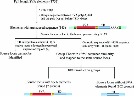
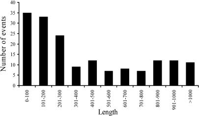
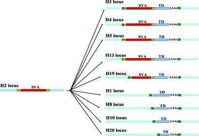
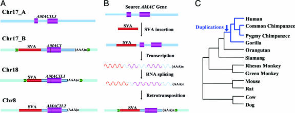
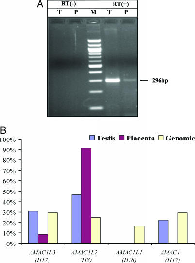
Gene duplication is one of the most important mechanisms for creating new genes and generating genomic novelty. Retrotransposon-mediated sequence transduction (i.e., the process by which a retrotransposon carries flanking sequence during its mobilization) has been proposed as a gene duplication mechanism. L1 exon shuffling potential has been reported in cell culture assays, and two potential L1-mediated exon shuffling events have been identified in the genome. SVA is the youngest retrotransposon family in primates and is capable of 3' flanking sequence transduction during retrotransposition. In this study, we examined all of the full-length SVA elements in the human genome to assess the frequency and impact of SVA-mediated 3' sequence transduction. Our results showed that approximately 53 kb of genomic sequences have been duplicated by 143 different SVA-mediated transduction events. In particular, we identified one group of SVA elements that duplicated the entire AMAC gene three times in the human genome through SVA-mediated transduction events, which happened before the divergence of humans and African great apes. In addition to the original AMAC gene, the three transduced AMAC copies contain intact ORFs in the human genome, and at least two are actively transcribed in different human tissues. The duplication of entire genes and the creation of previously undescribed gene families through retrotransposon-mediated sequence transduction represent an important mechanism by which mobile elements impact their host genomes.


Conflict of interest statement

The authors declare no conflict of interest.

Figures

Fig. 1.
Fig. 1.
Identification of SVA 3′ transduction events and their source elements. Shown are the schematic diagrams for the identification process. Flanking sequences of the source locus are shown as blue boxes; TSDs are shown as yellow and green arrows. SVA elements are depicted as red bars, and the transduced sequences are shown as blue bars and labeled “TD.” SVA element poly(A) tails are shown as “(AAA)n.” The numbers in parentheses correspond to the total number of SVA elements/groups identified in each step.
Fig. 2.
Fig. 2.
Length distribution of 3′ transduction events. The number of human SVA-mediated 3′ transduction events in each 100-bp size interval is shown.
Fig. 3.
Fig. 3.
SVA 3′ transduction events. One group of SVA 3′ transduction events (H3_186) is shown. Flanking sequences of the original locus are shown as blue boxes, and the flanking sequences of the transduced loci are shown as light blue boxes. TSDs are shown as yellow and green arrows. SVA elements are depicted as red bars, and the transduced sequences are shown as blue bars and labeled “TD.” SVA element poly(A) tails are shown as “(AAA)n.”
Fig. 4.
Fig. 4.
SVA transduction-mediated gene duplication. (A) Schematic diagram of the H17_76 transduction group in the human genome. Flanking sequences of the original locus are shown as blue boxes, and the flanking sequences of the transduced loci are shown as light blue boxes. TSDs are shown as yellow and green arrows. SVA elements are depicted as red bars, the transduced sequences are shown as blue bars, and coding regions are shown as purple bars. SVA element poly(A) tails are shown as “(AAA)n.” (B) Schematic diagrams for putative evolutionary scenarios of the SVA transduction-mediated gene duplications. Approximately 7 million to 14 million years ago, one active SVA element was inserted upstream of the original AMAC gene locus. Then, transcription of this active SVA element transduced the full-length AMAC gene sequence. During the retrotransposition process, the intron of the gene was removed by RNA processing machinery. Finally, the SVA element along with the intronless AMAC gene sequence retrotransposed into new genomic locations. The original retrotransposition-competent SVA element upstream of the source locus was eventually lost in the population. The predicted RNA transcripts are shown as curved lines. (C) The phylogenetic relationships among various species used in dN/dS analysis.
Fig. 5.
Fig. 5.
Expression analysis of AMAC gene duplicates in humans. (A) Agarose gel chromatograph of RT-PCR products derived from human testis (T) and placental (P) RNA templates. Negative controls with no reverse transcriptase (RT −) are on the left, a 100-bp marker (M) is in the middle, and reactions with reverse transcriptase (RT +) are on the right; the sizes of the correct fragments are indicated. (B) Relative expression levels of four human AMAC gene duplicates in human testis and placenta. Human genomic DNA (HeLa) amplification is the control for uniform amplification of all gene duplicates.

References

    1. Long M, Betran E, Thornton K, Wang W. Nat Rev Genet. 2003;4:865–875. - PubMed
    1. Ohno S. Evolution by Gene Duplication. Heidelberg: Springer; 1970.
    1. Johnson ME, Viggiano L, Bailey JA, Abdul-Rauf M, Goodwin G, Rocchi M, Eichler EE. Nature. 2001;413:514–519. - PubMed
    1. Marques AC, Dupanloup I, Vinckenbosch N, Reymond A, Kaessmann H. PLoS Biol. 2005;3:e357. - PMC - PubMed
    1. Vinckenbosch N, Dupanloup I, Kaessmann H. Proc Natl Acad Sci USA. 2006;103:3220–3225. - PMC - PubMed

Publication types

Substances

LinkOut - more resources