. 2007 Jan 2;104(1):139-44.
doi: 10.1073/pnas.0601947103. Epub 2006 Nov 13.

CARMA3/Bcl10/MALT1-dependent NF-kappaB activation mediates angiotensin II-responsive inflammatory signaling in nonimmune cells

Affiliations

CARMA3/Bcl10/MALT1-dependent NF-kappaB activation mediates angiotensin II-responsive inflammatory signaling in nonimmune cells

Linda M McAllister-Lucas et al. Proc Natl Acad Sci U S A. .

Abstract

Angiotensin II (Ang II) is a peptide hormone that, like many cytokines, acts as a proinflammatory agent and growth factor. After injury to the liver, the hormone assists in tissue repair by stimulating hepatocytes and hepatic stellate cells to synthesize extracellular matrix proteins and secrete secondary cytokines and by stimulating myofibroblasts to proliferate. However, under conditions of chronic liver injury, all of these effects conspire to promote pathologic liver fibrosis. Much of this effect of Ang II results from activation of the proinflammatory NF-kappaB transcription factor in response to stimulation of the type 1 Ang II receptor, a G protein-coupled receptor. Here, we characterize a previously undescribed signaling pathway mediating Ang II-dependent activation of NF-kappaB, which is composed of three principal proteins, CARMA3, Bcl10, and MALT1. Blocking the function of any of these proteins, through the use of either dominant-negative mutants, RNAi, or gene targeting, effectively abolishes Ang II-dependent NF-kappaB activation in hepatocytes. In addition, Bcl10(-/-) mice show defective hepatic cytokine production after Ang II treatment. Evidence also is presented that this pathway activates NF-kappaB through ubiquitination of IKKgamma, the regulatory subunit of the IkappaB kinase complex. These results elucidate a concrete series of molecular events that link ligand activation of the type 1 Ang II receptor to stimulation of the NF-kappaB transcription factor. These findings also uncover a function of the CARMA, Bcl10, and MALT1 proteins in cells outside the immune system.


Conflict of interest statement

The authors declare no conflict of interest.

Figures

Fig. 1.
Fig. 1.
CARMA3, Bcl10, and MALT1 are all expressed in Ang II-sensitive tissues. (AE) Total RNA was extracted from mouse tissues and used for real-time, quantitative RT-PCR. mRNA levels for the AT1R, CARMA3, Bcl10, MALT1, and CARMA1 were determined and then normalized against β-actin mRNA. For each gene, an mRNA expression value was arbitrarily set at 100 for the tissue with the greatest normalized mRNA level. Relative expression then was calculated for the remaining tissues. Results represent the mean ± SEM of three determinations. (F) Total protein was extracted from mouse spleen, thymus, and liver and subjected to Western blotting by using antisera to CARMA3, Bcl10, MALT1, or GAPDH. Results are representative of two independent experiments.
Fig. 2.
Fig. 2.
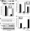
CARMA3 is required for Ang II-dependent NF-κB activation. (A Upper) A schematic depicting the domain structure of WT CARMA3 and the dominant-negative mutant of CARMA3 (CARMA3ΔCARD) is shown. (A Lower) HepG2 cells were transfected with an NF-κB-responsive luciferase reporter, a constitutively active Renilla reporter, and an AT1AR expression vector, either in the presence or absence of CARMA3ΔCARD. Twenty-four hours after transfection, cells were stimulated overnight with 500 nM Sar1-Ang II or 10 ng/ml TNFα, and NF-κB activation was determined by measuring the luciferase/Renilla ratio. Results represent the mean ± SEM for at least three experiments. (B) Hairpin sequence for the human-specific CARMA3 shRNA. (C) The CARMA3 shRNA vector was transfected into HepG2 cells in a dose-dependent fashion along with expression vectors for Flag-tagged CARMA3 and IKKγ. Forty-eight hours after transfection, cells were harvested and assayed for CARMA3 and IKKγ proteins by Western blot. In parallel experiments, cells were transfected with a vector containing an unrelated, control shRNA sequence. (D) HepG2 cells were transfected with either the CARMA3 or control shRNA vectors. Forty-eight hours later, cells were treated for an additional 12 h with either 500 nM Sar1-Ang II or 10 ng/ml TNFα, and NF-κB activation was assessed as described previously. Results represent the mean ± SEM for at least three experiments. (E) HepG2 cells were similarly transfected with vector encoding CARMA3 shRNA, with or without an expression vector encoding the WT mouse CARMA3 protein. Forty-eight hours later, cells were treated with 500 nM Sar1-Ang II for an additional 12 h. NF-κB activation was measured as above. Results represent the mean ± SEM of three to four determinations.
Fig. 3.
Fig. 3.
In hepatocytes, Bcl10 is essential for Ang II-dependent NF-κB activation and cytokine induction. (A Upper) A schematic depicting the domain structure of WT Bcl10 and the dominant-negative mutant of Bcl10 (Bcl10Δ107–119). (A Lower) HepG2 cells were transfected as described above with control vector or with expression vector encoding Bcl10Δ107–119. Forty-eight hours later, cells were stimulated with either 500 nM Sar1-Ang II or 10 ng/ml TNFα, and NF-κB activation was assessed. Results are expressed as the absolute luciferase/Renilla ratio and reflect the mean ± SEM of at least three determinations. (B) Primary hepatocytes were isolated from livers of WT and Bcl10−/− mice and cultured overnight in serum-free medium. Cells then were transfected with an NF-κB-responsive luciferase reporter, a constitutively active Renilla reporter, and an AT1AR expression vector. Cells were treated with either 500 nM Ang II or 10 ng/ml TNFα for an additional 16 h before NF-κB activation was measured. Results represent the mean ± SEM for 17–26 determinations; ∗∗, significance level of P < 0.001 (as analyzed by one-tailed Student's t test). (C) WT and Bcl10−/− mice received an i.p. injection of PBS or Ang II (6.4 μg/g of body weight). One hour after injection, mice were killed, and a sample of liver was obtained from each. Total RNA was isolated, and IL-6 mRNA levels were determined by real-time quantitative RT-PCR, normalizing for β-actin. Results are expressed as fold induction of IL-6 for mice receiving Ang II as compared with PBS and reflect the mean ± SEM of seven to nine mice. ∗∗∗, significance level of P < 0.03 (as analyzed by one-tailed Student's t test).
Fig. 4.
Fig. 4.
Ang II treatment induces IKKγ ubiquitination. (A) HepG2-AR cells were transfected with expression vectors encoding myc-tagged IKKγ and HA-tagged ubiquitin. Cells were treated overnight with 500 nM Ang II or 10 ng/ml TNFα before harvesting. IKKγ then was immunoprecipitated and subjected to Western blotting as indicated. (B) HepG2-AR cells were transfected and treated as described above but in the presence or absence of coexpressed CARMA3ΔCARD. IKKγ ubiquitination was assayed as above. (C) HepG2-AR cells were transfected with HA-ubiquitin only. After treatment with Ang II, endogenous IKKγ was immunoprecipitated and subjected to Western blotting as indicated. Results from all ubiquitination assays shown in A–C are representative of at least three separate experiments. (D) HepG2-AR cells were transfected transiently with or without 100 nM RNAi duplexes specifically targeting the human MALT1 RNA sequence. Forty-eight hours after transfection, cells were transfected again with an NF-κB-responsive luciferase reporter and a constitutively active Renilla reporter. Cells then were treated with either 500 nM Ang II or 10 ng/ml TNFα for 16 h, such that cell lysates could be prepared and assayed for NF-κB activation 96 h after the initial transfection with RNAi. Extracts used to measure NF-κB activation also were subjected to Western blotting as indicated, to assess effective knockdown of MALT1 protein (−, no RNAi; C, scrambled control RNAi; M, MALT1 RNAi). Results represent the mean ± SEM of five separate experiments.
Fig. 5.
Fig. 5.
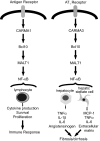
Model: A similar CARMA-Bcl10-MALT1 signaling pathway mediates NF-κB activation in response to antigen receptor stimulation in lymphocytes and AT1R stimulation in liver cells. (Left) In lymphocytes, antigen-dependent NF-κB activation leads to cytokine production and cellular proliferation. (Right) In liver, Ang II-dependent NF-κB activation leads to a variety of proinflammatory effects that are implicated in the development of hepatic fibrosis/cirrhosis.

References

    1. Phillips MI, Kagiyama S. Curr Opin Investig Drugs. 2002;3:569–577. - PubMed
    1. Bataller R, Sancho-Bru P, Gines P, Brenner DA. Antioxid Redox Signal. 2005;7:1346–1355. - PubMed
    1. Bataller R, Gines P, Nicolas JM, Gorbig MN, Garcia-Ramallo E, Gasull X, Bosch J, Arroyo V, Rodes J. Gastroenterology. 2000;118:1149–1156. - PubMed
    1. Shah BH, Alberto Olivares-Reyes J, Yesilkaya A, Catt KJ. Mol Endocrinol. 2002;16:610–620. - PubMed
    1. Bataller R, Brenner DA. J Clin Invest. 2005;115:209–218. - PMC - PubMed

Publication types

MeSH terms