. 2006 Nov 21;103(47):17885-90.
doi: 10.1073/pnas.0608344103. Epub 2006 Nov 13.

Evolution and expression of chimeric POTE-actin genes in the human genome

Affiliations

Evolution and expression of chimeric POTE-actin genes in the human genome

Yoomi Lee et al. Proc Natl Acad Sci U S A. .

Abstract

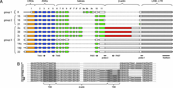
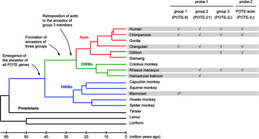
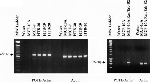
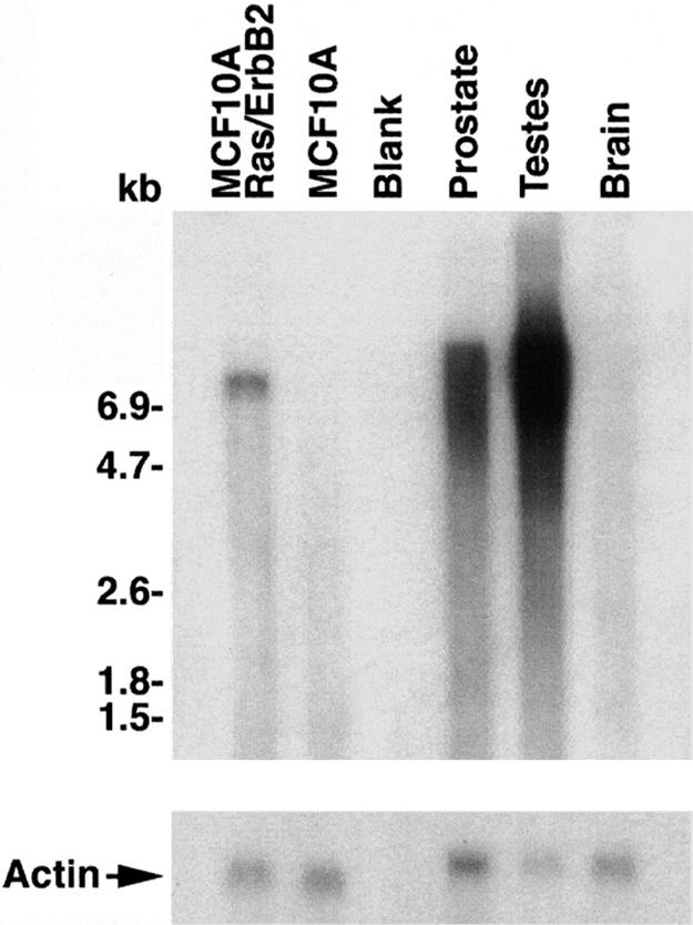
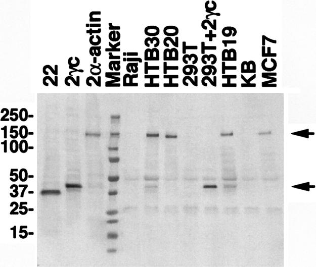
We previously described a primate-specific gene family, POTE, that is expressed in many cancers but in a limited number of normal organs. The 13 POTE genes are dispersed among eight different chromosomes and evolved by duplications and remodeling of the human genome from an ancestral gene, ANKRD26. Based on sequence similarity, the POTE gene family members can be divided into three groups. By genome database searches, we identified an actin retroposon insertion at the carboxyl terminus of one of the ancestral POTE paralogs. By Northern blot analysis, we identified the expected 7.5-kb POTE-actin chimeric transcript in a breast cancer cell line. The protein encoded by the POTE-actin transcript is predicted to be 120 kDa in size. Using anti-POTE mAbs that recognize the amino-terminal portion of the POTE protein, we detected the 120-kDa POTE-actin fusion protein in breast cancer cell lines known to express the fusion transcript. These data demonstrate that insertion of a retroposon produced an altered functional POTE gene. This example indicates that new functional human genes can evolve by insertion of retroposons.


Conflict of interest statement

The authors declare no conflict of interest.

Figures

Fig. 1.
Fig. 1.
Extended genomic organization of POTE genes. (A) Genomic organization of POTE paralogs are depicted. Domain organization and exon numbers are given at the top. Primer binding sites (arrows) and query sequences for the database search (probe 1 and probe 2) are marked at the bottom. Plain boxes indicate exons derived from the ancestral gene ANKRD26. The actin inserts and LINE elements are represented by stippled and hatched boxes, respectively. The ORFs are color-coded: orange, cysteine-rich domains (CRDs); blue, ankyrin repeats (ANKs); green, helices; and red, actin. (B) Actin insert and surrounding sequences are presented. The putative target site duplication (TSD) is marked. Dashes and dots indicate gaps and omitted bases, respectively.
Fig. 2.
Fig. 2.
Evolution of POTE genes. A tentative evolutionary history of POTE genes is summarized. A check mark indicates that a POTE gene fragment mostly similar to the corresponding probe sequence was observed in the given species (chimpanzee, Pan troglodytes; orangutan, Pongo pygmaeus; white-cheeked gibbon, Nomascus leucogenys; rhesus macaque, Macaca mulatta; Hamadryas baboon, Papio hamadryas; and white-tufted-ear marmoset, Callithrix jacchus). It is likely that the sole POTE gene fragment detected in marmoset (indicated by an asterisk) is ancestral to all of the POTE genes in OWMs and apes. The tree and estimated divergence times are given at the left (12).
Fig. 3.
Fig. 3.
RT-PCR analysis of POTE–actin fusion mRNA in human breast cancer cell lines. Total RNAs were isolated from different cell lines, and first-strand cDNAs were made by reverse transcriptase reaction. PCR on cDNA from breast cancer cell lines was performed with primers specific for POTE–actin fusion transcript (PA01 and PA07 shown in Fig. 1). The breast cancer cell lines MCF-7, HTB-30, HTB-19, and HTB-20 produce a specific 650-bp product. Primer specific for β-actin transcript was used to validate the integrity of the RNA samples.
Fig. 4.
Fig. 4.
Analysis of POTE–actin fusion transcript in a breast cancer cell line. Northern blot analysis showing expression and transcript sizes of POTE–actin fusion transcript in different samples. Approximately 2.5 μg of mRNAs from different samples was run on agarose gel and transferred to a nylon membrane. The 1.2-kb probe was generated by PCR and labeled with 32P by using the random priming extension method. Membrane was incubated for 2 h in hybridization buffer followed by addition of denatured probe and incubation for an additional 12 h. Membrane then was washed and subjected to autoradiography. A specific band of ≈7.5 kb in size was detected in the MCF-10A-Ras/ErbB2 lane, but in prostate and in testis the signal is somewhat diffuse over 6.5 kb to 9.0 kb in size, and there are no detectable bands in MCF-10A and in brain.
Fig. 5.
Fig. 5.
Detection of POTE–actin fusion protein in a breast cancer cell line. Cell lysates from different cell lines were subjected to IP by using HP8 antibodies. Immunoprecipitates were resolved in a 4–20% PAGE gel, transferred into a PVDF membrane and immunoblotted with PG5 antibody. A specific band of 140 kDa in size (upper arrow) was detected in MCF-7, HTB-19, HTB-20, HTB-30, and POTE-2α–actin-transfected 293T (positive control) lysates. No specific band was detected in KB, Raji, or 293T cell extracts. A smaller protein of 42 kDa in size (lower arrow) was detected in HTB-19 and in HTB-30, representing POTE protein without actin fusion. As positive controls for Western blot analysis, cell lysates prepared from 293T cells transfected with each POTE-encoding plasmid were loaded in the first three lanes without IP.
Fig. 6.
Fig. 6.
Localization of POTE–actin protein in transfected cells. MCF-7 cells were transfected with pcDNA3–POTE–actin plasmid, and, after 48 h, the cells were stained with the anti-POTE antibody PG5 and analyzed by confocal microscopy. Green represents POTE staining, and red represents F-actin staining. Nuclei (blue staining) were stained with 4,6-diamidino-2-phenylindole (DAPI).

References

    1. Bera TK, Zimonjic DB, Popescu NC, Sathyanarayana BK, Kumar V, Lee B, Pastan I. Proc Natl Acad Sci USA. 2002;99:16975–16980. - PMC - PubMed
    1. Bera TK, Huynh N, Maeda H, Sathyanarayana BK, Lee B, Pastan I. Gene. 2004;337:45–53. - PubMed
    1. Hahn Y, Bera TK, Pastan IH, Lee B. Gene. 2006;366:238–245. - PubMed
    1. Bera TK, Saint-Fleur A, Lee Y, Kydd A, Hahn Y, Popescu NC, Zimonjic DB, Lee BK, Pastan I. Cancer Res. 2006;66:52–56. - PubMed
    1. Long M, Betrán E, Thornton K, Wang W. Nat Rev Genet. 2003;4:865–875. - PubMed

Publication types

MeSH terms

LinkOut - more resources