. 2007 Jan 12;282(2):929-37.
doi: 10.1074/jbc.M607646200. Epub 2006 Nov 13.

Micromolar intracellular hydrogen peroxide disrupts metabolism by damaging iron-sulfur enzymes

Affiliations

Micromolar intracellular hydrogen peroxide disrupts metabolism by damaging iron-sulfur enzymes

Soojin Jang et al. J Biol Chem. .

Abstract

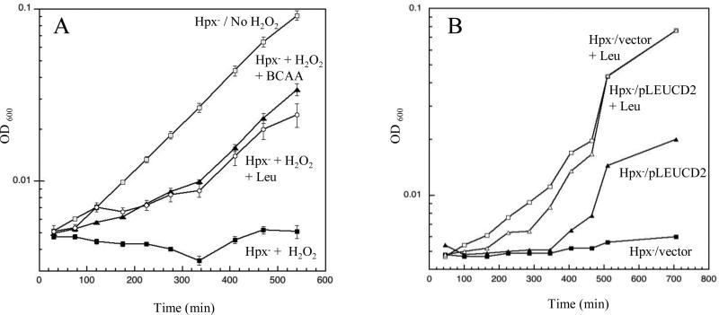
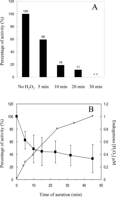
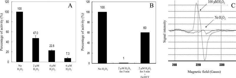
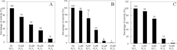
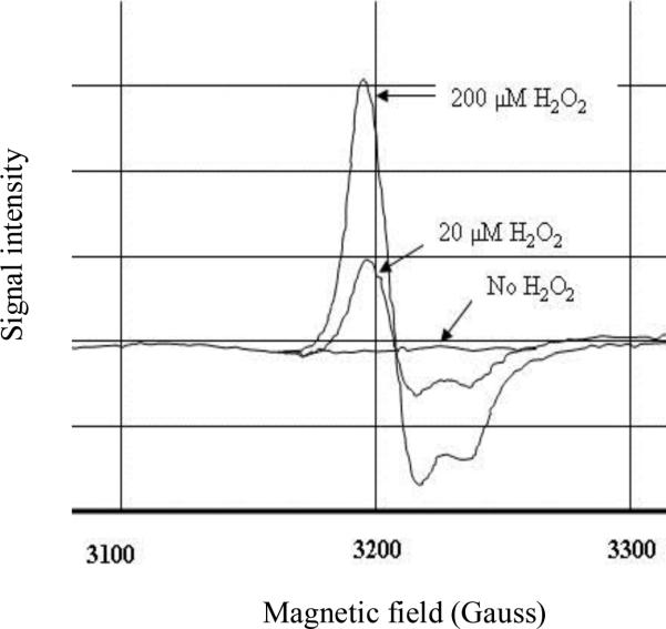
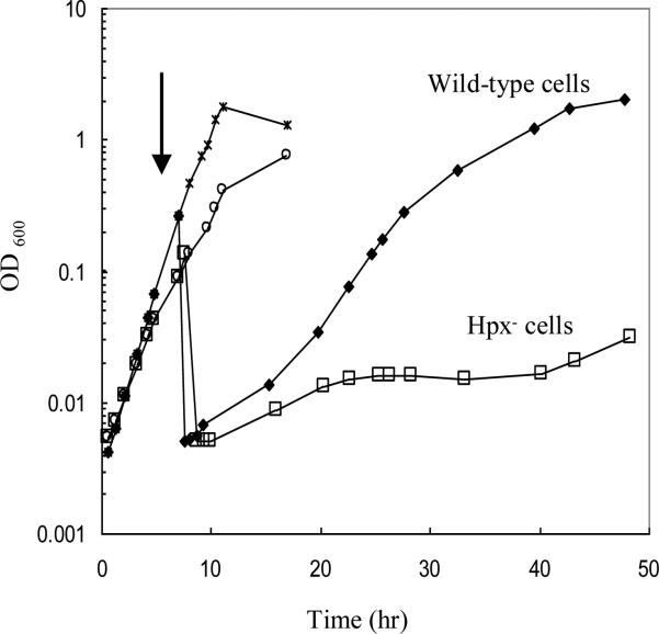
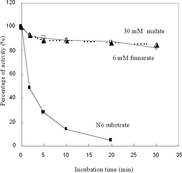
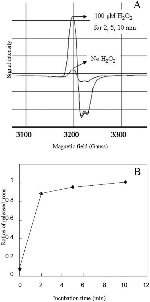
An Escherichia coli strain that cannot scavenge hydrogen peroxide has been used to identify the cell processes that are most sensitive to this oxidant. Low micromolar concentrations of H2O2 completely blocked the biosynthesis of leucine. The defect was tracked to the inactivation of isopropylmalate isomerase. This enzyme belongs to a family of [4Fe-4S] dehydratases that are notoriously sensitive to univalent oxidation, and experiments confirmed that other members were also inactivated. In vitro and in vivo analyses showed that H2O2 directly oxidized their solvent-exposed clusters in a Fenton-like reaction. The oxidized cluster then degraded to a catalytically inactive [3Fe-4S] form. Experiments indicated that H2O2 accepted two consecutive electrons during the oxidation event. As a consequence, hydroxyl radicals were not released; the polypeptide was undamaged; and the enzyme was competent for reactivation by repair processes. Strikingly, in scavenger-deficient mutants, the H2O2 that was generated as an adventitious by-product of metabolism (<1 microm) was sufficient to damage these [4Fe-4S] enzymes. This result demonstrates that aerobic organisms must synthesize H2O2 scavengers to avoid poisoning their own pathways. The extreme vulnerability of these enzymes may explain why many organisms, including mammals, deploy H2O2 to suppress microbial growth.


Figures

Figure 1
Figure 1. Low concentrations of H2O2 block leucine biosynthesis
(A) Hpx cells were cultured in aerobic glucose medium. Where indicated, 8 μM H2O2, branched-chain amino acids (BCAA: Leu, Ile, and Val), and/or leucine were added. (B) Hpx cells containing either pLEUCD2 (overproducing isopropylmalate isomerase) or pWKS30 (an empty vector) were cultured in lactose minimal medium with or without leucine supplementation. All media included histidine and aromatic amino acids. The pLEUCD2 plasmid boosted the isopropylmalate isomerase activity of anaerobic cultures about three-fold.
Figure 2
Figure 2. H2O2 rapidly inactivates isopropylmalate isomerase in vivo
(A) Hpx cells were anaerobically cultured to an OD600 of 0.2, and 8 μM H2O2 was then added to the cultures. At intervals catalase was added and residual enzyme activity was determined. (B) Hpx cells were cultured in anaerobic medium and then aerated starting at time zero. Isopropylmalate isomerase activity (squares) and H2O2 concentrations (circles) were monitored.
Figure 3
Figure 3. H2O2 inactivates isopropylmalate isomerase by converting its [4Fe-4S]2+ cluster to a [3Fe-4S]+ cluster in vitro
(A) An extract of anaerobically grown Hpx cells was exposed to the indicated concentrations of H2O2 for 2 min at room temperature. Catalase then was added to remove H2O2. The experiment was conducted anaerobically. (B) Isopropylmalate isomerase from anaerobically cultured Hpx cells was inactivated with 2 μM H2O2 for 5 min. Catalase was added to remove H2O2, and then 50 μM Fe(NH4)2(SO4)2 and 2.5 mM dithiothreitol were added. The activity was measured after 3 min incubation. (C) Hpx cells that overproduce IPMI were harvested when the cells were OD600 of 0.2. The cell pellets were resuspended in 1/500 of original culture volume of 10% glycerol. The resuspended cells were incubated with 100 μM H2O2 for 1 min at 37°C. The cell suspension (250 μl) then was transferred into an EPR tube and frozen.
Figure 4
Figure 4. H2O2 inactivates other [4Fe-4S] dehydratases
(A) 6-phosphogluconate dehydratase. (B) Fumarase A. (C) Fumarase B. Lysates were prepared from anaerobic cultures of LC106, SJ37, and SJ20, respectively, and the indicated concentrations of H2O2 were added for 5 min. Catalase was added to terminate the stress, and residual activity was determined.
Figure 5
Figure 5. H2O2 inactivates fumarase A by converting its [4Fe-4S]2+ cluster to a [3Fe-4S]+ cluster in vivo
SJ37 cells that overproduce fumarase A were harvested at an OD600 of 0.2, washed, and resuspended at 1/500 of original culture volume in 10 % glycerol. The resuspended cells were incubated with 200 or 20 μM H2O2 for 1 min at 37°C. The cell suspension (250 μl) then was transferred into an EPR tube and frozen.
Figure 6
Figure 6. The Hpx mutant is defective at using malate as the sole carbon source
Wild-type (stars) and Hpx (open circles) cells were cultured in aerobic glucose medium containing to 0.15 – 0.25 (OD600). At the arrow aliquots were removed, washed, and resuspended in aerobic malate medium. All media included histidine and aromatic amino acids.
Figure 7
Figure 7. Substrates protect fumarase against H2O2
Purified fumarase A (5 nM) was exposed to 1 μM H2O2 at 0 °C. Where indicated 30 mM malate or 6 mM fumarate were included in the buffer. At time points, aliquots were removed, catalase was added, and residual activity was measured.
Figure 8
Figure 8. H2O2 degrades the [4Fe-4S]2+ cluster of purified fumarase A to a [3Fe-4S]+ cluster with loss of one iron atom
Purified fumarase A (18 μM) was inactivated by 100 μM H2O2 in 50 mM Tris-Cl/10 mM Mg2+ containing 1 mM desferrioxamine. At time points, aliquots were removed, catalase was added, and the reaction mixture was frozen. The residual cluster (panel A) and released ferric iron (panel B) were analyzed by EPR spectroscopy.

References

    1. Imlay JA. Ann. Rev. Microbiol. 2003;57:395–418. - PubMed
    1. Seaver LC, Imlay JA. J. Bacteriol. 2001;183:7182–7189. - PMC - PubMed
    1. Griffiths SW, Cooney CL. Biochemistry. 2002;41:6245–6252. - PubMed
    1. Winterbourn CC, Metodiewa D. Free Rad. Biol. Med. 1999;27:322–328. - PubMed
    1. Halliwell B, Gutteridge JMC. Meth. Enzymol. 1990;186:1–85. - PubMed

Publication types

MeSH terms

LinkOut - more resources