Review
. 2007 Feb 5;85(2):115-30.
doi: 10.1002/bip.20627.

Biological consequences of tightly bent DNA: the other life of a macromolecular celebrity

Affiliations
Review

Biological consequences of tightly bent DNA: the other life of a macromolecular celebrity

Hernan G Garcia et al. Biopolymers. .

Abstract

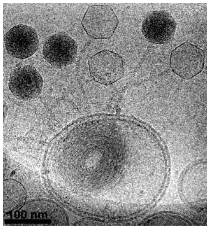
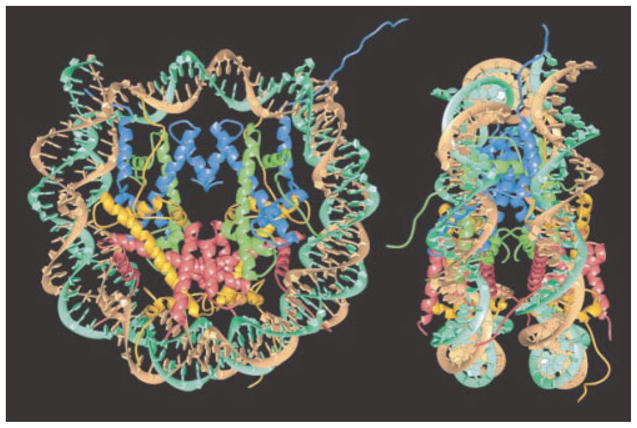
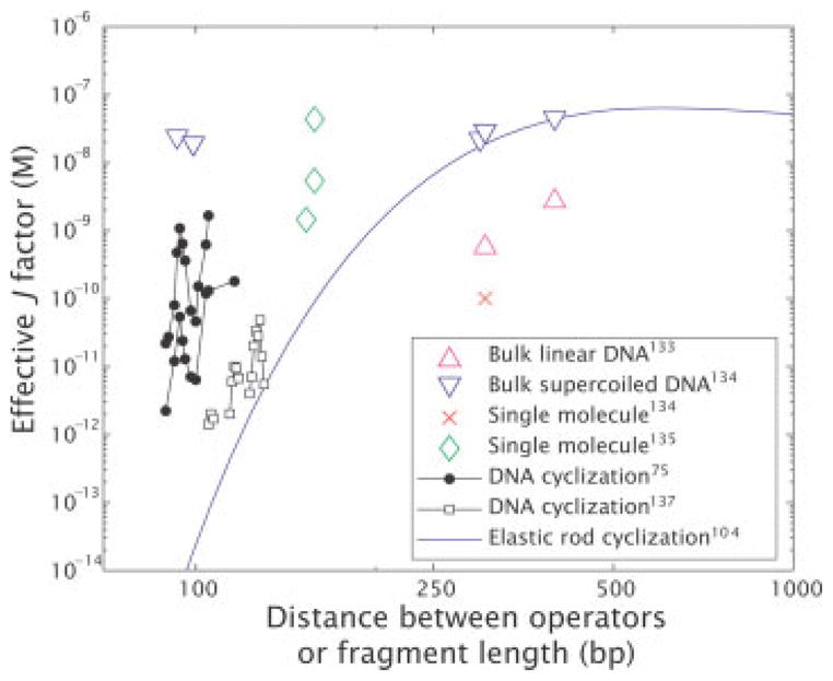
The mechanical properties of DNA play a critical role in many biological functions. For example, DNA packing in viruses involves confining the viral genome in a volume (the viral capsid) with dimensions that are comparable to the DNA persistence length. Similarly, eukaryotic DNA is packed in DNA-protein complexes (nucleosomes), in which DNA is tightly bent around protein spools. DNA is also tightly bent by many proteins that regulate transcription, resulting in a variation in gene expression that is amenable to quantitative analysis. In these cases, DNA loops are formed with lengths that are comparable to or smaller than the DNA persistence length. The aim of this review is to describe the physical forces associated with tightly bent DNA in all of these settings and to explore the biological consequences of such bending, as increasingly accessible by single-molecule techniques.


Figures

FIGURE 1
FIGURE 1
Biological examples of tightly bent DNA. (A) Transcription factor mediated DNA looping, (B) DNA packing in the nucleosome, (C) DNA packing in bacterial viruses. (Courtesy: David Goodsell, Scripps Research Institute, La Jolla, CA).
FIGURE 2
FIGURE 2
Images of packaged viral DNA. This figure shows two recent reconstructions using cryo electron microsopy of the packaged DNA. (A) Phage ε15 DNA from Jiang et al.—reconstruction without symmetry (reprinted by permission from Macmillan Publishers Ltd). The size of the scale bar is 10 nm. (B) Phage P22 DNA, with the portal shown in red (courtesy: Gabriel Lander and Jack Johnson). This view is looking into the capsid at the portal (the entry site for DNA) and the green hoops reflect density corresponding to the packed DNA.
FIGURE 3
FIGURE 3
Images of DNA ejected into a lipid bilayer vesicle. Empty capsids are distinguishable from their full counterparts because the full capsids are much darker (reprinted with permission from Elsevier).
FIGURE 4
FIGURE 4
Structure of the nucleosome. Two orthogonal views of the nucleosome showing the wrapping of the DNA around an octameric histone protein core (reprinted by permission from Macmillan Publishers Ltd). The core histone proteins are colored yellow, red, blue and green for histone H2A, H2B, H3, and H4, respectively. There are two copies of each histone in the core histone octamer. The two strands of the double helix are colored cyan and brown. The diameter of the nucleosome is roughly 11 nm and its height is roughly 6 nm.
FIGURE 5
FIGURE 5
Configurational equilibrium constant. Measured values of equilibrium accessibility and corresponding results from the model of nucleosome energetics. The inset shows a schematic of the coordinate system used to define the burial depth of the binding site of interest.
FIGURE 6
FIGURE 6
Free energy of cyclization and nucleosome formation. Difference free energies for wrapping of different 94 bp DNAs around the core histone H32H42 tetramer are plotted against the difference free energies of cyclization for these same DNAs. The line illustrates the least-squares fit to the data. The slope of the line is one, implying that the entirety of the difference in affinity for wrapping around histones can be explained by the difference in the ability to cyclize.
FIGURE 7
FIGURE 7
In vivo DNA looping by Lac repressor and the in vitro challenge. (A) Data from Müller et al. showing repression as a function of distance between operators. (B) Change in looping free energy obtained from the Müller-Hill data (black) and theoretical prediction of the energy of cyclization of a DNA molecule based on the worm like chain model and assuming a volume for E. coli of Vcell ≈ 1 μm3 such that ΔFcyclization = −ln(Jcyclization Vcell). Note that the minima in the two curves do not coincide, suggesting that the effective looping free energy in vivo is not the same as the bare looping free energy deduced from in vitro cyclization measurements. In addition, there is an overall shift in the scales in the two cases. (Inset, B) Difference in the magnitude of the twist modulation between the looping energy obtained from the Becker et al. data and the theoretical cyclization energy based on harmonic deformations of the base steps.
FIGURE 8
FIGURE 8
Effective J-factors for in vitro DNA looping. The graph is constructed by using a variety of different in vitro measurements to derive an effective looping J-factor, even in those cases where there was no direct measurement of J itself. The derived values were obtained from: (i) bulk linear DNA, (ii) bulk super-coiled DNA, (iii) single molecule measurements,, (iv) DNA cyclization, and the blue curve is a theoretical curve for cyclization corresponding to an extrapolation of the elastic rod model.
FIGURE 9
FIGURE 9
Illustration of TPM method. Schematics of both the unlooped and looped states which show how the effective tether length is a reporter of the state of looping. Typical tethers have a length of 1000 bp and typical bead sizes are 0.2–1.0 μm.

Similar articles

Cited by

References

    1. Boal DH. Mechanics of the Cell. Cambridge University Press; Cambridge, England: 2002.
    1. Nelson P. Biological Physics: Energy, Information, Life. Freeman; New York: 2004.
    1. McCauley MJ, Williams MC. 2007;85:154–168. - PubMed
    1. Wiggins PA, Heijde Tvd, Moreno-Herrero F, Spakowitz A, Phillips R, Widom J, Dekker C, Nelson PC. Nat Nanotechnol. 2006;1:137–141. - PubMed
    1. Olson WK, Gorin AA, Lu XJ, Hock LM, Zhurkin VB. Proc Natl Acad Sci USA. 1998;95:11163–11168. - PMC - PubMed

Publication types

LinkOut - more resources