. 2007 Jan;13(1):55-64.
doi: 10.1261/rna.139607. Epub 2006 Nov 14.

The CRM domain: an RNA binding module derived from an ancient ribosome-associated protein

Affiliations

The CRM domain: an RNA binding module derived from an ancient ribosome-associated protein

Alice Barkan et al. RNA. 2007 Jan.

Abstract

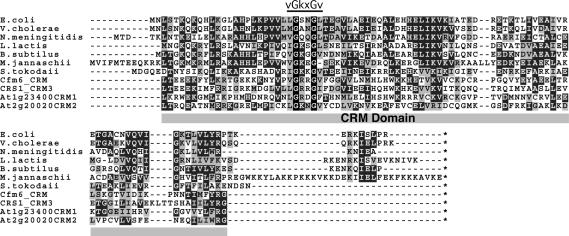
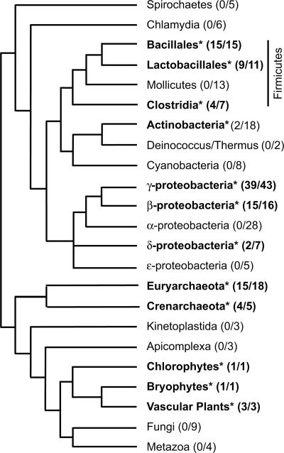
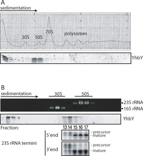
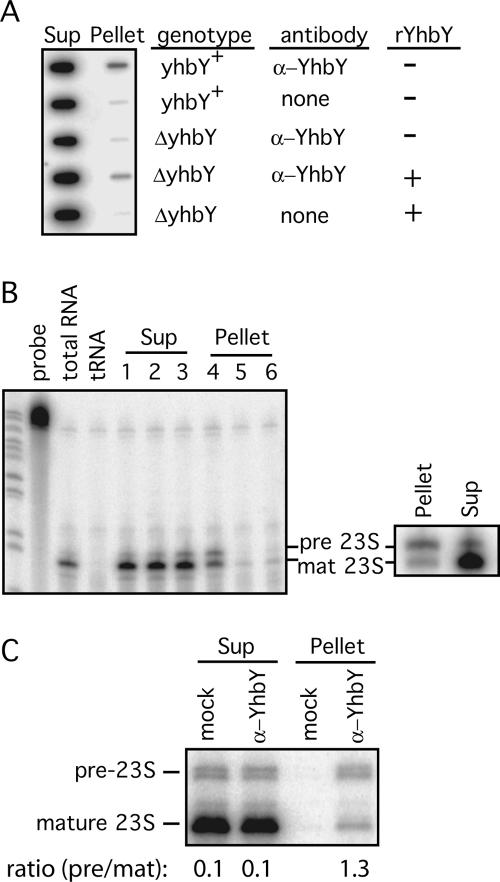
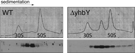
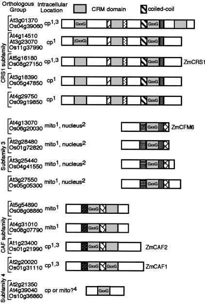
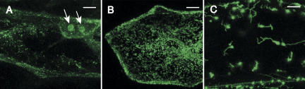
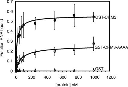
The CRS1-YhbY domain (also called the CRM domain) is represented as a stand-alone protein in Archaea and Bacteria, and in a family of single- and multidomain proteins in plants. The function of this domain is unknown, but structural data and the presence of the domain in several proteins known to interact with RNA have led to the proposal that it binds RNA. Here we describe a phylogenetic analysis of the domain, its incorporation into diverse proteins in plants, and biochemical properties of a prokaryotic and eukaryotic representative of the domain family. We show that a bacterial member of the family, Escherichia coli YhbY, is associated with pre-50S ribosomal subunits, suggesting that YhbY functions in ribosome assembly. GFP fused to a single-domain CRM protein from maize localizes to the nucleolus, suggesting that an analogous activity may have been retained in plants. We show further that an isolated maize CRM domain has RNA binding activity in vitro, and that a small motif shared with KH RNA binding domains, a conserved "GxxG" loop, contributes to its RNA binding activity. These and other results suggest that the CRM domain evolved in the context of ribosome function prior to the divergence of Archaea and Bacteria, that this function has been maintained in extant prokaryotes, and that the domain was recruited to serve as an RNA binding module during the evolution of plant genomes.


Figures

FIGURE 1.
FIGURE 1.
Alignment of representative YhbY orthologs and plant CRM domains. The complete sequences of seven prokaryotic YhbY orthologs are aligned with four CRM domains excerpted from larger plant proteins. The domain boundary was chosen according to the structural core of E. coli, Haemophilus influenzae, and Staphylococcus aureus YhbY (Ostheimer et al. 2002; Willis et al. 2002; Liu and Wyss 2004). Identical residues are shaded in black, and similar residues in gray (similarity threshold of 0.4 for shading). CFM6 and CRS1 are maize proteins with one and three CRM domains, respectively. At1g23400 and At2g20020 are Arabidopsis proteins with two CRM domains each; the position of the CRM domain in the multi-CRM proteins is stated in the domain name. The conserved vGkxGv motif, which is similar to a motif found in KH RNA binding domains, is indicated.
FIGURE 2.
FIGURE 2.
Phyletic distribution of YhbY orthologs and CRM domains. The organismal tree is a composite based on trees in Daubin et al. (2002) and Pennisi (2003). Taxa with CRM domains are indicated with bold text and an asterisk. The number of species within each group harboring CRM domains are indicated as the fraction of the number of fully sequenced genomes that were analyzed for the presence of the domain.
FIGURE 3.
FIGURE 3.
YhbY cosediments with pre-50S ribosomal subunits. (A) E. coli lysates prepared under conditions that maintain polysome integrity were sedimented through sucrose gradients. The A260 profile and an immunoblot of gradient fractions probed with anti-YhbY antibody are shown in the upper and lower panels, respectively. (B) E. coli lysates were sedimented through sucrose gradients under conditions that dissociate 70S ribosomes into 30S and 50S subunits, and that increase resolution in the 30S to 50S range. RNA extracted from gradient fractions was analyzed by agarose gel electrophoresis and ethidium bromide staining (upper panel). YhbY was detected in gradient fractions by probing an immunoblot with anti-YhbY antibody (middle panel). The termini of 23S rRNA in fractions 13 through 17 were mapped with RNAse-protection assays (bottom panel).
FIGURE 4.
FIGURE 4.
Pre-50S ribosomal subunits coimmunoprecipitate with YhbY. (A) Anti-YhbY antibody was used in immunoprecipitation reactions with cytoplasmic extract of yhbY+ or ΔyhbY E. coli cells. RNA extracted from the immunoprecipitation pellets and supernatants (Sup) was applied to a slot blot, and hybridized with a probe for the 23S rRNA. An equal proportion of the total pellet and supernatant RNA was analyzed in each slot. Addition of recombinant YhbY (rYhbY) to ΔyhbY extract reconstituted the coimmunoprecipitation of 23S rRNA, indicating that rYhbY can associate with a 50S subunit-related particle present in ΔyhbY cells. Similar results were obtained in four repetitions of this experiment involving different extract preparations (data not shown). (B) RNAse-protection analysis of the 5′ ends of 23S rRNA in the coimmunoprecipitation samples. Lanes 1 and 4: RNA from yhbY + extract immunoprecipitated with anti-YhbY serum; lanes 2 and 5: RNA from a mock immunoprecipitation with yhbY + extract; lanes 3 and 6: RNA from ΔyhbY extract immunoprecipitated with α-YhbY serum. The panel on the right shows the results of a replicate experiment with yhbY + extract and α-YhbY serum. Pre-23S and mat-23S are the products of protection by the precursor and mature 23S rRNAs, respectively. (C) Quantification of the ratio of precursor to mature 23S rRNA in YhbY immunoprecipitations. 5′ Termini were mapped by primer extension, using a radiolabeled primer complementary to the 5′ region of mature 23S rRNA; comparison to a sequencing ladder showed the bands to correspond to the established 5′ termini of mature and immature 23S rRNA (data not shown). The ratios of precursor to mature 5′ termini were quantified with a PhosphorImager and are shown below. This experiment involved a different immunoprecipitation reaction than those in B to illustrate the reproducible enrichment of pre-23S rRNA in YhbY coimmunoprecipitates. The mock assay was performed in parallel and lacked antiserum.
FIGURE 5.
FIGURE 5.
Aberrant ribosome accumulation in ΔyhbY mutant. Lysates of wild-type and ΔyhbY cells were resolved in sucrose gradients under conditions that dissociate 70S ribosomes into 30S and 50S subunits. Recombinant YhbY was added to the mutant lysate prior to sedimentation (right panels). Upper panels show A260 profiles; lower panels show immunoblots of gradient fractions probed with anti-YhbY antibody. The A260 profile of the mutant lysate was unchanged by the addition of recombinant YhbY (data not shown). The immunoblot signal in the mutant lysate derives only from the added recombinant YhbY, as the antibody detected no protein in unsupplemented mutant lysate (Supplemental Fig. 3B; http://rna.uoregon.edu/crm/BarkanSuppData.pdf; data not shown).
FIGURE 6.
FIGURE 6.
The CRM domain family in plants. Orthologous groups were assigned based on the results of mutual best hit BLAST comparisons among the complete proteomes of rice and Arabidopsis, and are supported by the phylogram shown as Supplemental Figure 5 (http://rna.uoregon.edu/crm/BarkanSuppData.pdf). The orthologous groups corresponding to maize (Zm) CRS1, CAF1, CAF2, and CFM6 are indicated. The proteins are grouped into four subfamilies based on their domain organization and their clustering in the phylogram. CRM domains are indicated by gray boxes, with other regions of similarity represented by distinct pattern fills. CRM domains harboring the “GxxG” motif are indicated and are conserved in both species, except where shown in parentheses. The coiled-coil motif that is characteristic of the CRS1 subfamily is shown, and is the only functional motif detected in these proteins other than the CRM domain itself. Intracellular locations were based on consensus predictions and/or experimental data, according to the following key: 1Prediction with TargetP (Emanuelsson and Heijne, 2001) and/or Predotar (Small et al. 2004) in both rice and Arabidopsis; 2Prediction with two of the three nuclear predictors PredictNLS (Cokol et al. 2000), NucPred (http://www.sbc.su.se/~maccallr/nucpred/), or PSORTII (http://psort.ims.u-tokyo.ac.jp/) in both rice and Arabidopsis; 3Maize ortholog established to be in chloroplast (data not shown; Till et al. 2001; Ostheimer et al. 2003); 4Weak predictions that differ between species.
FIGURE 7.
FIGURE 7.
Nucleolar localization of CFM6–GFP in onion epidermal cells. (A) Full-length maize CFM6 (40 kDa) was fused at its carboxy-terminus to GFP and transiently expressed in onion root epidermal cells. The arrows show nucleolar localization of GFP. The speckled fluorescence is similar to that shown in B for mitochondrial-targeted GFP, and is therefore likely to be mitochondrion-localized CFM6–GFP. (B) Mitochondrial targeting of GFP fused to the targeting peptide of mitochondrial FDH. (C) Chloroplast targeting of GFP fused to the targeting peptide of chloroplast RecA. Bars = 20 μm.
FIGURE 8.
FIGURE 8.
RNA binding activity of an isolated CRM domain. Filter binding assays were performed with a trace amount of 32P-labeled atpF intron RNA and increasing concentrations of GST–CRM3, GST–CRM3–AAAA, or GST. Values represent the means, ±1 standard deviation, of nine experiments involving four different protein preparations. Single-site binding isotherms were fit to the data using the equation: Fraction RNA bound = (maximum RNA bound*[protein])/(Kd+[protein]). GST–CRM3 and GST–CRM3–AAAA bound the atpF intron RNA with apparent Kds of 21.4 ± 4.3 and 79.3 ± 14.4 nM, respectively.

Similar articles

Cited by

References

    1. Aravind, L., Iyer, L.M., Anantharaman, V. The two faces of Alba: The evolutionary connection between proteins participating in chromatin structure and RNA metabolism. Genome Biol. 2003;4:R64. - PMC - PubMed
    1. Ban, N., Nissen, P., Hansen, J., Moore, P., Steitz, T. The complete atomic structure of the large ribosomal subunit at 2.4 A resolution. Science. 2000;289:905–920. - PubMed
    1. Barkan, A., Walker, M., Nolasco, M., Johnson, D. A nuclear mutation in maize blocks the processing and translation of several chloroplast mRNAs and provides evidence for the differential translation of alternative mRNA forms. EMBO J. 1994;13:3170–3181. - PMC - PubMed
    1. Bremer, H., Dennis, P. Modulation of chemical composition and other parameters of the cell by growth rate. In: Neidhardt F.C., editor. Escherichia coli and Salmonella: Cellular and molecular biology. Vol. 2. ASM Press; Washington, DC: 1996. pp. 1553–1569.
    1. Bugl, H., Fauman, E., Staker, B., Zheng, F., Kushner, S., Saper, M., Bardwell, J., Jakob, U. RNA methylation under heat shock control. Mol. Cell. 2000;6:349–360. - PubMed

Publication types

MeSH terms

Associated data

LinkOut - more resources