. 2006 Nov 14;12(42):6818-27.
doi: 10.3748/wjg.v12.i42.6818.

Promoted differentiation of cynomolgus monkey ES cells into hepatocyte-like cells by co-culture with mouse fetal liver-derived cells

Affiliations

Promoted differentiation of cynomolgus monkey ES cells into hepatocyte-like cells by co-culture with mouse fetal liver-derived cells

Ko Saito et al. World J Gastroenterol. .

Abstract

Aim: To explore whether a co-culture of cynomolgus monkey embryonic stem (cES) cells with embryonic liver cells could promote their differentiation into hepatocytes.

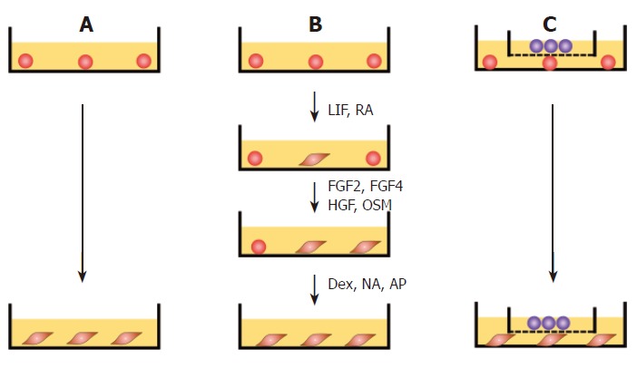
Methods: Mouse fetal liver-derived cells (MFLCs) were prepared as adherent cells from mouse embryos on embryonic d (ED) 14, after which undifferentiated cES cells were co-cultured with MFLCs. The induction of cES cells along a hepatic lineage was examined in MFLC-assisted differentiation, spontaneous differentiation, and growth factors (GF) and chemicals-induced differentiations (GF-induced differentiation) using retinoic acid, leukemia inhibitory factor (LIF), FGF2, FGF4, hepatocyte growth factor (HGF), oncostatin M (OSM), and dexamethasone.

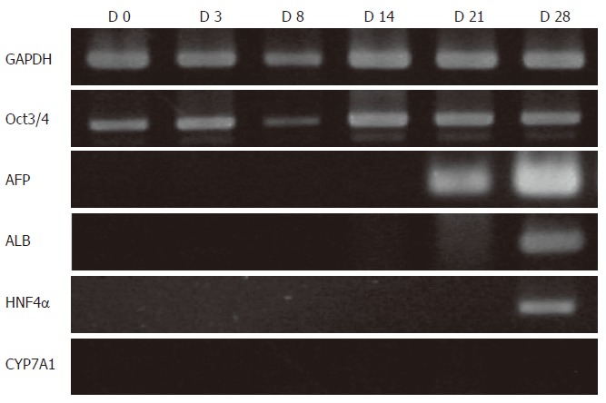
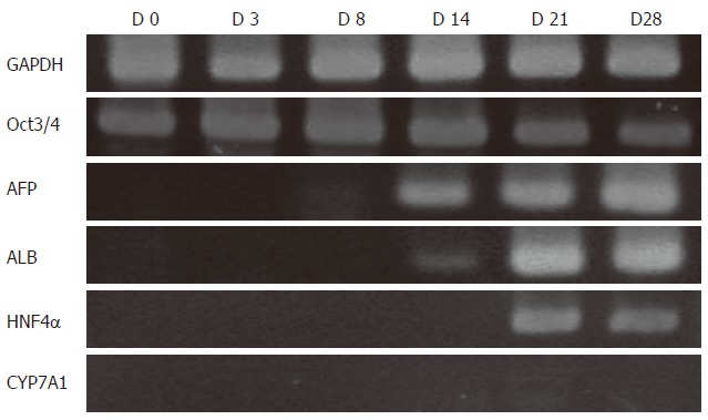
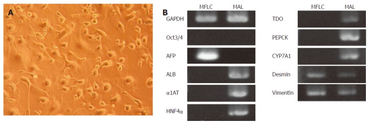
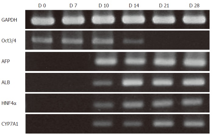
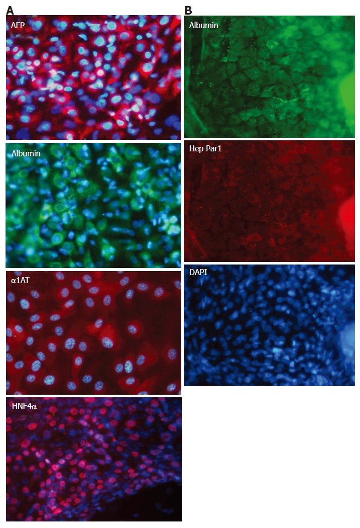
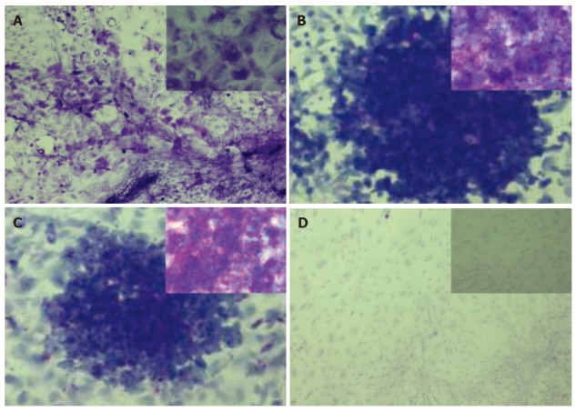
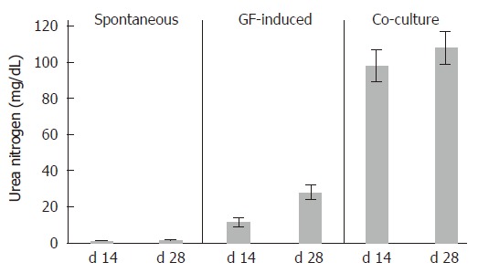
Results: The mRNA expression of alpha-fetoprotein, albumin (ALB), alpha-1-antitrypsin, and hepatocyte nuclear factor 4alpha was observed earlier in the differentiating cES cells co-cultured with MFLCs, as compared to cES cells undergoing spontaneous differentiation and those subjected to GF-induced differentiation. The expression of cytochrome P450 7a1, a possible marker for embryonic endoderm-derived mature hepatocytes, was only observed in cES cells that had differentiated in a co-culture with MFLCs. Further, the disappearance of Oct3/4, a representative marker of an undifferentiated state, was noted in cells co-cultured with MFLCs, but not in those undergoing spontaneous or GF-induced differentiation. Immunocytochemical analysis revealed an increased ratio of ALB-immunopositive cells among cES cells co-cultured with MFLCs, while glycogen storage and urea synthesis were also demonstrated.

Conclusion: MFLCs showed an ability to induce cES cells to differentiate toward hepatocytes. The co-culture system with MFLCs is a useful method for induction of hepatocyte-like cells from undifferentiated cES cells.


Figures

Figure 1
Figure 1
Protocol for differentiation of cES cells. A: Spontaneous differentiation. Undifferentiated cES cells were cultured in gelatin-coated dishes in DMEM basic medium for 28 d without additional feeders or growth factors; B: GF-induced differentiation. Undifferentiated cES cells were cultured in DMEM basic medium containing LIF (100 units/mL) and RA (10-8 mol/L) for 3 d, followed by a 5-d culture in DMEM basic medium containing FGF2 (10 mg/L), FGF4 (20 mg/L), HGF (25 mg/L), and OSM (10 mg/L). Thereafter, the cells were cultured for 20 d in DMEM basic medium containing 10-7 mol/L dexamethasone (Dex), 0.2 mmol/L, L-ascorbic-2-phosphate (AP), and 10 mmol/L nicotinamide (NA); C: Promoted differentiation by co-culture with MFLCs. Undifferentiated cES cells were plated onto 60-mm dishes and co-cultured with MFLCs in a 35-mm culture insert across a 0.4-μm Millicell CM membrane for 28 d.
Figure 2
Figure 2
RT-PCR analysis of spontaneous differentiation of cES cells. AFP mRNA expression was not observed prior to d 14, while ALB was detected on d 21. On d 28, ALB and HNF4α expression became detectable. CYP7A1 was not detected throughout the experimental period, while Oct3/4 was distinctly detected at all time points.
Figure 3
Figure 3
RT-PCR analysis of GF-induced differentiation of cES cells. AFP mRNA expression was not detected following a 3-d treatment with RA and LIF, though it was detected on d 8, and at the end of 5 d of treatment with FGF2, FGF4, and HGF. ALB and HNF4α were detected after the differentiating cES cells were exposed to OSM and Dex. Although ALB and HNF4α were strongly detected on d 21 and 28, no expression of CYP7A1 was observed. Oct3/4 mRNA expression remained up to d 28.
Figure 4
Figure 4
Characterization of MFLCs. A: Optical microscope image of MFLCs (× 100). MFLCs were prepared from E14.0 mouse livers and cultured for 48 h. After the floating cell fraction was discarded from the culture, resting adherent cells were further cultured until semi-confluent. MFLCs showed various morphologies, including cuboidal and stellate-shaped cells; B: RT-PCR analysis of MFLCs. AFP, desmin, and vimentin were expressed, whereas ALB was not. MAL: Mouse adult liver cells.
Figure 5
Figure 5
RT-PCR analysis of cES cells co-cultured with MFLCs. cES cells co-cultured with MFLCs across a membrane with 0.4-µm sized pores for 28 d were analyzed. AFP, ALB, HNF4α, and CYP7A1 mRNA expression was detected on d 10. Oct3/4 expression was faint on d 14 and disappeared by d 21.
Figure 6
Figure 6
Immunocytochemical analysis of cES cells co-cultured with MFLCs. A: After being co-cultured with MFLCs for 28 d, cES cells were immunostained with anti-AFP, anti-ALB, anti-alpha-1-antitrypsin, and anti-HNF4α antibodies; B: Immunoreactivity to the anti-human hepatocyte antibody Hep Par1 was shown by the albumin-positive cells. Original magnification × 100.
Figure 7
Figure 7
Histochemical staining of glycogen. Periodic acid Schiff (PAS) staining was performed on cES cells co-cultured with MFLCs for 28 d (A) and undifferentiated cES cells (B), as well as those following treatment with α-amylase (C and D, respectively). Both the undifferentiated cES cells and those co-cultured with MFLCs showed violet staining (A, B). Violet staining was apparent in the undifferentiated cES cells (C), while such staining did not appear in the differentiated cES cells pretreated with α-amylase(D). Original magnification ×100 (A-D), × 400 (insets in A-D).
Figure 8
Figure 8
Urea synthesis by differentiated cES cells. The level of urea nitrogen in the culture medium of cES cells cultured with MFLCs was greater than that in the medium of cES cells subjected to GF-induction, though amounts of urea synthesized by cES cells co-cultured with MFLCs scarcely increased from d 14 to 28. Spontaneously differentiated cES cells showed nearly no ability to synthesize urea.

Similar articles

Cited by

References

    1. Evans MJ, Kaufman MH. Establishment in culture of pluripotential cells from mouse embryos. Nature. 1981;292:154–156. - PubMed
    1. Martin GR. Isolation of a pluripotent cell line from early mouse embryos cultured in medium conditioned by teratocarcinoma stem cells. Proc Natl Acad Sci USA. 1981;78:7634–7638. - PMC - PubMed
    1. Shiroi A, Yoshikawa M, Yokota H, Fukui H, Ishizaka S, Tatsumi K, Takahashi Y. Identification of insulin-producing cells derived from embryonic stem cells by zinc-chelating dithizone. Stem Cells. 2002;20:284–292. - PubMed
    1. Shiroi A, Ueda S, Ouji Y, Saito K, Moriya K, Sugie Y, Fukui H, Ishizaka S, Yoshikawa M. Differentiation of embryonic stem cells into insulin-producing cells promoted by Nkx2.2 gene transfer. World J Gastroenterol. 2005;11:4161–4166. - PMC - PubMed
    1. Yamada T, Yoshikawa M, Takaki M, Torihashi S, Kato Y, Nakajima Y, Ishizaka S, Tsunoda Y. In vitro functional gut-like organ formation from mouse embryonic stem cells. Stem Cells. 2002;20:41–49. - PubMed

Publication types

MeSH terms

Substances