Comparative Study
. 2007 Mar;81(5):2472-84.
doi: 10.1128/JVI.02073-06. Epub 2006 Nov 15.

The Old World and New World alphaviruses use different virus-specific proteins for induction of transcriptional shutoff

Affiliations
Comparative Study

The Old World and New World alphaviruses use different virus-specific proteins for induction of transcriptional shutoff

Natalia Garmashova et al. J Virol. 2007 Mar.

Abstract

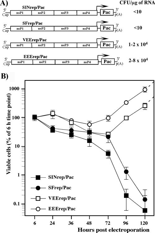
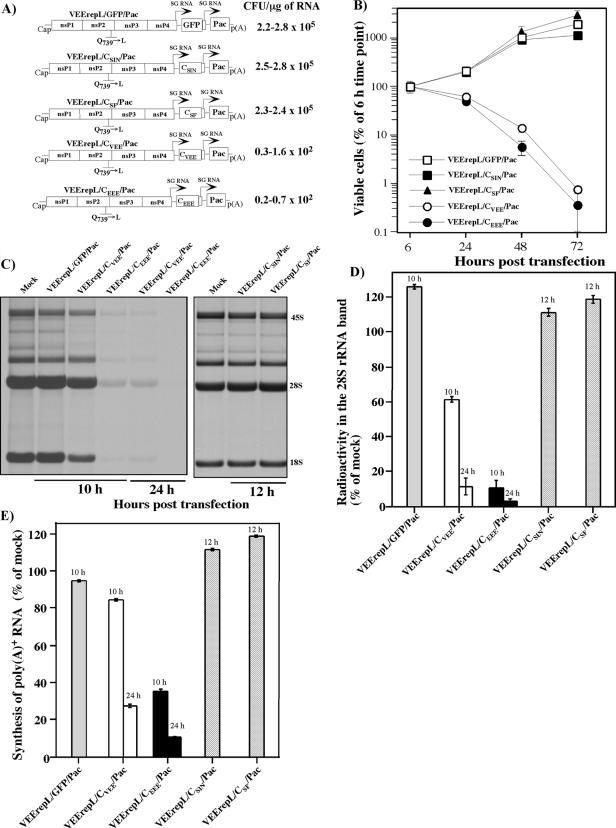
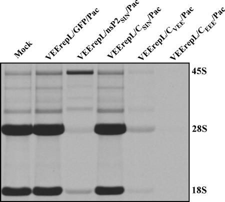
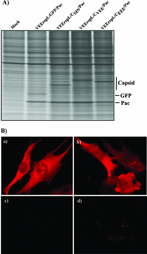
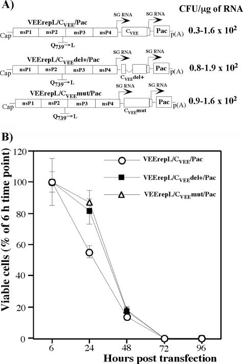
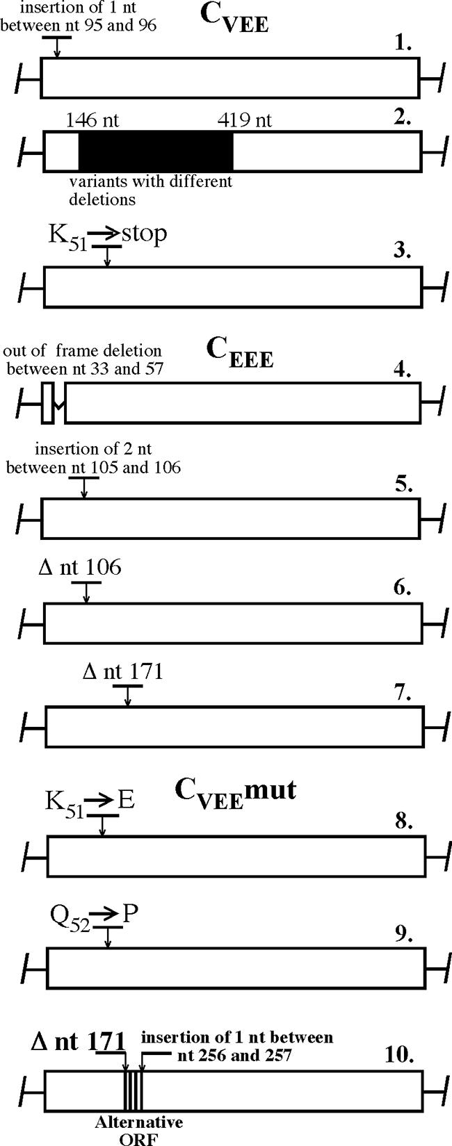
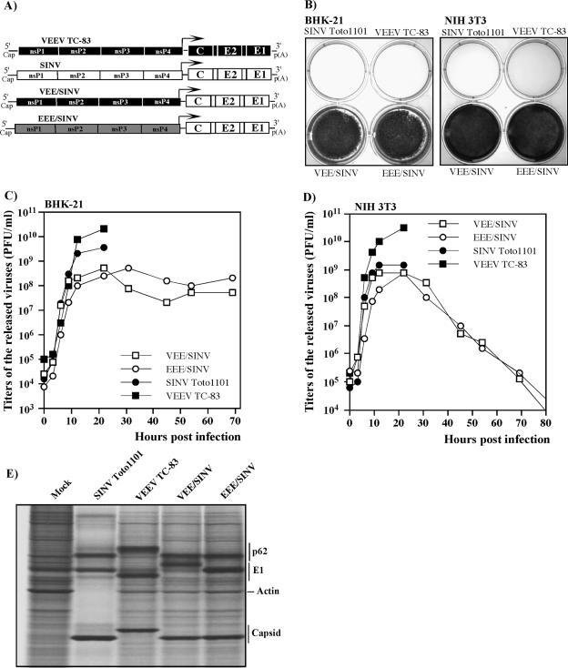
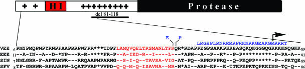
Alphaviruses are widely distributed throughout the world. During the last few thousand years, the New World viruses, including Venezuelan equine encephalitis virus (VEEV) and eastern equine encephalitis virus (EEEV), evolved separately from those of the Old World, i.e., Sindbis virus (SINV) and Semliki Forest virus (SFV). Nevertheless, the results of our study indicate that both groups have developed the same characteristic: their replication efficiently interferes with cellular transcription and the cell response to virus replication. Transcriptional shutoff caused by at least two of the Old World alphaviruses, SINV and SFV, which belong to different serological complexes, depends on nsP2, but not on the capsid protein, functioning. Our data suggest that the New World alphaviruses VEEV and EEEV developed an alternative mechanism of transcription inhibition that is mainly determined by their capsid protein, but not by the nsP2. The ability of the VEEV capsid to inhibit cellular transcription appears to be controlled by the amino-terminal fragment of the protein, but not by its protease activity or by the positively charged RNA-binding domain. These data provide new insights into alphavirus evolution and present a plausible explanation for the particular recombination events that led to the formation of western equine encephalitis virus (WEEV) from SINV- and EEEV-like ancestors. The recombination allowed WEEV to acquire capsid protein functioning in transcription inhibition from EEEV-like virus. Identification of the new functions in the New World alphavirus-derived capsids opens an opportunity for developing new, safer alphavirus-based gene expression systems and designing new types of attenuated vaccine strains of VEEV and EEEV.


Figures

FIG. 1.
FIG. 1.
Analysis of cytotoxicity of the alphavirus replicons. (A) Schematic representation of the New World and the Old World alphavirus replicons. Arrows indicate positions of the subgenomic promoters. Pac indicates the puromycin acetyltransferase gene. RNA transfections and puromycin selection was performed as described in Materials and Methods. Purr colonies were stained with crystal violet, and the results are presented in CFU per μg of RNA used for transfection. (B) Analysis of cell growth and cell death at different times posttransfection. Equal numbers of cells were seeded into six-well Costar plates. Puromycin selection was performed between 6 and 48 h posttransfection and then cells were incubated in puromycin-free medium. The number of viable cells was counted at the indicated time points. The data were normalized based on the number of viable adherent cells determined at 6 h posttransfection. Error bars indicate variations between parallel samples.
FIG. 2.
FIG. 2.
Analysis of effects of nsP2 expression on cellular transcription and cell viability. (A) Schematic representation of VEE genome-based replicons expressing SINV and SFV nsP2 and analysis of their ability to establish persistent replication and develop Purr foci. Arrows indicate the positions of the subgenomic promoters. Ubi indicates a ubiquitin sequence fused in frame with the SINV and SFV nsP2 genes. (B) Analysis of growth of cells carrying VEEV replicons expressing GFP or SINV- and SFV-derived nsP2. (C) Inhibition of transcription in BHK-21 cells transfected with VEEV replicons expressing SINV or SFV nsP2. Cells were electroporated with 5 μg of in vitro-synthesized RNAs. At 10 and 24 h posttransfection, cellular RNAs were labeled with [3H]uridine in the absence of ActD for 3 h and analyzed by RNA gel electrophoresis under the conditions described in Materials and Methods. (D) For quantitative analysis of transcription inhibition, aliquots of RNA samples used for the gel shown in panel C were washed on Whatman 3MM filters with trichloroacetic acid as described in Materials and Methods, and the radioactivity was measured by liquid scintillation counting. (E) Another aliquot of each sample was used for isolation of the poly(A)+ RNA as described in Materials and Methods, and the radioactivity was measured by liquid scintillation counting. One of three reproducible experiments is presented; error bars indicate variations between parallel samples.
FIG. 3.
FIG. 3.
Effects of capsid expression on cellular transcription and cell growth. (A) Schematic representation of VEEV replicons expressing SINV, SFV, VEEV, and EEEV capsids. Arrows indicate positions of the subgenomic promoters. Different dilutions of the electroporated cells were seeded into 100-mm tissue culture dishes. Puromycin selection was performed as described in Materials and Methods. Purr cell colonies were stained with crystal violet at days 4 to 9 posttransfection, depending on their growth rates. The results are presented in CFU per μg of RNA used for transfection. The ranges indicate variations between the experiments. (B) Analysis of growth of cells transfected with VEEV replicons expressing GFP and different capsids. Equal numbers of cells were seeded into six-well Costar plates. Puromycin selection (10 μg/ml) was performed between 6 and 48 h posttransfection. Then, cells were incubated in puromycin-free medium, and viable cells were counted at the indicated times. The data were normalized based on the number of viable adherent cells determined at 6 h posttransfection. (C and D) Analysis of cellular transcription. RNA labeling was performed with [3H]uridine at the indicated times posttransfection for 2 h. RNA samples were analyzed by gel electrophoresis under the conditions described in Materials and Methods (C). For quantitative analysis, the rRNA bands were excised from the 2,5-diphenyloxazole-impregnated gels (C), and the radioactivity was measured by liquid scintillation counting (D). (E) Equal aliquots of each sample were used for isolation of the poly(A)+ RNA as described in Materials and Methods, and the radioactivity was measured by liquid scintillation counting. One of three reproducible experiments is presented; error bars indicate variations between parallel samples.
FIG. 4.
FIG. 4.
RNA synthesis in cells transfected with VEEV replicons expressing GFP, SINV nsP2, or different alphavirus capsids. BHK-21 cells were transfected with replicons expressing different proteins. RNAs were labeled with [3H]uridine in the absence of ActD for 5 h at 24 h posttransfection and analyzed as described in Materials and Methods.
FIG. 5.
FIG. 5.
Synthesis and distribution of alphavirus capsids in cells transfected with VEEV replicons. (A) Cell were labeled with [35S]methionine at 10 h posttransfection as described in Materials and Methods, and equal amounts of proteins were separated by sodium dodecyl sulfate-10% polyacrylamide gel electrophoresis followed by autoradiography. (B) For analysis of capsid distribution, BHK-21 cells were electroporated with in vitro-synthesized RNAs of VEErepL/CSIN/Pac and VEErepL/CVEE/Pac and then, at 20 h posttransfection, stained with mouse anti-SINV (a and c) and anti-VEEV (b and d) antibodies and goat anti-mouse immunoglobulin G-Alexa Fluor 546-labeled secondary antibodies (Molecular Probes). Cells: (a) VEErepL/CSIN/Pac-transfected cells, (b) VEErepL/CVEE/Pac, and (c and d) mock transfection.
FIG. 6.
FIG. 6.
Analysis of cytotoxicity of VEEV capsid with mutated protease or deleted RNA-binding domain. (A) Schematic representation of VEEV genome-based replicons expressing VEEV capsid containing an S226→A mutation or deletion of aa 81 to 118 and analysis of their abilities to establish persistent replication and develop Purr foci. (B) Survival of cells transfected with the replicons expressing wt capsid or capsid with the indicated mutations. The data were normalized based on the number of viable adherent cells determined at 6 h posttransfection.
FIG. 7.
FIG. 7.
Accumulation of mutations in VEEV and EEEV capsids encoded by VEErepL replicon. Individual Purr cell colonies that formed after transfection of VEErepL/CVEE/Pac, VEErepL/CEEE/Pac, and VEErepL/CVEEmut/Pac replicons were randomly selected, and the genome fragment encoding capsid was sequenced. Positions of the mutations are indicated. Colony number 2 contained replicons with multiple deletions and/or insertions in the capsid-coding sequence that produced multiple sequences in the indicated fragment during the direct sequencing of the PCR fragment.
FIG. 8.
FIG. 8.
Replication of viruses expressing homologous and heterologous structural and nonstructural proteins. (A) Schematic representation of the viral genomes. SINV-, VEEV-, and EEEV-specific sequences are indicated by white, black, and gray, respectively. (B) BHK-21 and NIH 3T3 cells were infected with the indicated viruses at an MOI of 10 PFU/cell. Cells were stained with crystal violet at day 3 (BHK-21) and day 5 (NIH 3T3) postinfection. (C and D) The media were replaced as described in Materials and Methods at 0, 3, 6, 9, 12 and 22 h postinfection (for all of the viruses), and VEE/SINV- and EEE/SINV-containing samples were also harvested later, at the indicated times. Virus titers were determined as described in Materials and Methods. (E) BHK-21 cells (5 × 10⁵ cells in 35-mm dishes) were infected with SINV Toto1101, VEEV TC-83, VEE/SINV, and EEE/SINV at an MOI of 10 PFU/cell. At 16 h postinfection, proteins were pulse-labeled with [35S]methionine as described in Materials and Methods and analyzed on sodium dodecyl sulfate-10% polyacrylamide gels. The gels were dried and autoradiographed. The E1 proteins of VEE/SINV and EEE/SINV have differing mobilities on the gel because the SINV structural genes were derived from SINV TE12 and SINV Toto1101 strains, respectively, in which E1 differs by two amino acids (27, 37).
FIG. 9.
FIG. 9.
Schematic representation of VEEV capsid and sequence alignment with other alphavirus capsids. VEEV, EEEV, SINV, and SFV sequences are derived from references , , , and , respectively. All of the mutations identified in the capsids of replicons incapable of causing CPE (see variants 8, 9, and 10 in Fig. 7) are indicated in blue. Helix I sequences are indicated in red. Residues identical to those in the VEEV sequence are indicated by dashes. Stars indicate positions of the deletions introduced for better alignment of the sequences. The arrow indicates the beginning of the deletion made in the capsid of VEErepL/CVEEdel+/Pac mutant.

References

    1. Basler, C. F., A. Mikulasova, L. Martinez-Sobrido, J. Paragas, E. Muhlberger, M. Bray, H. D. Klenk, P. Palese, and A. Garcia-Sastre. 2003. The Ebola virus VP35 protein inhibits activation of interferon regulatory factor 3. J. Virol. 77:7945-7956. - PMC - PubMed
    1. Basler, C. F., X. Wang, E. Muhlberger, V. Volchkov, J. Paragas, H. D. Klenk, A. Garcia-Sastre, and P. Palese. 2000. The Ebola virus VP35 protein functions as a type I IFN antagonist. Proc. Natl. Acad. Sci. USA 97:12289-12294. - PMC - PubMed
    1. Billecocq, A., M. Spiegel, P. Vialat, A. Kohl, F. Weber, M. Bouloy, and O. Haller. 2004. NSs protein of Rift Valley fever virus blocks interferon production by inhibiting host gene transcription. J. Virol. 78:9798-9806. - PMC - PubMed
    1. Bouloy, M., C. Janzen, P. Vialat, H. Khun, J. Pavlovic, M. Huerre, and O. Haller. 2001. Genetic evidence for an interferon-antagonistic function of rift valley fever virus nonstructural protein NSs. J. Virol. 75:1371-1377. - PMC - PubMed
    1. Bredenbeek, P. J., I. Frolov, C. M. Rice, and S. Schlesinger. 1993. Sindbis virus expression vectors: packaging of RNA replicons by using defective helper RNAs. J. Virol. 67:6439-6446. - PMC - PubMed

Publication types

MeSH terms

LinkOut - more resources