. 2007 Feb;81(4):1701-13.
doi: 10.1128/JVI.01467-06. Epub 2006 Nov 15.

A severe acute respiratory syndrome coronavirus that lacks the E gene is attenuated in vitro and in vivo

Affiliations

A severe acute respiratory syndrome coronavirus that lacks the E gene is attenuated in vitro and in vivo

Marta L DeDiego et al. J Virol. 2007 Feb.

Abstract

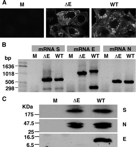
A deletion mutant of severe acute respiratory syndrome coronavirus (SARS-CoV) has been engineered by deleting the structural E gene in an infectious cDNA clone that was constructed as a bacterial artificial chromosome (BAC). The recombinant virus lacking the E gene (rSARS-CoV-DeltaE) was rescued in Vero E6 cells. The recovered deletion mutant grew in Vero E6, Huh-7, and CaCo-2 cells to titers 20-, 200-, and 200-fold lower than the recombinant wild-type virus, respectively, indicating that although the E protein has an effect on growth, it is not essential for virus replication. No differences in virion stability under a wide range of pH and temperature were detected between the deletion mutant and recombinant wild-type viruses. Although both viruses showed the same morphology by electron microscopy, the process of morphogenesis seemed to be less efficient with the defective virus than with the recombinant wild-type one. The rSARS-CoV-DeltaE virus replicated to titers 100- to 1,000-fold lower than the recombinant wild-type virus in the upper and lower respiratory tract of hamsters, and the lower viral load was accompanied by less inflammation in the lungs of hamsters infected with rSARS-CoV-DeltaE virus than with the recombinant wild-type virus. Therefore, the SARS-CoV that lacks the E gene is attenuated in hamsters, might be a safer research tool, and may be a good candidate for the development of a live attenuated SARS-CoV vaccine.


Figures

FIG. 1.
FIG. 1.
Rescue of SARS-CoV-ΔE from cDNA in Vero E6 cells. (A) Genetic organization of the rSARS-CoV-ΔE virus. The 142-nt deletion inside the E gene is indicated in a white box (Δ142). The mutated core sequence (CS-E) within the TRS and the mutated start codon are shown in a box and underlined, respectively. The mutations introduced to change the CS-E and the ATG codon of the E gene are shown in italics. The deleted ORF-E is shown in a box. Restriction sites BamHI, NheI, and RsrII used to delete E gene are indicated. Letters and numbers indicate the viral genes. CMV, cytomegalovirus promoter; L, leader sequence; An, poly(A) tail; Rz, hepatitis delta virus ribozyme; BGH, bovine growth hormone termination and polyadenylation sequences. (B) Cytopathic effect and plaque morphology produced by the indicated viruses on Vero E6 cells.
FIG. 2.
FIG. 2.
Characterization of defective virus proteins and mRNAs. Vero E6 cells were mock infected (M) or infected with the rSARS-CoV-ΔE (ΔE) or the recombinant wild-type (WT) viruses. (A) Viral protein expression was analyzed by indirect immunofluorescence 24 h postinfection using a SARS-CoV-specific human polyclonal antibody followed by Cy5-labeled goat anti-human antibody. (B) Viral mRNA expression was analyzed by RT-PCR using the oligonucleotides specific for sgmRNAs of S, E, and N genes. (C) Western blot analysis of infected cell lysates using S, N, and E protein-specific polyclonal antibodies followed by peroxidase-labeled goat anti-rabbit antibody.
FIG. 3.
FIG. 3.
Growth kinetics of rSARS-CoV-ΔE in different cell lines. Vero E6 (A), Huh-7 (B), and CaCo-2 (C) cells were infected at a MOI of 0.5 with either the rSARS-CoV-ΔE or the recombinant wild-type virus. At different times postinfection, virus titers were determined by plaque assay on Vero E6 cells. Error bars represent standard deviations of the mean of results from three experiments.
FIG. 4.
FIG. 4.
Effect of temperature and pH changes on rSARS-CoV-ΔE virus infectivity. Supernatants containing recombinant SARS-CoV and SARS-CoV-ΔE viruses were incubated for 30 min at the indicated temperature (A) or pH (B), and virus infectivity was evaluated by titration of culture supernatants on Vero E6 cells. Error bars represent standard deviations of the mean from three experiments. The lower limit of detection is 20 PFU/ml.
FIG. 5.
FIG. 5.
Ultrastructural analysis of rSARS-CoV-ΔE-infected Vero E6 cells. Vero E6 cells were infected at a MOI of 1 with rSARS-CoV and rSARS-CoV-ΔE viruses, and at 24 h postinfection, the cells were processed for electron microscopy of ultrathin sections. (A) Cytoplasm of an infected cell filled with swollen Golgi sacs containing virus particles (dark arrows). (B) Cytoplasm of infected cells showing the sites of budding of the nucleocapsid into the lumen of swollen Golgi sacs (thick dark arrows). Dense material in the cytoplasm of SARS-CoV-ΔE-infected cells is indicated with dark arrows. (C) Mature virus particles found in swollen Golgi sacs that appeared as large vacuoles (dark arrows). Pictures representing rSARS-CoV-infected cells or rSARS-CoV-ΔE-infected cells are displayed on the left and right sides, respectively. Bars, 2 μm (A) and 200 nm (B and C).
FIG. 6.
FIG. 6.
Morphology of rSARS-CoV-ΔE virions released from infected Vero E6 cells. (A) Electron micrographs of ultrathin sections showing extracellular viruses lining the cell surface. (B) Supernatants of rSARS-CoV- and rSARS-CoV-ΔE-infected cells were concentrated in an airfuge and analyzed by electron microscopy following negative staining with sodium phosphotungstate. Pictures on the left represent rSARS-CoV-infected cells, while those on the right represent rSARS-CoV-ΔE-infected cells. Bars, 200 nm (A) and 100 nm (B).
FIG. 7.
FIG. 7.
In vivo growth kinetics of the defective virus. Hamsters were inoculated with 103 TCID50 of rSARS-CoV or rSARS-CoV-ΔE. Animals were sacrificed, and tissues were harvested at different times postinfection. Viral titers in the lungs (A) and nasal turbinates (B) were determined in Vero E6 cell monolayers. The nonparametric Mann-Whitney U statistical method was used for ascertaining the significance of observed differences. Statistical significance was indicated by asterisks (P value < 0.05). The dotted line indicates the lower limit of detection.
FIG. 8.
FIG. 8.
Lung pathology caused by rSARS-CoV and rSARS-CoV-ΔE infection in hamsters. Hamsters were inoculated with 103 TCID50 of rSARS-CoV or rSARS-CoV-ΔE. Animals were sacrificed 2 days after infection, and lungs were inflated with and fixed in 10% formalin and processed for histopathological examination and immunohistochemistry. (A) Hematoxylin and eosin staining of a lung section shows prominent mononuclear inflammatory infiltrates in peribronchiolar, interstitial, and alveolar spaces. (B) Hematoxylin and eosin staining of a lung section shows scant mononuclear inflammatory infiltrates. (C) Immunoalkaline phosphatase staining shows abundant viral antigens in bronchiolar epithelial cells and alveolar pneumocytes. (D) Scattered immunohistochemical staining of viral antigens in bronchiolar epithelial cells and alveolar pneumocytes. Panels A and C show lung tissues from rSARS-CoV-infected animals. Panels B and D show lung tissues from rSARS-CoV-ΔE-infected animals. Bar, 100 μm.

References

    1. Almazán, F., M. L. DeDiego, C. Galan, D. Escors, E. Alvarez, J. Ortego, I. Sola, S. Zuñiga, S. Alonso, J. L. Moreno, A. Nogales, C. Capiscol, and L. Enjuanes. 2006. Construction of a SARS-CoV infectious cDNA clone and a replicon to study coronavirus RNA synthesis. J. Virol. 80:10900-10906. - PMC - PubMed
    1. Arbely, E., Z. Khattari, G. Brotons, M. Akkawi, T. Salditt, and I. T. Arkin. 2004. A highly unusual palindromic transmembrane helical hairpin formed by SARS coronavirus E protein. J. Mol. Biol. 341:769-779. - PMC - PubMed
    1. Baudoux, P., C. Carrat, L. Besnardeau, B. Charley, and H. Laude. 1998. Coronavirus pseudoparticles formed with recombinant M and E proteins induce alpha interferon synthesis by leukocytes. J. Virol. 72:8636-8643. - PMC - PubMed
    1. Brierley, I., P. Digard, and S. C. Inglis. 1989. Characterization of an efficient coronavirus ribosomal frameshifting signal: requirement for an RNA pseudoknot. Cell 57:537-547. - PMC - PubMed
    1. Chu, C. M., L. L. Poon, V. C. Cheng, K. S. Chan, I. F. Hung, M. M. Wong, K. H. Chan, W. S. Leung, B. S. Tang, V. L. Chan, W. L. Ng, T. C. Sim, P. W. Ng, K. I. Law, D. M. Tse, J. S. Peiris, and K. Y. Yuen. 2004. Initial viral load and the outcomes of SARS. Can. Med. Assoc. J. 171:1349-1352. - PMC - PubMed

Publication types

Substances

LinkOut - more resources