. 2007 Feb;81(3):1492-501.
doi: 10.1128/JVI.01843-06. Epub 2006 Nov 15.

Cholesterol-depleting statin drugs protect postmitotically differentiated human neurons against ethanol- and human immunodeficiency virus type 1-induced oxidative stress in vitro

Affiliations

Cholesterol-depleting statin drugs protect postmitotically differentiated human neurons against ethanol- and human immunodeficiency virus type 1-induced oxidative stress in vitro

Edward Acheampong et al. J Virol. 2007 Feb.

Abstract

The majority of human immunodeficiency virus type 1 (HIV-1)-infected individuals are either alcoholics or prone to alcoholism. Upon ingestion, alcohol is easily distributed into the various compartments of the body, particularly the brain, by crossing through the blood-brain barrier. Both HIV-1 and alcohol induce oxidative stress, which is considered a precursor for cytotoxic responses. Several reports have suggested that statins exert antioxidant as well as anti-inflammatory pleiotropic effects, besides their inherent cholesterol-depleting potentials. In our studies, postmitotically differentiated neurons were cocultured with HIV-1-infected monocytes, T cells, or their cellular supernatants in the presence of physiological concentrations of alcohol for 72 h. Parallel cultures were pretreated with statins (atorvastatin and simvastatin) with the appropriate controls, i.e., postmitotically differentiated neurons cocultured with uninfected cells and similar cultures treated with alcohol. The oxidative stress responses in the presence/absence of alcohol in these cultures were determined by the production of the well-characterized oxidative stress markers, 8-isoprostane-F2-alpha, total nitrates as an indicator for various isoforms of nitric oxide synthase activity, and heat shock protein 70 (Hsp70). An in vitro culture of postmitotically differentiated neurons with HIV-1-infected monocytes or T cells as well as supernatants from these cells enhanced the release of 8-isoprostane-F2-alpha in the conditioned medium six- to sevenfold (monocytes) and four- to fivefold (T cells). It was also observed that coculturing of HIV-1-infected primary monocytes over a time period of 72 h significantly elevated the release of Hsp70 compared with that of uninfected controls. Cellular supernatants of HIV-1-infected monocytes or T cells slightly increased Hsp70 levels compared to neurons cultured with uninfected monocytes or T-cell supernatants (controls). Ethanol (EtOH) presence further elevated Hsp70 in both infected and uninfected cultures. The amount of total nitrates was significantly elevated in the coculture system when both infected cells and EtOH were present. Surprisingly, pretreatment of postmitotic neurons with clinically available inhibitors of HMG-coenzyme A reductase (statins) inhibited HIV-1-induced release of stress/toxicity-associated parameters, i.e., Hsp70, isoprostanes, and total nitrates from HIV-1-infected cells. The results of this study provide new insights into HIV-1 neuropathogenesis aimed at the development of future HIV-1 therapeutics to eradicate viral reservoirs from the brain.


Figures

FIG. 1.
FIG. 1.
Characterization of postmitotic neurons and other cells. Postmitotically differentiated neurons derived from neuronal cell line NT2 upon treatment with RA, followed by selective isolation. (A) Isolated neurons. (B and C) Immunofluorescence staining for the expression of cellular MAP2 and CD14 marker for postmitotic neurons and monocytes, respectively.
FIG. 2.
FIG. 2.
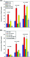
HIV-1-infected CD14+ monocytes and CD3+ T cells significantly induced the secretion of Hsp70 from postmitotically differentiated neurons. (A) The release of Hsp70 from postmitotic neurons cocultured with macrophage tropic HIV-1, YU2-infected CD14+ monocytes (Mono), or uninfected controls was determined. Parallel coculture setup of the same experiment with 0.1% and 0.3% EtOH was also carried out for the determination of Hsp70 release. The secretion of Hsp70 in the cocultured conditioned cellular supernatant (Sup) was quantified using the StressXpress ELISA (Stressgen, Victoria, BC, Canada). (B) The HIV-1-infected, NL4-3-infected, and uninfected CD3+ T lymphocytes and their supernatants were then cocultured with postmitotic neurons either in the presence or in the absence of 0.1% or 0.3% EtOH. Hsp70 release from conditioned cellular supernatants was quantified as described above. Results are the mean values for duplicate samples ± standard errors of the means. Experiments were carried out at least two times.
FIG. 3.
FIG. 3.
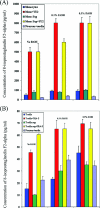
HIV-1-infected CD14+ monocytes and CD3+ T lymphocytes significantly induced the secretion of 8-isoprostaglandin-F2-α from postmitotically differentiated neurons. (A) Postmitotically differentiated neurons were cocultured with either HIV-1-infected CD14+ monocytes (Mono) or their cellular conditioned supernatants (Sup) with and without EtOH. (B) The CD3+ T lymphocytes infected with HIV-1 NL4-3 and uninfected controls were cocultured with postmitotically differentiated neurons. Neurons were also cultured in the presence of cellular supernatants obtained from HIV-1-infected and uninfected CD3+ T cells. The levels of 8-isoprostaglandin-F2-α in the conditioned media were determined using the StressXpress ELISA (Stressgen, Victoria, BC, Canada). The effects of EtOH either in the presence or in the absence of HIV-1-infected CD3+ T cells and their cellular supernatants were also investigated. Results are the mean values for duplicate samples ± standard errors of the means. The data presented are averages for two independent experiments.
FIG. 4.
FIG. 4.
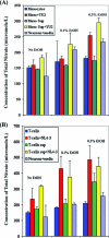
HIV-1-infected CD14+ monocytes and CD3+ T lymphocytes significantly induce the secretion of total nitrates from postmitotically differentiated neurons. (A) The levels of total nitrates (an indicator of the activity of various isoforms of nitric oxide synthase) in the cellular supernatants (Sup) of postmitotic neurons cocultured with HIV-1-infected/uninfected CD14+ monocytes (Mono) in the presence or absence of various concentrations of EtOH were determined after 72 h. (B) HIV-1-infected and uninfected CD3+ T lymphocytes and their cellular supernatants were added to postmitotic neurons either in the presence or in the absence of EtOH. After 72 h, total nitrate secretion in conditioned media was quantified using the StressXpress ELISA (Stressgen, Victoria, BC, Canada). Results are the mean values for duplicate samples ± standard errors of the means. Experiments were carried out at least two times.
FIG. 5.
FIG. 5.
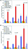
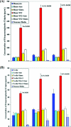
The statins atorvastatin and simvastatin significantly block the induction of Hsp70 by HIV-1-infected CD14+ monocytes (A) and CD3+ T lymphocytes (B) from postmitotically differentiated neurons. HIV-1-infected CD14+ monocytes (Mono) and CD3+ T lymphocytes were cocultured with postmitotic neurons pretreated (24 h before coculturing) with 10 μM statins (Ator, atorvastatin; Simva, simvastatin). The levels of statins were adjusted and maintained for 72 h during the coculturing studies. Cellular supernatants were subjected to quantification of Hsp70 secretion as described above. Postmitotic neurons were washed and subjected to Western blot analyses for the determination of intracellular Hsp70 expression (see Fig. 7). Results are the mean values for duplicate samples ± standard errors of the means. Experiments were repeated at least two times.
FIG. 6.
FIG. 6.
The statins atorvastatin and simvastatin significantly block the induction of 8-isoprostaglandin-F2-α by HIV-1-infected primary CD14+ monocytes (A) and CD3+ T lymphocytes (B) from postmitotically differentiated neurons. Postmitotically differentiated neurons were cocultured with HIV-1-infected primary human monocytes (Mono) and T cells in the presence of two concentrations of EtOH (0.1 and 0.3%). A negative control consisted of postmitotic neurons cocultured with HIV-1-infected and uninfected T cells without EtOH. To study the effects of statins on isoprostane production, cells were treated with 10 μM of one of two statins, i.e., atorvastatin (Ator) or simvastatin (Simva). The data presented in the graphs are representative of two independent experiments.
FIG. 7.
FIG. 7.
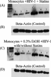
Statin-mediated inhibition of HIV-1- and EtOH-induced production of Hsp70 in postmitotically differentiated neurons. Postmitotically differentiated neurons were cocultured with HIV-1-infected monocytes either in the presence or in the absence of 0.3% EtOH for 72 h. The neurons were washed to remove primary monocytes. Cellular lysate protein (25 μg/lane) was loaded onto a sodium dodecyl sulfate-polyacrylamide gel and electrophoresed, followed by a transfer onto a nitrocellulose membrane. These blots were then probed with antibody specific for Hsp70. (A and B) Expression of Hsp70 under conditions without EtOH and with 0.3% EtOH, respectively. Lanes: 1, protein marker; 2, nontreated monocytes (control); 3, monocytes pretreated with simvastatin (10 μM); 4, monocytes pretreated with atorvastatin (10 μM); 5, monocytes infected with R5 HIV-1 strain YU2; 6, monocytes infected with R5 HIV-1 strain YU2 in the presence of simvastatin (10 μM); and 7, monocytes infected with R5 HIV-1 strain YU2 in the presence of atorvastatin (10 μM). Panel B represents the human β-actin control for the various lanes in panel A, and panel D represents the control for samples shown in panel C.

References

    1. Acheampong, E., M. Mukhtar, Z. Parveen, N. Ngoubilly, N. Ahmad, C. Patel, and R. J. Pomerantz. 2002. Ethanol strongly potentiates apoptosis induced by HIV-1 proteins in primary human brain microvascular endothelial cells. Virology 304:222-234. - PubMed
    1. Agnew, L. L., M. Kelly, J. Howard, S. Jeganathan, M. Batterham, R. A. Ffrench, J. Gold, and K. Watson. 2003. Altered lymphocyte heat shock protein 70 expression in patients with HIV disease. AIDS 17:1985-1988. - PubMed
    1. Akaike, T. 2001. Role of free radicals in viral pathogenesis and mutation. Rev. Med. Virol. 11:87-101. - PMC - PubMed
    1. Ancuta, P., A. Moses, and D. Gabuzda. 2004. Transendothelial migration of CD16+ monocytes in response to fractalkine under constitutive and inflammatory conditions. Immunobiology 209:11-20. - PubMed
    1. Apel, K., and H. Hirt. 2004. Reactive oxygen species: metabolism, oxidative stress, and signal transduction. Annu. Rev. Plant Biol. 55:373-399. - PubMed

Publication types

MeSH terms

Substances