. 2007 Feb;81(3):1220-9.
doi: 10.1128/JVI.01515-06. Epub 2006 Nov 15.

Severe acute respiratory syndrome coronavirus protein 6 accelerates murine coronavirus infections

Affiliations

Severe acute respiratory syndrome coronavirus protein 6 accelerates murine coronavirus infections

Chandra Tangudu et al. J Virol. 2007 Feb.

Abstract

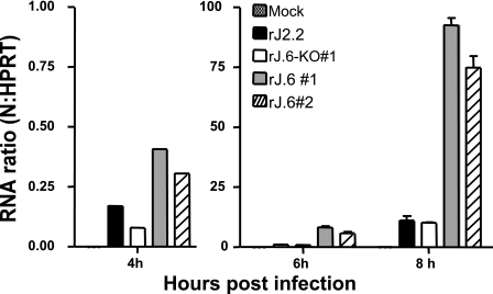
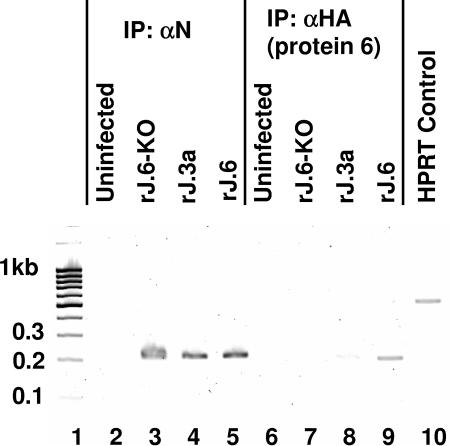
One or more of the unique 3'-proximal open reading frames (ORFs) of the severe acute respiratory syndrome (SARS) coronavirus may encode determinants of virus virulence. A prime candidate is ORF6, which encodes a 63-amino-acid membrane-associated peptide that can dramatically increase the lethality of an otherwise attenuated JHM strain of murine coronavirus (L. Pewe, H. Zhou, J. Netland, C. Tangudu, H. Olivares, L. Shi, D. Look, T. Gallagher, and S. Perlman, J. Virol. 79:11335-11342, 2005). To discern virulence mechanisms, we compared the in vitro growth properties of rJ.6, a recombinant JHM expressing the SARS peptide, with isogenic rJ.6-KO, which has an inactive ORF containing a mutated initiation codon and a termination codon at internal position 27. The rJ.6 infections proceeded rapidly, secreting progeny about 1.5 h earlier than rJ.6-KO infections did. The rJ.6 infections were also set apart by early viral protein accumulation and by robust expansion via syncytia, a characteristic feature of JHM virus dissemination. We found no evidence for protein 6 operating at the virus entry or assembly stage, as virions from either infection were indistinguishable. Rather, protein 6 appeared to operate by fostering viral RNA and protein synthesis, as RNA quantifications by reverse transcription-quantitative PCR revealed viral RNA levels in the rJ.6 cultures that were five to eight times higher than those lacking protein 6. Furthermore, protein 6 coimmunoprecipitated with viral RNAs and colocalized on cytoplasmic vesicles with replicating viral RNAs. The SARS coronavirus encodes a novel membrane protein 6 that can accelerate replication of a related mouse virus, a property that may explain its ability to increase in vivo virus virulence.


Figures

FIG. 1.
FIG. 1.
Time course analysis of secreted virion infectivities. HeLa-MHVR5 cells were infected for 1 h at 0.01 PFU/cell with rJ.6#1 or rJ.6.KO#1, then rinsed, and replaced with fresh growth media. Infectious virus in the media collected at the indicated time points were quantified by plaque assay on 17cl1 indicator cells. The horizontal line indicates the sensitivity of the plaque assays (4 PFU/ml). Similar results were obtained in three independent experiments.
FIG. 2.
FIG. 2.
Immunofluorescence detection of infected cells at 8 and 10 h postinfection. HeLa-MHVR5 cells were infected with rJ.6-KO or rJ.6 at 0.01 PFU/cell, fixed with paraformaldehyde at the indicated times, and then permabilized with methanol. Cells were immunostained for M protein (MAb J.1.3), and nuclei were recognized by incubation with Hoescht 33258. The panels in this figure are representative of ∼20 panels from each culture that were evaluated to obtain the percentage of IFA-positive cells stated in the text.
FIG. 3.
FIG. 3.
Quantification of syncytial expansions in rJ.6- and rJ.6-KO-infected cultures. vTF7.3-infected HeLa-MHVR cells were cocultivated with pEMC-T7-luc-transfected HeLa-MHVR cells that had been infected 8 h earlier with the indicated recombinant viruses. At the indicated times after cocultivation, cells were dissolved, mixed with luciferin substrate, and evaluated in a luminometer for light emissions. Data are plotted in relative light units (RLU).
FIG. 4.
FIG. 4.
Time course analysis of virion protein accumulations. (A) HeLa-MHVR5 cells were infected with the indicated viruses at 0.01 PFU/cell, and individual cultures were dissolved at hourly intervals. A total of 105 cell equivalents were electrophoresed, and virion proteins S, N, and M were detected by Western blotting. All Western blots were incubated together in the same antibody solutions and then exposed equally via chemiluminescence detection methods. The positions of molecular mass standards (in kilodaltons) are listed to the left of the gels. S-unc, uncleaved S; S2, C-terminal S posttranslation product. (B) HeLa-MHVR cells were infected with the indicated viruses at 0.01 PFU/cell. At 3 hpi, rabbit anti-MHVR antiserum (1:50) was added to infected cultures at 3 hpi to block syncytial developments. Cell lysates were then collected at the indicated times postinfection and evaluated by Western blotting for S, N, and M proteins. The positions of molecular mass standards (in kilodaltons) are listed to the left of the gel.
FIG. 5.
FIG. 5.
Time course analysis of viral RNA accumulations. HeLa-MHVR cells were infected with the indicated recombinant viruses at a multiplicity of infection of 0.01 PFU/cell, and total cellular RNAs were harvested from individual cultures at 4, 6 and 8 h postinfection. MHV N gene-specific RNAs were then quantified by RT-qPCR, normalizing the level of N gene amplicons to that of HeLa cell HPRT amplicons, as described in Materials and Methods.
FIG. 6.
FIG. 6.
Coimmunoprecipitation of protein 6 with JHM-specific RNAs. Cytoplasmic extracts were prepared by needle extrusion of rJHM-infected HeLa-MHVR5 cells and then incubated with the indicated antibody-coated magnetic beads. Coimmunoprecipitating RNAs were eluted from the harvested beads and used to act as templates in RT-PCR mixtures designed to amplify both viral nucleocapsid and cellular HPRT sequences. PCR amplification products were separated by electrophoresis and imaged by ethidium bromide staining. Molecular size standards in kilobase pairs are illustrated in lane 1. HPRT amplicons templated by total uninfected cell RNAs are revealed in lane 10. IP: αN, immunoprecipitation with anti-N.
FIG. 7.
FIG. 7.
Intracellular localizations of protein 6 relative to replicating viral RNAs and nsp3 and M proteins. HeLa-MHVR cells infected with rJ.6 viruses were pulse-labeled with BrUTP in the presence of actinomycin D (top two rows of panels) or incubated without BrUTP labeling (bottom two rows). All cells were then fixed, permeabilized, and incubated with fluorescent antibodies recognizing incorporated BrU (top two rows), MHV nsp3 (third row), MHV M (bottom row), and the C-terminal HA epitopes were appended to protein 6 (all panels). The images in the top row were taken with a Leica DM-IRB epifluorescence microscope; all remaining images were taken with a Zeiss model 510 laser-scanning confocal microscope. Images depicting the subcellular locations of BrUTP-labeled RNAs, nsp3, and M are shown in the leftmost column, images of ORF6-HA are shown in the middle column, and superimposed images (Merge) are shown in the rightmost column.

References

    1. Baric, R. S., B. Yount, L. Hensley, S. A. Peel, and W. Chen. 1997. Episodic evolution mediates interspecies transfer of a murine coronavirus. J. Virol. 71:1946-1955. - PMC - PubMed
    1. Chan, W. S., C. Wu, S. C. Chow, T. Cheung, K. F. To, W. K. Leung, P. K. Chan, K. C. Lee, H. K. Ng, D. M. Au, and A. W. Lo. 2005. Coronaviral hypothetical and structural proteins were found in the intestinal surface enterocytes and pneumocytes of severe acute respiratory syndrome (SARS). Mod. Pathol. 18:1432-1439. - PMC - PubMed
    1. Choi, K. S., P. Huang, and M. M. Lai. 2002. Polypyrimidine-tract-binding protein affects transcription but not translation of mouse hepatitis virus RNA. Virology 303:58-68. - PubMed
    1. Chow, S. C., C. Y. Ho, T. T. Tam, C. Wu, T. Cheung, P. K. Chan, M. H. Ng, P. K. Hui, H. K. Ng, D. M. Au, and A. W. Lo. 2006. Specific epitopes of the structural and hypothetical proteins elicit variable humoral responses in SARS patients. J. Clin. Pathol. 59:468-476. - PMC - PubMed
    1. de Haan, C. A., and P. J. Rottier. 2005. Molecular interactions in the assembly of coronaviruses. Adv. Virus Res. 64:165-230. - PMC - PubMed

Publication types

MeSH terms

Substances