. 2006 Nov 15;26(46):11844-9.
doi: 10.1523/JNEUROSCI.3295-06.2006.

The development of cortical multisensory integration

Affiliations

The development of cortical multisensory integration

Mark T Wallace et al. J Neurosci. .

Abstract

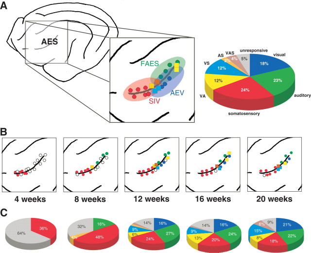
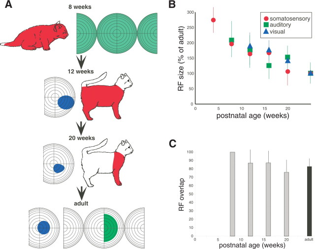
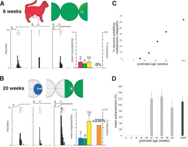
Although there are many perceptual theories that posit particular maturational profiles in higher-order (i.e., cortical) multisensory regions, our knowledge of multisensory development is primarily derived from studies of a midbrain structure, the superior colliculus. Therefore, the present study examined the maturation of multisensory processes in an area of cat association cortex [i.e., the anterior ectosylvian sulcus (AES)] and found that these processes are rudimentary during early postnatal life and develop only gradually thereafter. The AES comprises separate visual, auditory, and somatosensory regions, along with many multisensory neurons at the intervening borders between them. During early life, sensory responsiveness in AES appears in an orderly sequence. Somatosensory neurons are present at 4 weeks of age and are followed by auditory and multisensory (somatosensory-auditory) neurons. Visual neurons and visually responsive multisensory neurons are first seen at 12 weeks of age. The earliest multisensory neurons are strikingly immature, lacking the ability to synthesize the cross-modal information they receive. With postnatal development, multisensory integrative capacity matures. The delayed maturation of multisensory neurons and multisensory integration in AES suggests that the higher-order processes dependent on these circuits appear comparatively late in ontogeny.


Figures

Figure 1.
Figure 1.
The developmental chronology of sensory and multisensory responses in AES cortex. A, Schematic drawing of the lateral surface of the cat cortex showing the location of the AES in the adult. Inset shows the modality distributions of neurons within the three major subdivisions of AES: SIV, FAES, and AEV. Colored circles represent penetrations in which only unisensory neurons (red, somatosensory; green, auditory; blue, visual) were encountered, and colored squares represent penetrations in which multisensory neurons [light blue, auditory–somatosensory (AS); yellow, visual–auditory (VA); orange, visual–somatosensory (VS); brown, visual–auditory–somatosensory (VAS)] were found. Data are from a single animal in which AES was extensively sampled. Pie graph shows the modality distributions in the adult and is pooled for the three animals. B, Emergence of sensory-responsive neuron types as a function of postnatal age. For details, see Results. Note that, in contrast to A, data has been pooled across animals to provide a more comprehensive view of the modality distributions in AES at the different ages (for how these data were merged, see Materials and Methods). C, Modality distributions in AES at the sampled ages. Pie graphs represent the pooled data from all animals at each age.
Figure 2.
Figure 2.
Receptive fields of multisensory AES neurons decline in size as a function of postnatal age. A, Example neurons illustrate the shrinkage of receptive fields (RF) (colored shading) and the emergence of good receptive field overlap with maturation. B, The decline in mean receptive field size as a function of age. Note that all values are shown as a percentage of adult values. C, Average receptive field overlap as a function of age. Error bars in B and C show SDs.
Figure 3.
Figure 3.
Multisensory integration is absent in the earliest AES neurons and appears and matures during postnatal life. A, Shown at the top are the receptive fields (shading) and stimulus locations (icons) used in sensory testing of this auditory–somatosensory AES neuron in an 8-week-old animal. Rasters and peristimulus time histograms show the neuron responses to somatosensory (S), auditory (A), and combined (SA) stimulation. Bar graph at the right summarizes these responses and shows the lack of any multisensory interaction. Scale bar at the bottom of the histograms represents 100 ms. B, Multisensory integration in a visual–auditory neuron at 20 weeks of age. A, Auditory stimulation; V, visual stimulation; VA, combined visual–auditory stimulation. Conventions are the same as in A. *p < 0.01, t test. C, Growth in the integrating multisensory population as a function of age. D, As soon as it appears (i.e., at 12 weeks), multisensory integration is of similar magnitude to the adult. Error bars show the SDs.

References

    1. Barth DS, Kithas J, Di S. Anatomic organization of evoked potentials in rat parietotemporal cortex: somatosensory and auditory responses. J Neurophysiol. 1993;69:1837–1849. - PubMed
    1. Birch H, Lefford A. Intersensory development in children. Monogr Soc Res Child Dev. 1963;28:1–47. - PubMed
    1. Bower TGR. Development in infancy. San Francisco: Freeman; 1974.
    1. Burgess PR. Cutaneous mechanoreceptors. In: Carterette EC, Friedman MP, editors. Handbook of perception. New York: Academic; 1973. pp. 219–249.
    1. Clarey JC, Irvine DR. Auditory response properties of neurons in the anterior ectosylvian sulcus of the cat. Brain Res. 1986;386:12–19. - PubMed

Publication types

MeSH terms