. 2006 Nov 15;26(46):11850-6.
doi: 10.1523/JNEUROSCI.2354-06.2006.

Massive and specific dysregulation of direct cortical input to the hippocampus in temporal lobe epilepsy

Affiliations

Massive and specific dysregulation of direct cortical input to the hippocampus in temporal lobe epilepsy

Chyze W Ang et al. J Neurosci. .

Abstract

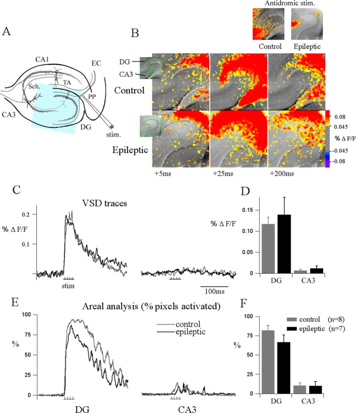
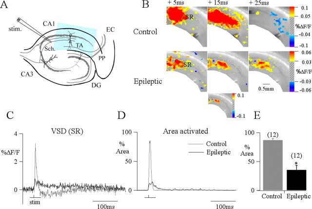
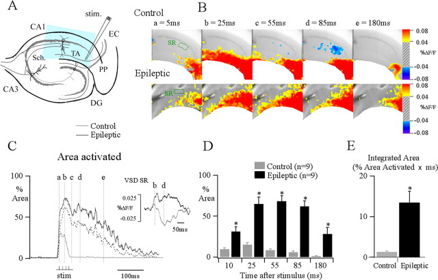
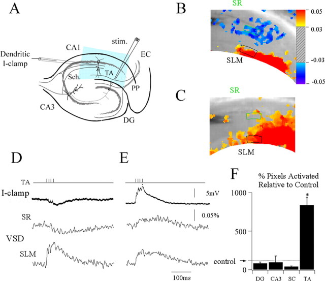
Epilepsy affects 1-2% of the population, with temporal lobe epilepsy (TLE) the most common variant in adults. Clinical and experimental studies have demonstrated hippocampal involvement in the seizures underlying TLE. However, identification of specific functional deficits in hippocampal circuits associated with possible roles in seizure generation remains controversial. Significant attention has focused on anatomic and cellular alterations in the dentate gyrus. The dentate gyrus is a primary gateway regulating cortical input to the hippocampus and, thus, a possible contributor to the aberrant cortical-hippocampal interactions underlying the seizures of TLE. Alternate cortical pathways innervating the hippocampus might also contribute to seizure initiation. Despite this potential importance in TLE, these pathways have received little study. Using simultaneous voltage-sensitive dye imaging and patch-clamp recordings in slices from animals with epilepsy, we assessed the relative degree of synaptic excitation activated by multiple cortical inputs to the hippocampus. Surprisingly, dentate gyrus-mediated regulation of the relay of cortical input to the hippocampus is unchanged in epileptic animals, and input via the Schaffer collaterals is actually decreased despite reduction in Schaffer-evoked inhibition. In contrast, a normally weak direct cortical input to area CA1 of hippocampus, the temporoammonic pathway, exhibits a TLE-associated transformation from a spatially restricted, highly regulated pathway to an excitatory projection with >10-fold increased effectiveness. This dysregulated temporoammonic pathway is critically positioned to mediate generation and/or propagation of seizure activity in the hippocampus.


Figures

Figure 1.
Figure 1.
Dentate gyrus (DG) gate function is retained in slices prepared from epileptic animals. A, The schematic of the hippocampus illustrates the major afferent pathways to the hippocampus and the position of the stimulation electrode that is used to activate the perforant path (stim.). The blue area indicates the typical area imaged with our camera. TA, Temporoammonic pathway; EC, entorhinal cortex. B, Gray scale images with green ROI (region of interest) overlays show images of hippocampal slices from control (top right) and epileptic animals (bottom right), containing most of each dentate blade and CA3. The time series shows that in both conditions, a burst stimulus applied to the perforant path generates a strong voltage response in the dentate gyrus that is minimally transmitted as excitation in CA3. The snapshot in the inset (A, bottom) shows the response 25 ms after antidromic stimulation of the Schaffer collaterals (Sch.) in the same slices depicted in B, demonstrating that CA3 pyramidal neurons are viable and can be activated to generate an EPSP. VSD signals averaged over time from each of the ROIs form the traces in C from control (gray) and epileptic slices (black), demonstrating little difference in the averaged DG or CA3 voltage responses, consistent with the group data in D. A more sensitive assay of activation is shown in E; traces are the number of pixels in the dentate gyrus and area CA3 ROIs that exhibited a significant depolarization after perforant path (PP) stimulation (>0.04% ΔF/F, corresponding to a response ≥3 SDs over noise levels) plotted against time. Unlike the averaged pixel values in C and D, this analysis is able to resolve some CA3 activity from both control (gray) and epileptic tissue (black). F, Nevertheless, although CA3 activity is now resolved, in both the representative and group data, only a small and equivalent percent of pixels for each condition are activated in CA3 after dentate activation (p = 0.94).
Figure 2.
Figure 2.
Impairment in Schaffer collateral-evoked responses in epilepsy. A, The schematic of the hippocampus illustrates the major afferent pathways to the hippocampus and the position of the stimulation electrode that is used to activate the Schaffer collaterals (stim.). The blue area indicates the typical area imaged with our camera for CA1 studies. B, Snapshots of VSD recordings from control and epileptic slices at 5, 15, and 25 ms after the response to a stimulus (200 μs pulses at 50–100 μA) applied at stratum radiatum to activate the Schaffer collaterals (Sch.). To capture the responses that were smaller in slices from epileptic animals (supplemental Fig. 1, available at www.jneurosci.org as supplemental material), we mapped the snapshots to a smaller color scale; for comparison, the inset (B) shows peak response mapped to control color bar. Note the powerful excitatory activation of the Schaffer collaterals in control and the reduced Schaffer response in epileptic tissue. C, Traces showing VSD signals averaged from regions of interest in CA1 stratum radiatum (SR; green box) from control and epileptic are superimposed, contrasting a strong EPSP–IPSP sequence in control and the weak EPSP in epileptic. Focusing on the excitation generated in both conditions, the percentage of the total area in CA1 activated by Schaffer collateral stimulation is plotted against time. For the purposes of analysis, this area was delineated as a wedge shape between the alveus and hippocampal fissure, with sides drawn perpendicular to both curved boundaries to generate a consistent region of interest in all slices. Because simultaneous current clamp records show reduced ΔF/F per membrane voltage change in recordings from epileptic slices compared with controls (supplemental Fig. 1, available at www.jneurosci.org as supplemental material), to reduce the chance of underestimating responses from slices derived from epileptic animals, pixels showing depolarizations of greater than a calculated 5 mV change (0.05% ΔF/F for control and 0.035% ΔF/F for epileptic) were counted. D, This also demonstrated a large amplitude, brief response in controls compared with a prolonged but more limited amplitude response from epileptic animals. E, In group data this is illustrated: more then 70% of pixels in the region of interest are activated in control slices after Schaffer stimulation, whereas a significantly more limited and variable activation is seen in slices from epileptic animals (ANOVA, *p ≤ 0.05). PP, Perforant path; DG, dentate gyrus; EC, entorhinal cortex; TA, temporoammonic pathway.
Figure 3.
Figure 3.
Excitation evoked by activation of the temporoammonic pathway is increased by an order of magnitude in area CA1. A, The schematic of the hippocampus illustrates the major afferent pathways to the hippocampus and the position of the stimulation electrode that is used to activate the temporoammonic pathway (stim.). The blue area indicates typical area imaged with our camera. B, VSD recording snapshots of control and epileptic responses at 5, 25, 55, 85, and 180 ms after response to a burst stimulation (four stimuli at 100 HZ, 200 μs pulses at 50–100 μA) applied to the temporoammonic pathway at the angular bundle. This stimulus produces spatially restricted temporoammonic activity in control. In contrast, in epileptic tissue, excitation is propagated throughout the stratum radiatum and pyramidale. This is demonstrated in C, by comparing the percentage of the total CA1 area activated by temporoammonic stimulation in control (gray) and epileptic (black) plotted against time. Pixels showing depolarizations of >0.05% ΔF/F for control and 0.035% ΔF/F for epileptic are counted, normalized to the total CA1 area imaged (for comparison dotted line shows epileptic response when threshold is set at 0.05% ΔF/F). This reveals a greater area and prolonged period of activation in epileptic tissue compared with controls, inset traces depicting percent change in fluorescence shows that this is caused in part by loss of inhibition, with an IPSP present in the stratum radiatum (SR) in control animals, which is transformed into an EPSP in epileptic animals. D, Summary data illustrates both the differences in peak amplitude and time course of area activated for control (gray) compared against epileptic (black) for time points of 10, 25, 55, 85, and 180 ms. An asterisk indicates that epileptic animals demonstrate a significant increase in the maximum area activated by temporoammonic stimulation over control for the selected time points. E, These results can be summarized as a simple comparison by integrating the area under the percent pixel activated traces, capturing both the number and duration of pixels activated, which illustrates a >10-fold increase in activation in slices from epileptic animals compared with controls (ANOVA, *p ≤ 0.05; n = 12). PP, Perforant path; DG, dentate gyrus; TA, temporoammonic pathway; EC, entorhinal cortex.
Figure 4.
Figure 4.
Transformation of the temporoammonic pathway from an inhibitory, spatially restricted pathway to a powerful excitatory projection in epileptic animals. A, The schematic of the hippocampus illustrates the major afferent pathways to the hippocampus and the position of the stimulation electrode that is used to activate the temporoammonic pathway (stim.) as well as the position of the patch electrode used for the dendritic whole-cell records. The blue area indicates the typical area imaged with our camera. B, The control is a VSD recording snapshot of EPSP activation at 30 ms evoked by burst stimulation (four stimuli at 100 HZ, 200 μs pulses at 50–100 μA) applied to the temporoammonic pathway in a slice prepared from a control animal, showing distal depolarization (red) and concurrent more proximal inhibition (blue). Sch., Schaffer collaterals. I-clamp dendritic shows the whole-cell recording from the apical dendrite of a CA1 pyramidal cell in stratum radiatum (SR), which exhibited a small IPSP. VSD traces, SR and SLM are the local VSD signals quantified from regions of interest in stratum radiatum (green box) and stratum lacunosum moleculare (black box) respectively. C, Epileptic. A VSD recording snapshot of the EPSP at 30 ms evoked by temporoammonic stimulation in a slice prepared from an epileptic animal is shown. Note that the temporoammonic-evoked EPSP is not spatially restricted to the distal dendrites in stratum lacunosum moleculare but propagates to stratum radiatum, depolarizing the proximal dendrites. D, E, A dendritic whole-cell recorded response (control, D; epileptic, E; top trace), shows a concurrent 9.3 mV EPSP during the temporoammonic VSD EPSP in SR and SLM (middle and bottom traces). F, Summary data for all hippocampal pathways tested comparing maximal area of activation in slices from epileptic animals relative to control in the dentate gyrus (DG) and CA3 in response to perforant path stimulation, in CA1 in response to Schaffer collaterals (SC) and temporoammonic pathway stimulation demonstrating the dramatic increase in excitability is limited to TA activation of area CA1 (n = 8 for DG and CA3; n = 12 for SC and TA; ANOVA, *p ≤ 0.05). TA, Temporoammonic pathway; EC, entorhinal cortex.

Comment in

References

    1. Ang CW, Carlson GC, Coulter DA. Hippocampal CA1 circuitry dynamically gates direct cortical inputs preferentially at theta frequencies. J Neurosci. 2005;25:9567–9580. - PMC - PubMed
    1. Barbarosie M, Louvel J, Kurcewicz I, Avoli M. CA3-released entorhinal seizures disclose dentate gyrus epileptogenicity and unmask a temporoammonic pathway. J Neurophysiol. 2000;83:1115–1123. - PubMed
    1. Behr J, Lyson KJ, Mody I. Enhanced propagation of epileptiform activity through the kindled dentate gyrus. J Neurophysiol. 1998;79:1726–1732. - PubMed
    1. Bernard C, Anderson A, Becker A, Poolos NP, Beck H, Johnston D. Acquired dendritic channelopathy in temporal lobe epilepsy. Science. 2004;305:532–535. - PubMed
    1. Bertram EH. Functional anatomy of spontaneous seizures in a rat model of epilepsy. Epilepsia. 1997;38:95–105. - PubMed

Publication types

MeSH terms

LinkOut - more resources