. 2006 Nov 15;26(46):11893-902.
doi: 10.1523/JNEUROSCI.2044-06.2006.

Cranial visceral afferent pathways through the nucleus of the solitary tract to caudal ventrolateral medulla or paraventricular hypothalamus: target-specific synaptic reliability and convergence patterns

Affiliations

Cranial visceral afferent pathways through the nucleus of the solitary tract to caudal ventrolateral medulla or paraventricular hypothalamus: target-specific synaptic reliability and convergence patterns

Timothy W Bailey et al. J Neurosci. .

Abstract

Cranial visceral afferents activate central pathways that mediate systemic homeostatic processes. Afferent information arrives in the brainstem nucleus of the solitary tract (NTS) and is relayed to other CNS sites for integration into autonomic responses and complex behaviors. Little is known about the organization or nature of processing within NTS. We injected fluorescent retrograde tracers into two nuclei to identify neurons that project to sites involved in autonomic regulation: the caudal ventrolateral medulla (CVLM) or paraventricular nucleus of the hypothalamus (PVN). We found distinct differences in synaptic connections and performance in the afferent path through NTS to these neurons. Anatomical studies using confocal and electron microscopy found prominent, primary afferent synapses directly on somata and dendrites of CVLM-projecting NTS neurons identifying them as second-order neurons. In brainstem slices, afferent activation evoked large, constant latency EPSCs in CVLM-projecting NTS neurons that were consistent with the precise timing and rare failures of monosynaptic contacts on second-order neurons. In contrast, most PVN-projecting NTS neurons lacked direct afferent input and responded to afferent stimuli with highly variable, intermittently failing synaptic responses, indicating polysynaptic pathways to higher-order neurons. The afferent-evoked EPSCs in most PVN-projecting NTS neurons were smaller and unreliable but also often included multiple, convergent polysynaptic responses not observed in CVLM-projecting neurons. A few PVN-projecting NTS neurons had monosynaptic EPSC characteristics. Together, we found that cranial visceral afferent pathways are structured distinctly within NTS depending on the projection target. Such, intra-NTS pathway architecture will substantially impact performance of autonomic or neuroendocrine reflex arcs.


Figures
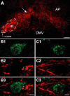
Figure 1.
Figure 1.
Confocal micrographs show close appositions between anterogradely labeled vagal afferents and retrogradely labeled projection neurons within medial NTS. A, An epifluorescent micrograph shows BDA labeling (red) in medial NTS after injection into the nodose ganglion. Labeling was most intense in dorsomedial NTS (arrow) between the area postrema (AP) and the solitary tract (st; dashed line) but was rarely seen in the dorsal motor nucleus of the vagus (DMV). Dorsal and medial orientations are indicated by arrows (bottom left). B, C, High-magnification confocal micrographs illustrate vagal afferents (red) and retrogradely labeled projection neurons (green) to CVLM (B) and PVN (C). B1, A single FluoroGold-labeled CVLM-projecting NTS neuron. B2, BDA-labeled vagal afferents within the same optical plane. B3, Overlay shows appositions (arrowheads) between varicosities in vagal afferents and the CVLM-projecting neuron. C1, A single FluoroGold-labeled PVN-projecting NTS neuron. C2, BDA-labeled vagal afferents from the same optical section. C3, Overlay shows vagal axons bypassing the PVN-projecting neuron with no varicosities or appositions. Scale bars: A, 100 μm; B, C, 5 μm.
Figure 2.
Figure 2.
Electron micrographs show BDA-labeled terminals contacting the somata and dendrites of FluoroGold-labeled CVLM-projecting neurons. A, Two BDA-labeled axon terminals (BDA-t1, BDA-t2) form asymmetric synapses (curved arrows) with a FluoroGold-labeled somata (FGold-p). Immunogold labeling for FluoroGold (arrowheads) was found throughout the cytoplasm. G, Golgi apparatus. B, A FluoroGold-labeled dendrite (FGold-d) received asymmetric synapses (curved arrows) from two BDA-labeled axon terminals (BDA-t1, BDA-t2), as well as from an unlabeled axon terminal (ut). C, A FluoroGold-labeled dendrite (FGold-d) received an asymmetric synapse (curved arrow) from a BDA-labeled axon terminal (BDA-t) and received an apposition from an unlabeled axon terminal (ut). Axosomatic contacts were found on 10 different neurons, in addition to 22 axodendritic contacts. Scale bars, 500 nm.
Figure 3.
Figure 3.
Fluorescent retrograde label injected into either CVLM or PVN marked NTS projection neurons in nearly horizontal slices of brainstem. Low-power fluorescence images of representative horizontal NTS slices with previous tracer injection into CVLM (A; rhodamine) or PVN (B; DiI). Labeled neurons were distributed throughout the medial NTS. CVLM injections generally labeled more NTS neurons than did PVN injections. C, IRDIC low-power image of a typical horizontal NTS slice preparation showing typical landmarks such as solitary tract and fourth ventricle (4V). Stimulating electrode on ST and typical recording electrode placement are also shown. D, Line drawing shows the relative location of recorded NTS neurons: monosynaptic CVLM-projecting (black triangles; n = 22), monosynaptic PVN-projecting (red circles; n = 5), and polysynaptic PVN-projecting (green squares; n = 19). Scale bars, 200 μm.
Figure 4.
Figure 4.
CVLM-projecting NTS neurons exhibit large invariant ST synaptic responses consistent with direct monosynaptic connections from cranial visceral afferents. A, Overlaid traces of ST-evoked EPSCs (for clarity, limited to 10 traces) in a representative CVLM-projecting NTS neuron. ST-evoked EPSCs had very few failures and consistent latency (3.2 ms). The low variability in EPSC latency (45 μs jitter) is clearly evident for the first EPSC in the expanded timescale below. B, Top shows neuron in A under fluorescence illumination (arrow). Note the accumulation of retrograde tracer (rhodamine beads) throughout the cell body and proximal process. Scale bar, 10 μm. Bottom shows IRDIC image of the same neuron with recording pipette (arrow).
Figure 5.
Figure 5.
PVN-projecting NTS neurons exhibit highly variable synaptic responses to ST consistent with afferent transmission from cranial visceral afferents through polysynaptic pathways. A, Overlaid traces of ST-evoked EPSCs (for clarity, limited to 10 traces) in a representative PVN-projecting NTS neuron. PVN-projecting neurons exhibited ST-evoked EPSCs that were very inconsistent in latency and amplitude. In this neuron, many ST shocks produced no EPSC, i.e., synaptic failures indicated by flat ST-synched trace. The mean latency was 5.2 ms but initiation of the EPSC was highly variable, as highlighted in the expanded timescale (bottom) of the first EPSC traces (707 μs jitter). These ST-evoked EPSC characteristics are consistent with an indirect path from ST afferents through polysynaptic excitatory connections. B, Top shows a fluorescence image of a recorded neuron (arrow) showing the accumulation of retrograde tracer (rhodamine beads) throughout the cell body and proximal processes. The bottom shows the same cell under IRDIC with recording pipette (arrow). Scale bar, 10 μm.
Figure 6.
Figure 6.
Some PVN-projecting NTS neurons display low-jitter ST-EPSC responses indicating direct, monosynaptic connections from cranial visceral afferents. A, Overlaid current traces of ST-evoked EPSCs (for clarity, limited to 10 traces) in a representative PVN-projecting NTS neuron strongly resemble those typical in NTS neurons projecting to CVLM (see Fig. 4). Latencies of ST-evoked EPSCs had low jitter, consistent with a direct path from ST. B, Top is a fluorescence image of the neuron recorded in A showing the accumulation of retrograde tracer (rhodamine beads) throughout the cell body and proximal processes. Bottom shows the same neuron under IRDIC with recording pipette (arrow). Scale bar, 10 μm.
Figure 7.
Figure 7.
CVLM-projecting NTS neurons were monosynaptically coupled to ST, whereas PVN-projecting neurons were primarily coupled through polysynaptic pathways via NTS interneurons. A, In a representative CVLM-projecting NTS neuron, ST stimuli always evoked an EPSC (top left). The EPSC latency was highly consistent, as highlighted in traces in which the first EPSC in the burst is shown on an expanded timescale (top right). A histogram of EPSC latencies shows a narrow distribution of values, with a total range of <500 μs and a calculated jitter of 63 μs. B, In a representative higher-order PVN-projecting NTS neuron (top left), ST shocks evoked EPSCs at highly variable latencies and variable amplitudes, and note that a large proportion of ST shocks failed to evoke synaptic events (top right). The latency histogram for this neuron showed a very broad distribution of response latencies that covered a range of over 2000 μs with a jitter of 432 μs. C, The summary relationship between jitter and latency for CVLM-projecting (black triangles) and those for second-order (red circles) and higher-order (green squares) PVN-projecting NTS neurons shows that, although absolute latency ranges were fairly similar, high jitters were found only in the PVN-projecting neurons. All CVLM-projecting neurons plus five PVN-projecting neurons had the low jitter values (<200 μs, below dashed line) that were consistent with direct, monosynaptic ST input. Most PVN-projecting neurons had very high jitters and were thus coupled through complex polysynaptic pathways to afferent inputs.
Figure 8.
Figure 8.
Summary of the EPSC amplitude changes to the burst of five ST stimuli clearly distinguished second from higher-order NTS neurons, whether projecting to CVLM or to PVN. A, Second-order CVLM-projecting (n = 22) and PVN-projecting (n = 5) NTS neurons expressed similar amplitudes throughout the ST burst from EPSC1 to EPSC5. Higher-order PVN-projecting NTS neurons (n = 19) had significantly smaller EPSC1 amplitudes compared with CVLM-projecting and second-order PVN-projecting neurons (p < 0.035). B, FDD comparisons between higher-order and second-order neurons are even clearer in plots of these relationships normalized to the EPSC1 amplitude. Higher-order PVN-projecting NTS neurons showed no FDD. This minimal change in EPSC amplitude within burst is different than the robust FDD changes in second-order CVLM-projecting and second-order PVN-projecting neurons (p < 0.01). C, Failure rates were quite low in second-order PVN and CVLM-projecting neurons and significantly greater in the higher-order PVN-projecting NTS neuron group at first and second positions within the burst of five shocks (p < 0.018). All statistical comparisons are post hoc comparisons (Fisher's PLSD test) after repeated measures or one-way ANOVA when appropriate.
Figure 9.
Figure 9.
Higher-order, PVN-projecting NTS neurons received variable-latency EPSCs, and often multiple distinct ST inputs indicated convergence on these single neurons. A, In a representative higher-order, PVN-projecting neuron, a single ST shock elicited two, high-jitter synaptic responses that were distinctly separated by latency. As was typical of these polysynaptically coupled neurons, multiple individual EPSCs could be discerned at different latencies in each sweep (A, inset). The upper set of current traces shows seven overlaid responses to ST-EPSC bursts (current traces with synaptic failures have been removed for clarity). The inset expands the set of traces to overlay the responses only to the initial ST shock of each burst of five ST shocks (full response displayed above). Ovals mark the regions of analysis for the two unique synaptic inputs to this neuron. The histogram in the bottom portion of A displays the distributions for the latency values detected for the shorter-latency ST response (open oval) and the longer-latency response (gray oval). Note that this analysis produced two discrete clusters of latency values that reflect the two independent inputs. The jitters for multiple inputs to higher-order PVN neurons were always in the polysynaptic range (>200 μs). B, Summary of failure rates and the relationship to synaptic jitter for all NTS projection neurons. Failure rate is expressed as the percentage of ST shocks failing to evoke a synaptic response. Second-order PVN- and CVLM-projecting NTS neurons expressed very low rates of synaptic failure (inset). Failure rates increased with jitter in higher-order neurons, and thus more complex pathways unreliably transmit afferent signals to higher-order PVN-projecting neurons.

References

    1. Aicher SA, Kurucz OS, Reis DJ, Milner TA. Nucleus tractus solitarius efferent terminals synapse on neurons in the caudal ventrolateral medulla that project to the rostral ventrolateral medulla. Brain Res. 1995;693:51–63. - PubMed
    1. Aicher SA, Saravay RH, Cravo S, Jeske I, Morrison SF, Reis DJ, Milner TA. Monosynaptic projections from the nucleus tractus solitarii to C1 adrenergic neurons in the rostral ventrolateral medulla: comparison with input from the caudal ventrolateral medulla. J Comp Neurol. 1996;373:62–75. - PubMed
    1. Aicher SA, Sharma S, Cheng PY, Pickel VM. The N-methyl-d-aspartate (NMDA) receptor is postsynaptic to substance P-containing axon terminals in the rat superficial dorsal horn. Brain Res. 1997;772:71–81. - PubMed
    1. Aicher SA, Sharma S, Pickel VM. N-methyl-d-aspartate receptors are present in vagal afferents and their dendritic targets in the nucleus tractus solitarius. Neuroscience. 1999;91:119–132. - PubMed
    1. Aicher SA, Milner TA, Pickel VM, Reis DJ. Anatomical substrates for baroreflex sympathoinhibition in the rat. Brain Res Bull. 2000;51:107–110. - PubMed

Publication types

MeSH terms

Substances

LinkOut - more resources