. 2006 Nov 15;26(46):11915-22.
doi: 10.1523/JNEUROSCI.3821-06.2006.

Alpha-synuclein overexpression in PC12 and chromaffin cells impairs catecholamine release by interfering with a late step in exocytosis

Affiliations

Alpha-synuclein overexpression in PC12 and chromaffin cells impairs catecholamine release by interfering with a late step in exocytosis

Kristin E Larsen et al. J Neurosci. .

Abstract

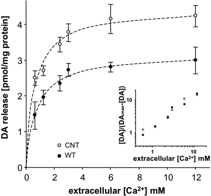
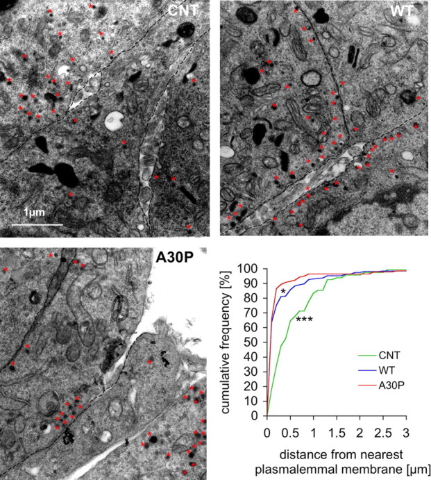
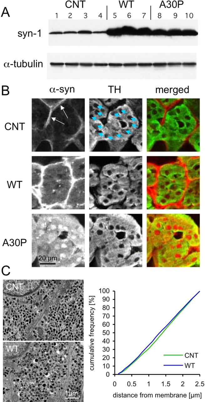
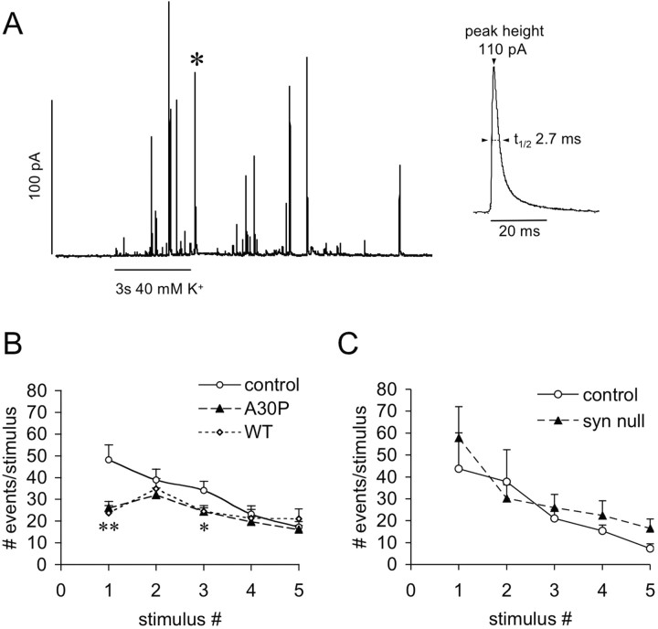
Alpha-synuclein (alpha-syn), a protein implicated in Parkinson's disease pathogenesis, is a presynaptic protein suggested to regulate transmitter release. We explored how alpha-syn overexpression in PC12 and chromaffin cells, which exhibit low endogenous alpha-syn levels relative to neurons, affects catecholamine release. Overexpression of wild-type or A30P mutant alpha-syn in PC12 cell lines inhibited evoked catecholamine release without altering calcium threshold or cooperativity of release. Electron micrographs revealed that vesicular pools were not reduced but that, on the contrary, a marked accumulation of morphologically "docked" vesicles was apparent in the alpha-syn-overexpressing lines. We used amperometric recordings from chromaffin cells derived from mice that overexpress A30P or wild-type (WT) alpha-syn, as well as chromaffin cells from control and alpha-syn null mice, to determine whether the filling of vesicles with the transmitter was altered. The quantal size and shape characteristics of amperometric events were identical for all mouse lines, suggesting that overexpression of WT or mutant alpha-syn did not affect vesicular transmitter accumulation or the kinetics of vesicle fusion. The frequency and number of exocytotic events per stimulus, however, was lower for both WT and A30P alpha-syn-overexpressing cells. The alpha-syn-overexpressing cells exhibited reduced depression of evoked release in response to repeated stimuli, consistent with a smaller population of readily releasable vesicles. We conclude that alpha-syn overexpression inhibits a vesicle "priming" step, after secretory vesicle trafficking to "docking" sites but before calcium-dependent vesicle membrane fusion.


Figures

Figure 1.
Figure 1.
α-syn-mediated reduction of stimulated dopamine release in PC12 cell lines. Clonal PC12 cell lines were generated expressing empty vector, EGFP, WT, or A30P mutant (A30P) α-syn. A, Examples of PC12 cell cultures from control, WT, and A30P-expressing cell lines immunolabeled for α-syn. B, Stimulated DA release in control and α-syn-overexpressing PC12 cell lines. Cell cultures were stimulated for 1 min with high K+ medium (80 mm), and the superfusate was analyzed by HPLC. Evoked DA release was reduced in the α-syn-overexpressing lines (n = 6 cultures for each line; means ± SEM; **p < 0.001, ANOVA with Tukey's post hoc test).
Figure 2.
Figure 2.
Calcium sensitivity of DA release from PC12 cell lines. Control and WT α-syn-overexpressing cell cultures were stimulated with high-potassium solution in the presence of varying extracellular Ca2+ concentrations. DA release was significantly reduced from the α-syn-overexpressing cell line at all Ca2+ concentrations tested (n = 6–7, 3–4 dishes from 2 independent experiments; p < 0.001, ANOVA with Tukey's post hoc test). The inset shows a double logarithmic plot of normalized DA release and Ca2+ concentration, indicating that there was no difference in Ca2+ sensitivity (threshold) or cooperativity (slope) of release between the cell lines (Sorensen et al., 2002).
Figure 3.
Figure 3.
DCV distribution in PC12 cell lines. Electron micrographs of PC12 cells are shown from CNT, WT, and A30P α-syn-overexpressing lines with examples of DCVs labeled by red stars. A greater fraction of the DCVs were located in a docked position near plasmalemmal membranes (marked by dashed lines) in both overexpressing cells lines (cumulative frequency of controls was different from WT and A30P; *p < 0.05, ***p < 0.01, Kolmogorov–Smirnov test; n = 10–11 cell sections).
Figure 4.
Figure 4.
Chromaffin cells from WT and A30P α-syn transgenic mice. A, Western immunoblots of adrenal gland tissue indicate a 19-fold and 12-fold increase of α-syn level in WT and A30P transgenic mice, respectively, compared with nontransgenic littermates. Each lane corresponds to protein derived from one animal (4 nontransgenic, 3 WT, and 3 A30P α-syn transgenic mice). B, Immunohistochemistry for α-syn of adrenal medulla sections provided relatively strong labeling in the connective tissue surrounding groups of chromaffin cells (arrows) and in endothelial cells lining blood vessels, but the chromaffin cells, stained for tyrosine- hydroxylase (TH; blue arrows point to nuclei), showed very faint labeling. In contrast, both WT and A30P α-syn overexpression resulted in substantial cytoplasmic and nuclear immunostaining of chromaffin cells. C, Electron micrographs of chromaffin cells derived from CNT and WT mouse adrenal glands are shown. The cytosol of both CNT and WT is almost completely filled with DCVs. The cumulative frequency plot indicates the location of large DCVs with respect to the plasma membrane. For each condition, sections of adrenal glands from two animals were analyzed (CNT, n = 1871, 1670 vesicles; WT, n = 2574, 1420 vesicles). Because the cytosol of mature chromaffin cells are filled with large DCVs, the frequency distribution is nearly linear with no difference detected in the morphological docking or total number of DCVs between the genotypes (Kolmogorov–Smirnov test, p > 0.5).
Figure 5.
Figure 5.
Amperometric recordings of quantal catecholamine release from cultured chromaffin cells derived from control, α-syn null, WT, and A30P α-syn-overexpressing mice. A, A representative recording trace from a control chromaffin cell. Individual events (A, inset, magnified event marked in trace by asterisk) were analyzed for peak height (Imax), duration (t1/2), and number of molecules (see Table 1). B, Chromaffin cells were stimulated with 40 mm KCl five times with a 2 min period between stimuli. Quantal release was recorded for a period of 1 min after the 3 s high KCl stimulus. The number of events was reduced twofold in cells from WT and A30P α-syn-overexpressing mice during the first stimulus (**p < 0.01), whereas for the consecutive stimuli, the number of events was close to controls (significant difference only for the third stimulus; *p < 0.05, paired ANOVA, followed by Tukey's post hoc test). C, The number of events for control and α-syn null mice at all stimuli was not different.

Comment in

  • Prime time for alpha-synuclein.
    Gitler AD, Shorter J. Gitler AD, et al. J Neurosci. 2007 Mar 7;27(10):2433-4. doi: 10.1523/JNEUROSCI.0094-07.2007. J Neurosci. 2007. PMID: 17344380 Free PMC article. No abstract available.

References

    1. Abeliovich A, Schmitz Y, Farinas I, Choi-Lundberg D, Ho WH, Castillo PE, Shinsky N, Verdugo JM, Armanini M, Ryan A, Hynes M, Phillips H, Sulzer D, Rosenthal A. Mice lacking alpha-synuclein display functional deficits in the nigrostriatal dopamine system. Neuron. 2000;25:239–252. - PubMed
    1. Bayer TA, Jakala P, Hartmann T, Egensperger R, Buslei R, Falkai P, Beyreuther K. Neural expression profile of alpha-synuclein in developing human cortex. NeuroReport. 1999;10:2799–2803. - PubMed
    1. Bittner MA, Krasnoperov VG, Stuenkel EL, Petrenko AG, Holz RW. A Ca2+-independent receptor for α-latrotoxin, CIRL, mediates effects on secretion via multiple mechanisms. J Neurosci. 1998;18:2914–2922. - PMC - PubMed
    1. Cabin DE, Shimazu K, Murphy D, Cole NB, Gottschalk W, McIlwain KL, Orrison B, Chen A, Ellis CE, Paylor R, Lu B, Nussbaum RL. Synaptic vesicle depletion correlates with attenuated synaptic responses to prolonged repetitive stimulation in mice lacking α-synuclein. J Neurosci. 2002;22:8797–8807. - PMC - PubMed
    1. Chandra S, Gallardo G, Fernandez-Chacon R, Schluter OM, Sudhof TC. Alpha-synuclein cooperates with CSPalpha in preventing neurodegeneration. Cell. 2005;123:383–396. - PubMed

Publication types

MeSH terms

LinkOut - more resources