. 2007 Feb;18(2):369-79.
doi: 10.1091/mbc.e06-08-0751. Epub 2006 Nov 15.

Characterization of AtNUC-L1 reveals a central role of nucleolin in nucleolus organization and silencing of AtNUC-L2 gene in Arabidopsis

Affiliations

Characterization of AtNUC-L1 reveals a central role of nucleolin in nucleolus organization and silencing of AtNUC-L2 gene in Arabidopsis

Frederic Pontvianne et al. Mol Biol Cell. 2007 Feb.

Abstract

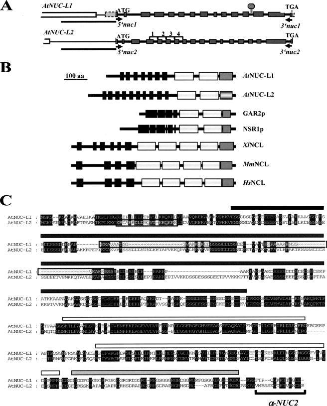
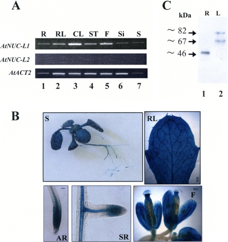
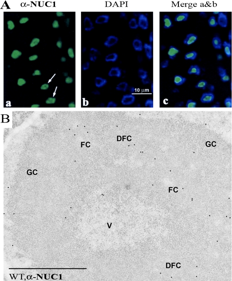
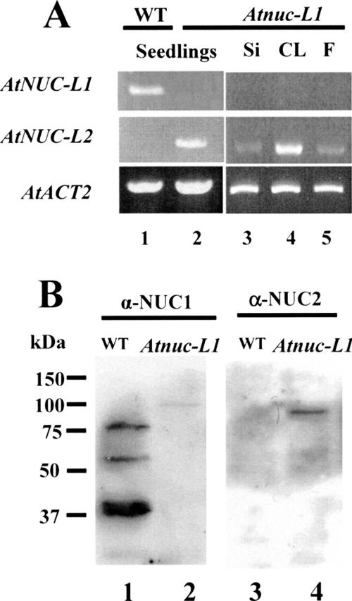
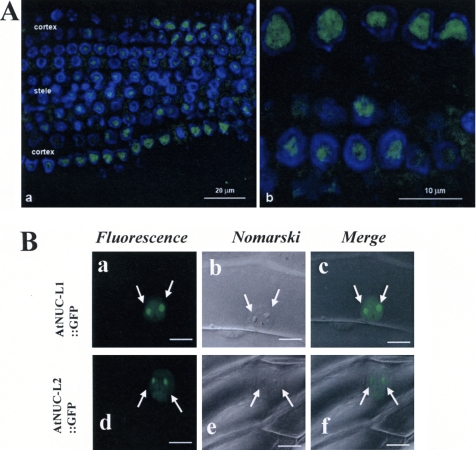
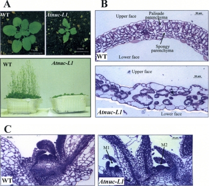
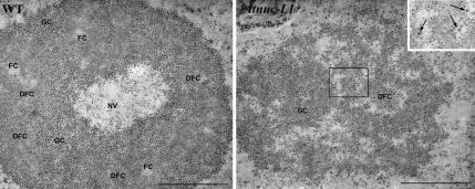
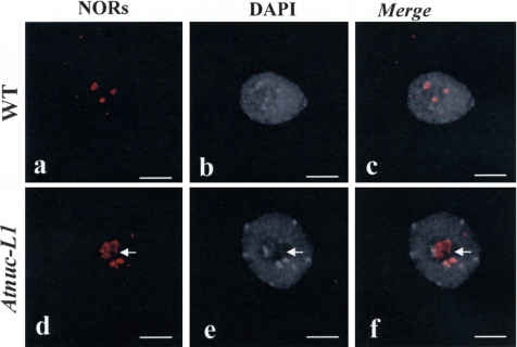
Nucleolin is one of the most abundant protein in the nucleolus and is a multifunctional protein involved in different steps of ribosome biogenesis. In contrast to animals and yeast, the genome of the model plant Arabidopsis thaliana encodes two nucleolin-like proteins, AtNUC-L1 and AtNUC-L2. However, only the AtNUC-L1 gene is ubiquitously expressed in normal growth conditions. Disruption of this AtNUC-L1 gene leads to severe plant growth and development defects. AtNUC-L1 is localized in the nucleolus, mainly in the dense fibrillar component. Absence of this protein in Atnuc-L1 plants induces nucleolar disorganization, nucleolus organizer region decondensation, and affects the accumulation levels of pre-rRNA precursors. Remarkably, in Atnuc-L1 plants the AtNUC-L2 gene is activated, suggesting that AtNUC-L2 might rescue, at least partially, the loss of AtNUC-L1. This work is the first description of a higher eukaryotic organism with a disrupted nucleolin-like gene and defines a new role for nucleolin in nucleolus structure and rDNA chromatin organization.


Figures

Figure 1.
Figure 1.
The A. thaliana genome encodes two nucleolin-like proteins. (A) Diagram of AtNUC-L1 and L2 genes from the ATG start to the TGA stop codons. Gray boxes correspond to exons separated by fourteen introns. The T-DNA insertion in the Atnuc-L1 plants is indicated by a gray diamond. Position of primers 5nuc1 and 3nuc1 used to detect AtNUC-L1 transcripts are indicated by black arrows. White boxes shows At1g48930 and At3g18600 genes, localized upstream of AtNUC-L1 and AtNUC-L2, respectively. Black bars under each gene show the 1.1- and 1.0-kbp DNA sequence fused to GUS reporter genes construct. Brackets 1–4 show tandemly repeated sequences in the AtNUC-L2 gene. (B) Schematic representation of nucleolin and nucleolin-like proteins from A. thaliana (AtNUC-L1 and L2), Schizosaccharomyces pombe (GAR2p), Saccharomyces cerevisiae (NSR1p), X. laevis (XlNCL), Mus musculus (MmNCL), and Homo sapiens (HsNCL). The black boxes correspond to the acidic regions in the N-terminal domain, the white boxes represent the RRM domains, and the dark gray boxes represent the GAR domain. The light gray box in the AtNUC-L2 sequence indicates the less conserved GAR domain. (C) Amino acid sequence alignment of AtNUC-L1 and AtNUC-L2 proteins. Conserved amino acids are black shaded. The black rectangle shows the acidic N-terminal domain, the white rectangles show the two RRM domains, and the gray rectangle the putative GAR domain. The putative nuclear localization signals of AtNUC-L1 and AtNUC-L2 are boxed. Specific antibodies α-NUC2, were prepared against the peptide sequence located in the C-terminal domain.
Figure 2.
Figure 2.
AtNUC-L1 gene and protein expression in A. thaliana plants. (A) RT-PCR analysis of AtNUC-L1 (top) and AtNUC-L2 (middle) gene expression in roots (R), rosettes leaves (RL), cauline leaves (CL), stem (ST), flowers (F), siliques (Si), and seeds (S). AtACT2 (bottom) gene expression was analyzed as a PCR control to evaluate the amount of cDNA used in each reaction. (B) Analysis of the AtNUC-L1 promoter activity in A. thaliana plants transformed with a AtNUC-L1:GUS construct. The GUS staining is visualized in 3-wk-old S, RL, apical root (AR), secondary root (SR), and F. (C) Western blot analyses of AtNUC-L1 protein expression in R (lane 1) and leaves using α-NUC-L1 antibodies.
Figure 3.
Figure 3.
Nucleolar localization of AtNUC-L1. (A) Immunofluorescence localization of AtNUC-L1 in A. thaliana root meristematic cells fixed with paraformaldehyde/dimethyl sulfoxide and incubated with α-NUC1 antibodies, observed with the confocal microscope. The images correspond to a single optical section, obtained with a Z-step of 0.3 μm. Labeling is shown concentrated in the nucleolus (a), but it does not seem evenly distributed through the whole nucleolar area (arrows). Nucleoplasm labeled by DAPI is observed as a ring around the dark unstained nucleolus (b), and the merged image shows that the immunofluorecent labeling localizes precisely in the nucleolus, unstained by DAPI (panel c). (B) Immunogold electron microscopic localization of AtNUC-L1 in A. thaliana root meristematic cells. AtNUC-L1 is observed to localize in the DFC, relatively near FCs. The interior of FCs seems devoid of gold particles, whereas the GC and the nucleolar vacuole (V) show a very scarce labeling, the same as nucleoplasm. Bar, 1 μm.
Figure 4.
Figure 4.
Disruption of AtNUC-L1 induces expression of AtNUC-L2. (A) RT-PCR reaction using cDNA prepared from RNA isolated from 15-d-old seedlings (lanes 1 and 2), siliques (Si; lane 3), cauline leaves (CL; lane 4), and flowers (F; lane 5) (B). Whole cell protein extract from WT and Atnuc-L1 plants were fractionated on SDS-PAGE and hybridized either with α-NUC1 (lanes 1 and 2) or with α-NUC2 antibodies (lanes 3 and 4).
Figure 5.
Figure 5.
AtNUC-L2 is a nucleolar protein. (A) Immunofluorescent localization of AtNUC-L2 in Atnuc-L1 plants. The images represent single optical sections, obtained using a Z-step of 0.3 μm. In a, a panoramic overview of the root is displayed, showing the differential distribution of the labeling in the different parts of the root, the cortex showing a more intense labeling, and the stele a fainter immunostaining. In b, a higher magnification is shown, with nucleoli being the major target of the antibody. (B) Nucleolar localization of AtNUC-L1::GFP and AtNUC-L2::GFP fusion protein in transfected onion epidermis cells. Arrows point the two nucleolus visualized by the GFP fluorescence (a and d). Nucleolus can be easily observed by Nomarski (b and e), and they colocalize with the GFP in the merge images (c and f).
Figure 6.
Figure 6.
AtNUC-L1 gene disruption affects growth and plant development. (A) A. thaliana WT and Atnuc-L1 plants grown on soil 4 (top) or 6 (bottom) wk under a 16:8 (L:D)-h cycle. (B and C) Transverse section of first leaves (B) and apical meristem (C) from WT and Atnuc-L1 plants stained with toluidine blue. Arrows M1 and M2 show two meristems in Atnuc-L1 plants. Bars, 50 μm.
Figure 7.
Figure 7.
AtNUC-L1 gene disruption affects ultrastructure of the nucleolus. The WT nucleolus is formed by some masses of DFC, which contain FCs in their interior and that are surrounded by GC. These components seem organized forming a cortex surrounding a central nucleolar “vacuole” (NV) in which granules similar to those of the GC can be identified. In Atnuc-L1 plants, this nucleolar organization is lost. Small masses of DFC can hardly be identified, and the bulk of the nucleolus is composed by a component reminiscent of the GC, but with the granules more loosely packed and embedded in a matrix. Numerous interstices occur, in some cases connected with the nucleoplasm. In the border between the GC-like component and the interstices, some granules can be observed. They are larger than the usual nucleolar GC granules and seem either isolated or forming small loose clusters (see in the inset a magnification of the area contained in the square). They have been identified as NPGs (arrows in the inset). Bar, 1 μm.
Figure 8.
Figure 8.
NOR condensation is affected in Atnuc-L1 plants. FISH in nuclei of WT (a–c) and Atnuc-L1 (d–f) seedlings. Left, FISH using rRNA gene probe shown in Figure 9A (a and d); middle, chromatin counterstained with DAPI (b and e); and right, superposition of a and b (c) and d and e (f) images. Arrows shows FISH signals that do not colocalize with heterochromatin. Bar, 5 μm.
Figure 9.
Figure 9.
AtNUC-L1 binds rDNA and affects rate of processing of pre-rRNA. (A) Diagram of an A. thaliana rDNA unit containing the 18S, 5.8S, and 25S rRNA genes. TIS at +1 and primary cleavage site (P) at +1275 are indicated. The A123B box (from +131 to +183), indicates the rDNA binding site of the U3snoRNP complex containing nucleolin-like protein in plants cruciferous. Blacks arrows show position of primers prom and 5ets used for ChIPs experiments; and primers tis and p used to measure RNA Pol I transcription and pre-rRNA processing respectively. (B) Chromatin isolated from WT and Atnuc-L1 plants was incubated either with protein A only (lanes 2 and 4) or with α-NUC1-conjugated to protein A (lanes 3 and 5) or with unrelated antibodies (lane 6). Immunoprecipitated DNA was analyzed by PCR to detect rDNA (top) or T24H24.15 control (bottom) genes. Lane 1 corresponds to PCR amplification using chromatin isolated from WT plants to verify amplification of T24H24.15 gene. (C) Primer extension experiments were performed using total RNA extracted from WT and Atnuc-L1 plants and primer tis (lanes 5 and 6) or p (lanes 12 and 13) or tis and p together (lanes 8 and 9). Lanes 7 and 10 and 11 correspond to control reactions using yeast tRNA. Lanes 1–4 and 14–17 show DNA sequencing reactions used to accurately map transcription initiation site and pre-RNA processing at the P site, respectively.

Similar articles

Cited by

References

    1. Allain F. H., Bouvet P., Dieckmann T., Feigon J. Molecular basis of sequence-specific recognition of pre-ribosomal RNA by nucleolin. EMBO J. 2000;19:6870–6881. - PMC - PubMed
    1. Angelov D., et al. Nucleolin is a histone chaperone with FACT-like activity and assists remodeling of nucleosomes. EMBO J. 2006;25:1669–1679. - PMC - PubMed
    1. Bogre L., Jonak C., Mink M., Meskiene I., Traas J., Ha D. T., Swoboda I., Plank C., Wagner E., Heberle-Bors E., Hirt H. Developmental and cell cycle regulation of alfalfa nucMs1, a plant homolog of the yeast Nsr1 and mammalian nucleolin. Plant Cell. 1996;8:417–428. - PMC - PubMed
    1. Bouche G., Caizergues-Ferrer M., Bugler B., Amalric F. Interrelations between the maturation of a 100 kDa nucleolar protein and pre rRNA synthesis in CHO cells. Nucleic Acids Res. 1984;12:3025–3035. - PMC - PubMed
    1. Bouvet P., Diaz J. J., Kindbeiter K., Madjar J. J., Amalric F. Nucleolin interacts with several ribosomal proteins through its RGG domain. J. Biol. Chem. 1998;273:19025–19029. - PubMed

Publication types

MeSH terms