. 2007 Feb;18(2):455-63.
doi: 10.1091/mbc.e06-08-0696. Epub 2006 Nov 15.

Misfolded proteins traffic from the endoplasmic reticulum (ER) due to ER export signals

Affiliations

Misfolded proteins traffic from the endoplasmic reticulum (ER) due to ER export signals

Margaret M Kincaid et al. Mol Biol Cell. 2007 Feb.

Abstract

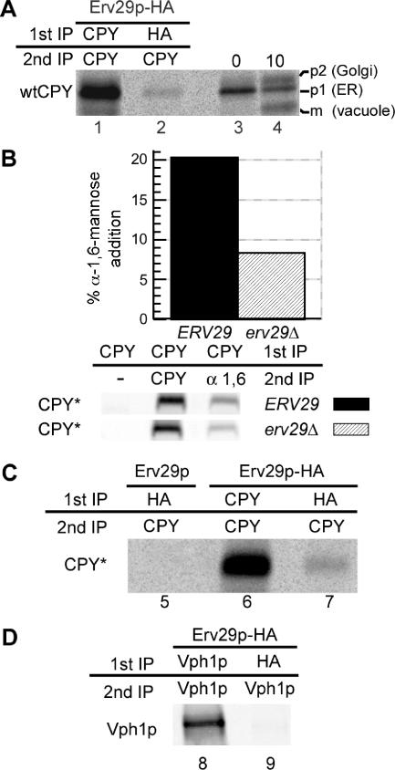
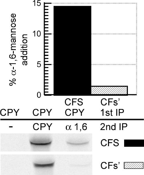
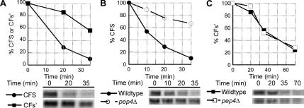
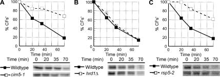
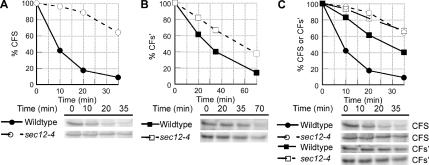
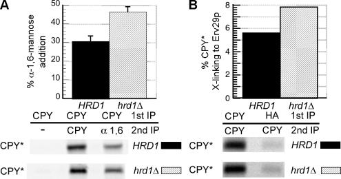
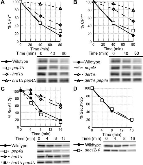
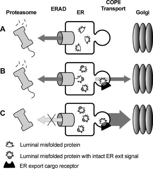
Most misfolded secretory proteins remain in the endoplasmic reticulum (ER) and are degraded by ER-associated degradation (ERAD). However, some misfolded proteins exit the ER and traffic to the Golgi before degradation. Using model misfolded substrates, with or without defined ER exit signals, we found misfolded proteins can depart the ER by continuing to exhibit the functional export signals present in the corresponding correctly folded proteins. Anterograde transport of misfolded proteins utilizes the same machinery responsible for exporting correctly folded proteins. Passive ER retention, in which misfolded proteins fail to exit the ER due to the absence of exit signals or the inability to functionally present them, likely contributes to the retention of nonnative proteins in the ER. Intriguingly, compromising ERAD resulted in increased anterograde trafficking of a misfolded protein with an ER exit signal, suggesting that ERAD and ER exit machinery can compete for binding of misfolded proteins. Disabling ERAD did not result in transport of an ERAD substrate lacking an export signal. This is an important distinction for those seeking possible therapeutic approaches involving inactivating ERAD in anticipation of exporting a partially active protein.


Figures

Figure 1.
Figure 1.
ER exit of both wtCPY and misfolded CPY* is dependent on the ER export cargo receptor Erv29p. (A) erv29Δ cells (KHY271) expressing wtCPY and Erv29p-HA (pAC530) were grown at 30°C and radiolabeled with 35S methionine/cysteine for 6 min, and spheroplasts were treated with the cross-linking agent, DSP. Cell lysates were immunoprecipitated with anti-CPY (lane 1) or anti-HA antibodies (lane 2), the cross-linker was cleaved, and proteins reimmunoprecipitated with anti-CPY antibodies. Immunoprecipitated proteins were separated by SDS-PAGE and exposed to a phosphor screen. As markers of p1, p2, and mature wtCPY, KHY271 cells expressing Erv29 (pAC505) were grown at 30°C, radiolabeled for 5 min, and chased with unlabeled methionine/cysteine. Samples were removed at indicated times, and cell extracts were prepared, and wtCPY was immunoprecipitated with anti-CPY antibodies (lanes 3 and 4). Proteins were resolved by SDS-PAGE. (B) ERV29 (KHY171) and erv29Δ cells (KHY279) expressing CPY* (pAC519) were radiolabeled for 20 min and chased for 5 min, and CPY* was immunoprecipitated. The samples were solubilized, reimmunoprecipitated with either anti-CPY or anti-α-1,6-mannose antibodies, and treated with Endo H followed by SDS-PAGE and quantification. Graphic representation of the α-1,6-mannose modification of CPY* in ERV29 and erv29Δ cells is shown. (C) erv29Δ (KHY270) cells expressing Erv29p (pAC505, lane 5) or Erv29p-HA (pAC530, lane 6 and 7) were radiolabeled for 10 min before cross-linking and immunoprecipitation with anti-HA or anti-CPY antibodies and then reimmunoprecipitated with anti-CPY antibodies. Samples were treated with Endo H before SDS-PAGE. (D) erv29Δ (KHY270) cells expressing Vph1p (pMM322) and Erv29p-HA (pAC530) were radiolabeled for 6 min before cross-linking and immunoprecipitation with anti-HA or anti-Vph1 antibodies and then reimmunoprecipitated with anti-Vph1 antibodies.
Figure 2.
Figure 2.
Delivery of misfolded proteins to the Golgi is dependent on ER exit signals. KHY517 cells expressing CFS (pAC812) or CFs′ (pAC815) were radiolabeled for 15 min and chased for 10 min, and CFS or CFs′ were immunoprecipitated with anti-CPY antibodies. Samples were solubilized, diluted, and reimmunoprecipitated with either anti-CPY or anti-α-1,6-mannose antibodies followed by treatment with Endo H. Graphic representation of the α-1,6-mannose modification of CFS and CFs′ is shown.
Figure 3.
Figure 3.
ER exit of misfolded proteins and subsequent vacuolar delivery and degradation depends on ER exit signal efficiency. (A) Wild-type (KHY298) cells expressing CFS (pAC812) or CFs′ (pAC815) were subjected to pulse-chase analysis as described. Graphic representations of the degradation of CFS and CFs′ are shown. (B and C) Wild-type (KHY298) and pep4Δ (KHY401) cells expressing CFS (pAC812) or CFs′ (pAC815) were subjected to pulse-chase analysis. Graphic representations of the degradation of CFS (B) and CFs′ (C) in wild-type and pep4Δ cells are shown.
Figure 4.
Figure 4.
CFs′ is an ERAD substrate and degraded by the proteasome. (A) Wild-type (KHY516) and cim5-1 (KHY517) cells expressing CFs′ (pAC815) were subjected to pulse-chase analysis as described. The degradation rate of CFs′ in wild-type and cim5-1 cells is shown. (B) Degradation of CFs′ was determined in wild-type (KHY298) and hrd1Δ (KHY299) cells. (C) Wild-type (KHY741) and rsp5-2 (KHY747) cells expressing CFs′ (pAC815) were grown at 24°C and shifted to the restrictive temperature (38°C) for 20 min before pulse-chase analysis. Graphic representations of the degradation of CFs′ in wild-type and rsp5-2 cells are shown.
Figure 5.
Figure 5.
ER exit of misfolded proteins depends on ER exit signal efficiency. (A and B) Wild-type (KHY298) and sec12-4 (KHY388) cells expressing CFS (pAC812) or CFs′ (pAC815) were grown at 24°C and shifted to the restrictive temperature (34.5°C) for 15 min before pulse-chase analysis. Samples were treated with Endo H before SDS-PAGE analysis. Graphic representations of the degradation of CFS (A) and CFs′ (B) in wild-type and sec12-4 cells are shown. (C) Overlay of graphic representations of the degradation of CFS and CFs′ in wild-type and sec12-4 cells from data shown in A and B.
Figure 6.
Figure 6.
Inactivation of ERAD results in increased Golgi delivery of CPY* by allowing greater interaction with Erv29p. (A) HRD1 (KHY252) and hrd1Δ cells (KHY265) expressing CPY* were radiolabeled for 20 min and chased for 15 min, and CPY* was immunoprecipitated. The samples were then reimmunoprecipitated with either anti-CPY or anti-α-1,6-mannose antibodies followed by treatment with Endo H. Graphic representation of the α-1,6-mannose modification of CPY* in HRD1 and hrd1Δ cells is shown. The data were plotted as mean values with SDs of three independent experiments, and representative gels are shown. (B) erv29Δ (KHY270) and erv29Δ hrd1Δ (KHY279) cells expressing Erv29p-HA (pAC530) were radiolabeled for 10 min before cross-linking and immunoprecipitation with anti-CPY or anti-HA antibodies followed by reimmunoprecipitation with anti-CPY antibodies. Graphic representation of the extent of CPY* binding to Erv29p in erv29Δ (KHY270) and erv29Δ hrd1Δ (KHY279) cells is shown.
Figure 7.
Figure 7.
Inactivation of ERAD results in increased trafficking of CPY* from the ER, but not Sec61-2p. (A) Wild-type (KHY163), pep4Δ (KHY252), hrd1Δ (KHY171), and hrd1Δ pep4Δ (KHY265) cells were grown at 30°C and radiolabeled for 10 min and chased. Samples were removed at the times indicated, cell extracts were prepared, and CPY* was immunoprecipitated. The immunoprecipitated protein was then digested with Endo H before SDS-PAGE analysis. Graphic representations of the degradation of CPY* are shown. (B) Wild-type (KHY163), pep4Δ (KHY252), der1Δ (KHY169), and der1Δ pep4Δ (KHY264) cells were grown at 30°C, radiolabeled, and chased. Aliquots were harvested at times indicated and treated as described above. Graphic representations of the degradation of CPY* are shown. (C) Wild-type (KHY163), pep4Δ (KHY583), hrd1Δ (KHY171), and hrd1Δ pep4Δ (KHY662) cells expressing Sec61-2p-HA (pAC460) were grown at 30°C, radiolabeled, and chased. Aliquots were harvested at times indicated and treated as described. Graphic representations of the degradation of Sec61-2p-HA are shown. (D) Wild-type (KHY163) and sec12-4 (KHY306) cells expressing Sec61-2p-HA (pAC460) were grown at 24°C and shifted to the restrictive temperature (34.5°C) for 15 min before radiolabeling and chase. Samples were removed at the times indicated, cell extracts were prepared, and Sec61-2p-HA was immunoprecipitated. Graphic representations of the degradation of Sec61-2p-HA in wild-type and sec12-4 cells are shown.
Figure 8.
Figure 8.
ERAD and ER exit can compete for misfolded substrates. (A) Misfolded proteins lacking ER exit signals remain in the ER and are degraded by ERAD. (B) Misfolded proteins with intact ER exit signals are engaged by ERAD machinery for dislocation and proteasomal degradation. However, these misfolded proteins can also exit the ER via interaction with ER exit receptors (for soluble substrates) or via direct interaction of cytosolic signals with COPII components (for membrane-spanning substrates). (C) Inactivating ERAD results in greater ER exit of misfolded proteins containing ER export signals.

References

    1. Aridor M., Weissman J., Bannykh S., Nuoffer C., Balch W. E. Cargo selection by the COPII budding machinery during export from the ER. J. Cell Biol. 1998;141:61–70. - PMC - PubMed
    1. Barlowe C. Signals for COPII-dependent export from the ER: what's the ticket out? Trends Cell Biol. 2003;13:295–300. - PubMed
    1. Bays N. W., Gardner R. G., Seelig L. P., Joazeiro C. A., Hampton R. Y. Hrd1p/Der3p is a membrane-anchored ubiquitin ligase required for ER-associated degradation. Nat. Cell Biol. 2001;3:24–29. - PubMed
    1. Belden W. J., Barlowe C. Role of Erv29p in collecting soluble secretory proteins into ER-derived transport vesicles. Science. 2001;294:1528–1531. - PubMed
    1. Bordallo J., Plemper R. K., Finger A., Wolf D. H. Der3p/Hrd1p is required for endoplasmic reticulum-associated degradation of misfolded lumenal and integral membrane proteins. Mol. Biol. Cell. 1998;9:209–222. - PMC - PubMed

Publication types

MeSH terms

Substances