. 2007 Feb;18(2):337-47.
doi: 10.1091/mbc.e06-06-0487. Epub 2006 Nov 15.

Internalization of large double-membrane intercellular vesicles by a clathrin-dependent endocytic process

Affiliations

Internalization of large double-membrane intercellular vesicles by a clathrin-dependent endocytic process

Michelle Piehl et al. Mol Biol Cell. 2007 Feb.

Abstract

Beyond its well-documented role in vesicle endocytosis, clathrin has also been implicated in the internalization of large particles such as viruses, pathogenic bacteria, and even latex beads. We have discovered an additional clathrin-dependent endocytic process that results in the internalization of large, double-membrane vesicles at lateral membranes of cells that are coupled by gap junctions (GJs). GJ channels bridge apposing cell membranes to mediate the direct transfer of electrical currents and signaling molecules from cell to cell. Here, we report that entire GJ plaques, clusters of GJ channels, can be internalized to form large, double-membrane vesicles previously termed annular gap junctions (AGJs). These internalized AGJ vesicles subdivide into smaller vesicles that are degraded by endo/lysosomal pathways. Mechanistic analyses revealed that clathrin-dependent endocytosis machinery-components, including clathrin itself, the alternative clathrin-adaptor Dab2, dynamin, myosin-VI, and actin are involved in the internalization, inward movement, and degradation of these large, intercellular double-membrane vesicles. These findings contribute to the understanding of clathrin's numerous emerging functions.


Figures
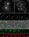
Figure 1.
Figure 1.
Internalization of GJs generates large cytoplasmic double-membrane vesicles. (A) Early after transfection (12 h), GJs assembled from Cx43-GFP are visible in the PM between transfected HeLa cells (labeled with arrows in all figures). In addition, secretory connexin cargo is visible in the Golgi region of the cell (labeled with asterisks). Later (24 h posttransfection) additional bright fluorescent vesicular structures (∼1–5 μm in diameter) are detectable primarily in the cytoplasm of one cell (marked with arrowheads in all figures). (B) Time-lapse recording of a GJ plaque that internalizes to form such a double-membrane GJ vesicle, previously termed AGJs (also see Supplemental Movie 1B). (C) DIC and fluorescence time-lapse microscopy demonstrate the formation, detachment, and cytoplasmic translocation of AGJ vesicles away from the PM into the cell body. Note the assembly of a new GJ plaque in the membrane (labeled with an arrow; also see Supplemental Movie 1C). (D) Spherical membranous structure of AGJ vesicles revealed by DiI staining and confocal z-sectioning. (E) AGJ vesicles are double-membrane structures that are composed of connexons (GJ half-channels) derived from the PMs of both coupled cells. This was demonstrated by growing stably transfected Cx43-CFP (green) and Cx43-YFP (red) HeLa cells in mixed cultures. GJ plaques between red and green cells look yellow. Also, yellow internalized AGJ vesicles are detectable primarily in one of the two coupled cells. (F and G) The lumen of AGJ vesicles corresponds to cytoplasm derived from the neighboring cell. This was demonstrated by microinjecting one cell of a coupled pair with red fluorescent quantum dots (GJ impermeable). AGJ vesicles internalized into the labeled cell have a black lumen (F), whereas AGJ vesicles that bud into a noninjected cell have a red lumen (G). (H) Ultrastructural analysis of Cx43-GFP–transfected HeLa cells show all stages of progressive GJ internalization and AGJ vesicle formation. N, nuclei. Bars in all figures, micrometers in fluorescence and nanometers in electron microscopic images.
Figure 1.
Figure 1.
Internalization of GJs generates large cytoplasmic double-membrane vesicles. (A) Early after transfection (12 h), GJs assembled from Cx43-GFP are visible in the PM between transfected HeLa cells (labeled with arrows in all figures). In addition, secretory connexin cargo is visible in the Golgi region of the cell (labeled with asterisks). Later (24 h posttransfection) additional bright fluorescent vesicular structures (∼1–5 μm in diameter) are detectable primarily in the cytoplasm of one cell (marked with arrowheads in all figures). (B) Time-lapse recording of a GJ plaque that internalizes to form such a double-membrane GJ vesicle, previously termed AGJs (also see Supplemental Movie 1B). (C) DIC and fluorescence time-lapse microscopy demonstrate the formation, detachment, and cytoplasmic translocation of AGJ vesicles away from the PM into the cell body. Note the assembly of a new GJ plaque in the membrane (labeled with an arrow; also see Supplemental Movie 1C). (D) Spherical membranous structure of AGJ vesicles revealed by DiI staining and confocal z-sectioning. (E) AGJ vesicles are double-membrane structures that are composed of connexons (GJ half-channels) derived from the PMs of both coupled cells. This was demonstrated by growing stably transfected Cx43-CFP (green) and Cx43-YFP (red) HeLa cells in mixed cultures. GJ plaques between red and green cells look yellow. Also, yellow internalized AGJ vesicles are detectable primarily in one of the two coupled cells. (F and G) The lumen of AGJ vesicles corresponds to cytoplasm derived from the neighboring cell. This was demonstrated by microinjecting one cell of a coupled pair with red fluorescent quantum dots (GJ impermeable). AGJ vesicles internalized into the labeled cell have a black lumen (F), whereas AGJ vesicles that bud into a noninjected cell have a red lumen (G). (H) Ultrastructural analysis of Cx43-GFP–transfected HeLa cells show all stages of progressive GJ internalization and AGJ vesicle formation. N, nuclei. Bars in all figures, micrometers in fluorescence and nanometers in electron microscopic images.
Figure 2.
Figure 2.
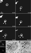
Internalized double-membrane GJ vesicles fragment into smaller vesicles. (A) Time-lapse recordings of Cx43-GFP–transfected HeLa cells demonstrate initial degradation of AGJ vesicles by successive budding of smaller AGJ vesicles from a newly internalized, larger AGJ vesicle (marked with arrowheads; also see Supplemental Movie 2A). (B) Confocal section of an AGJ vesicle cluster stained with DiI. (C and D) Ultrastructural composition of AGJ vesicles from Cx43-GFP–transfected HeLa cells. Note the “collapsed” AGJ vesicle with its two closely apposed double-membrane envelopes (D, asterisk).
Figure 3.
Figure 3.
Double-membrane GJ internalization is clathrin mediated. (A and B) Colocalization of clathrin patches with GJs and internalized AGJ vesicles in Cx43-GFP–transfected HeLa cells. (C–E) Defined patches of dense protein coats with the characteristic thickness and appearance of clathrin coats on the outside of internalized GJ vesicles (C and E), and on GJ plaques (D). (F) Significant reduction of clathrin heavy chain (CHC) expression by RNAi (Tub, β-tubulin; WT, wild type). (G) Significant reduction of internalized GJ numbers in KD cells (number of cells = wt [157] and KD ]189]; internalized GJ vesicles counted and grouped by size (wt [KD]): ≥2 μm: 68 [40]; ≥1.5 μm: 199 [134]; ≥1 μm: 645 [366]; and ≥0.5 μm 1759 [973]). Data shown are mean ± SEM. p values indicate significance. (H) Significant reduction (p = 0.04) of internalized GJ vesicle numbers in hypertonic medium treated cells (number of untreated cells, 125; number of treated cells, 99; Con, control cells; Tr, treated cells). Only AGJ vesicles ≥2 μm in diameter were counted. Data shown are mean ± SEM.
Figure 4.
Figure 4.
Colocalization of clathrin adaptors and dynamin with Cx43-based GJs and AGJ vesicles. GJs are marked with arrows. AGJ vesicles are marked with arrowheads. (A and B) The classical PM clathrin adaptor AP-2 shows minimal colocalization with GJs and AGJ vesicles. (C and D) The alternative potent clathrin adaptor Dab-2 colocalizes robustly with GJs and AGJ vesicles in Cx43-GFP–transfected HeLa cells. (E and F) Specificity of the Dab2/Cx43 colocalization was tested in COS-7 cells that express significantly lower levels of Dab2 (see immunoblot in K). Other known potent clathrin adaptor proteins CALM (G) or epsin1 (H) exhibited weak colocalization with Cx43-GFP based GJs and AGJ vesicles. Dynamin also colocalizes robustly with GJs and AGJ vesicles (I and J).
Figure 5.
Figure 5.
Myosin-VI colocalizes with invaginating plaques and AGJ vesicles, and actin/myosin-VI activity drives AGJ vesicle translocation. (A–D) Myosin-VI colocalizes specifically with internalizing GJ plaques and with newly internalized AGJ vesicles (marked with arrowheads) but not with planar GJs (marked with arrows), late AGJ vesicle degradation products, or Cx43-GFP–containing secretory cargo vesicles (marked with asterisks) as indicated by confocal colocalization studies of Cx43-GFP–transfected HeLa cells. (E–G) Actin filaments (stained with rhodamine-phalloidin in E and F) colocalize with GJs (E, arrows) and AGJ vesicles (F and G, arrowheads) in Cx43-GFP–transfected HeLa cells (confocal microscopy in E and F; ultrastructural analyses in G). To examine the involvement of actin/myosin-VI in AGJ vesicle translocation, actin filaments were either stabilized (by treating the cells with jasplakinolide [Jaspla]) or disrupted (by treating with latrunculin A [LatA]), or a GFP-tagged full length (Myo6FL) construct was coexpressed with Cx43-GFP and the mean velocity (H) and the maximum distance traveled (I) of AGJ vesicles was measured (number of cells [AGJ vesicles] tracked for DMSO: 7 [11]; jasplakinolide: 8 [21]; latrunculin A: 9 [20]; control cells: 20 [28]; and GFP-M6full–expressing cells: 22 [44]; ANOVA; p < 0.05).
Figure 6.
Figure 6.
Schematic model of GJ internalization and degradation based on protein components that were characterized in this work. The alternative potent clathrin adaptor Dab2 is recruited to Cx43-based GJs possibly through a direct interaction of its N-terminal phosphotyrosine binding (PTB) domain with a putative XPXY internalization motif located in the C-terminal tail of Cx43 and in a number of other connexin family members. The distal portion of Dab2 on its opposite end binds the globular N-terminal domain of clathrin heavy chains and triggers clathrin lattice assembly. The GTPase dynamin is also recruited to GJs, resulting in double-membrane protrusion/invagination, neck restriction, and double-membrane scission. Other proteins, such as AP-2 or epsin might be recruited transiently, or in smaller numbers either actively or passively through their interaction with Cx43, Dab2, and/or clathrin. Dab2 then associates through its C-terminal serine- and proline-rich region with the C-terminal globular tail of myosin-VI that binds through its N-terminal motor domain to actin filaments resulting in the inward translocation of the internalized double-membrane GJ vesicles. GJ vesicles then fragment into smaller vesicles that are degraded by endo/lysosomal pathways.

Similar articles

Cited by

References

    1. Aggeler J., Werb Z. Initial events during phagocytosis by macrophages viewed from outside and inside the cell: membrane-particle interactions and clathrin. J. Cell Biol. 1982;94:613–623. - PMC - PubMed
    1. Aschenbrenner L., Lee T., Hasson T. Myo6 facilitates the translocation of endocytic vesicles from cell peripheries. Mol. Biol. Cell. 2003;14:2728–2743. - PMC - PubMed
    1. Aschenbrenner L., Naccache S. N., Hasson T. Uncoated endocytic vesicles require the unconventional myosin, Myo6, for rapid transport through actin barriers. Mol. Biol. Cell. 2004;15:2253–2263. - PMC - PubMed
    1. Beardslee M. A., Laing J. G., Beyer E. C., Saffitz J. E. Rapid turnover of connexin43 in the adult rat heart. Circ. Res. 1998;83:629–635. - PubMed
    1. Berthoud V. M., Minogue P. J., Laing J. G., Beyer E. C. Pathways for degradation of connexins and gap junctions. Cardiovasc. Res. 2004;62:256–267. - PubMed

Publication types

MeSH terms