Morphological characterisation of portal myofibroblasts and hepatic stellate cells in the normal dog liver
- PMID: 17109742
- PMCID: PMC1660578
- DOI: 10.1186/1476-5926-5-7
Morphological characterisation of portal myofibroblasts and hepatic stellate cells in the normal dog liver
Abstract
Background: Hepatic fibrosis is a common outcome of hepatic injury in both man and dog. Activated fibroblasts which develop myofibroblastic characteristics play an essential role in hepatic fibrogenesis, and are comprised of three subpopulations: 1) portal or septal myofibroblasts, 2) interface myofibroblasts and 3) the perisinusoidally located hepatic stellate cells (HSC). The present study was performed to investigate the immunohistochemical characteristics of canine portal myofibroblasts (MF) and HSC in the normal unaffected liver as a basis for further studies on fibrogenesis in canine liver disease.
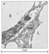
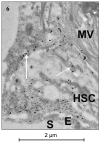
Results: In the formalin-fixed and paraffin embedded normal canine liver vimentin showed staining of hepatic fibroblasts, probably including MF in portal areas and around hepatic veins; however, HSC were in general negative. Desmin proved to react with both portal MF and HSC. A unique feature of these HSC was the positive immunostaining for alpha-smooth muscle actin (alpha-SMA) and muscle-specific actin clone HHF35 (HHF35), also portal MF stained positive with these antibodies. Synaptophysin and glial fibrillary acidic protein (GFAP) were consistently negative in the normal canine liver. In a frozen chronic hepatitis case (with expected activated hepatic MF and HSC), HSC were negative to synaptophysin, GFAP and NCAM. Transmission electron microscopy (TEM) immunogold labelling for alpha-SMA and HHF35 recognized the positive cells as HSC situated in the space of Disse.
Conclusion: In the normal formalin-fixed and paraffin embedded canine liver hepatic portal MF and HSC can be identified by alpha-SMA, HHF35 and to a lesser extent desmin immunostaining. These antibodies can thus be used in further studies on hepatic fibrosis. Synaptophysin, GFAP and NCAM do not seem suitable for marking of canine HSC. The positivity of HSC for alpha-SMA and HHF35 in the normal canine liver may eventually reflect a more active regulation of hepatic sinusoidal flow by these HSC compared to other species.
Figures


Similar articles
-
Hepatic stellate cell/myofibroblast subpopulations in fibrotic human and rat livers.J Hepatol. 2002 Feb;36(2):200-9. doi: 10.1016/s0168-8278(01)00260-4. J Hepatol. 2002. PMID: 11830331
-
Localization of liver myofibroblasts and hepatic stellate cells in normal and diseased rat livers: distinct roles of (myo-)fibroblast subpopulations in hepatic tissue repair.Histochem Cell Biol. 1999 Nov;112(5):387-401. doi: 10.1007/s004180050421. Histochem Cell Biol. 1999. PMID: 10603079
-
Characterization of glial fibrillary acidic protein (GFAP)-expressing hepatic stellate cells and myofibroblasts in thioacetamide (TAA)-induced rat liver injury.Exp Toxicol Pathol. 2013 Nov;65(7-8):1159-71. doi: 10.1016/j.etp.2013.05.008. Epub 2013 Jun 25. Exp Toxicol Pathol. 2013. PMID: 23806769
-
Cooperation of liver cells in health and disease.Adv Anat Embryol Cell Biol. 2001;161:III-XIII, 1-151. doi: 10.1007/978-3-642-56553-3. Adv Anat Embryol Cell Biol. 2001. PMID: 11729749 Review.
-
Wnt signaling in liver fibrosis: progress, challenges and potential directions.Biochimie. 2013 Dec;95(12):2326-35. doi: 10.1016/j.biochi.2013.09.003. Epub 2013 Sep 13. Biochimie. 2013. PMID: 24036368 Review.
Cited by
-
Establishment and characterization of rat portal myofibroblast cell lines.PLoS One. 2015 Mar 30;10(3):e0121161. doi: 10.1371/journal.pone.0121161. eCollection 2015. PLoS One. 2015. PMID: 25822334 Free PMC article.
-
An Overview of Epithelial-to-Mesenchymal Transition and Mesenchymal-to-Epithelial Transition in Canine Tumors: How Far Have We Come?Vet Sci. 2022 Dec 28;10(1):19. doi: 10.3390/vetsci10010019. Vet Sci. 2022. PMID: 36669020 Free PMC article. Review.
-
Pancreatic stellate/myofibroblast cells express G-protein-coupled melatonin receptor 1.Wien Med Wochenschr. 2008;158(19-20):575-8. doi: 10.1007/s10354-008-0599-7. Wien Med Wochenschr. 2008. PMID: 18998076
-
Lipid Dysmetabolism in Canine Chronic Liver Disease: Relationship Between Clinical, Histological and Immunohistochemical Features.Vet Sci. 2025 Mar 2;12(3):220. doi: 10.3390/vetsci12030220. Vet Sci. 2025. PMID: 40266905 Free PMC article.
-
Liver tissue remodeling following ablation with irreversible electroporation in a porcine model.Front Vet Sci. 2022 Nov 4;9:1014648. doi: 10.3389/fvets.2022.1014648. eCollection 2022. Front Vet Sci. 2022. PMID: 36406062 Free PMC article.
References
-
- Crawford JM. Liver cirrhosis. In: MacSween RNM, Burt AD, Portmann BC, Ishak KG, Scheuer PJ, Anthony PP, editor. Pathology of the Liver. Edinburgh: Churchill Livingstone; 2002. pp. 575–619.
-
- Knittel T, Kobold D, Piscaglia F, Saile B, Neubauer K, Mehde M, Timpl R, Ramadori G. Localization of liver myofibroblasts and hepatic stellate cells in normal and diseases rat livers: distinct roles of (myo-)fibroblast subpopulations in hepatic tissue repair. Histochem Cell Biol. 1999;112:387–401. doi: 10.1007/s004180050421. - DOI - PubMed
-
- Guyot C, Lepreux S, Combe C, Doudnikoff E, Bioulac-Sage P, Balabaud C, Desmouliere A. Hepatic fibrosis and cirrhosis: The (myo)fibroblastic cell subpopulations involved. Int J Bioch & Cell Biol. 2006;38:135–151. - PubMed
LinkOut - more resources
Full Text Sources
Research Materials
Miscellaneous
