. 2007 Jan 1;457(1):16-28.
doi: 10.1016/j.abb.2006.10.011. Epub 2006 Nov 2.

Angiotensin II induces phosphorylation of glucose-regulated protein-75 in WB rat liver cells

Affiliations

Angiotensin II induces phosphorylation of glucose-regulated protein-75 in WB rat liver cells

Sharath B Krishna et al. Arch Biochem Biophys. .

Abstract

Studies in vascular smooth muscle cells suggest that, angiotensin II (Ang II)-mediated cellular response requires transactivation of epidermal growth factor receptor (EGF-R), and involves tyrosine phosphorylation of caveolin-1. Here we demonstrate that, exposure of WB rat liver cells to Ang II does not cause transactivation of EGF-R, but did rapidly activate p42/p44 mitogen-activated protein (MAP) kinases suggesting that it activates MAP kinases independent of EGF-R transactivation. We observed that the phospho-specific anti-caveolin-1 antibody detected a tyrosine phosphorylated, 75kDa protein in Ang II-treated cells which we identified as glucose regulated protein-75 (GRP-75). Phosphoamino acid analysis showed that Ang II induced its phosphorylation at tyrosine, serine and threonine residues and was localized to the cytoplasm. The ability of Ang-II to induce GRP-75 phosphorylation suggests that it may play a role in the protection of cytoplasmic proteins from the damaging effect of oxidative stress known to be produced during Ang-II induced signaling.


Figures

FIG. 1
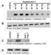
FIG. 1. Angiotensin II activates p42/p44 MAP kinases independent of EGF-R transactivation; phosphospecific anti-caveolin-1 antibody cross-reacts with a 75 kDa protein
A, shows that Ang II does not transactivate EGF-R. Lysates were prepared from cells untreated (control), or treated with Ang II (100 nM, 3 min), or EGF (100 ng/ml; 3 min). Equal amount of proteins were run on a SDS-polyacrylamide gel and immunoblotted with phospho-specific EGF-R antibody specific for Y1068. B, the blot in A was stripped and reprobed with EGF-R antibody. C, shows that Ang II and EGF activate p42/p44 MAP kinases. The samples representing A were run on a SDS-polyacrylamide gel and immunoblotted with phospho-specific p42/p44 anti-MAP kinase antibody. D, The blot in C was stripped and reprbed with p42/p44 anti-MAP kinase antibody. E, shows that Ang II does not induce tyrosine phosphorylation of caveolin-1; anti-phospho caveolin-1 antibody cross reacts with a 75 kDa protein. Serum starved cells were left untreated (control) (lane 1), or treated with Ang II (100 nM) for different times (lanes 2–5), and total extracts were prepared, run on a 8% SDS-polyacrylamide gel, and immunoblotted with phospho-specific anti-caveolin-1 antibody. F, The blot in E was stripped and reprobed with anti-caveolin-1 antibody. These blots are representative of four independent experiments. The position of the 75 kDa protein and p42/p44 MAP kinases are shown in an arrows. CR, cross-reactive.
FIG. 2
FIG. 2. The 75 kDa protein detected by phospho-specific anti-caveolin-1 antibody is tyrosine and serine phosphorylated in Ang II-treated cells
A and B show that Ang II-induces tyrosine phosphorylation of the 75 kDa protein. A, Serum starved cells were left untreated (control) or treated with Ang II (100 nM) for 15 min. Total cell lysates prepared and immunoprecipitated (IP) with anti-phospho-tyrosine antibody, immunocomplexes were run on a 8% SDS-polyacrylamide gel and immunoblotted with phosphospecific anti-caveolin-1 antibody. B, Lasates representing A were immunoprecipitated (IP) with phosphospecific anti-caveolin-1 antibody, immunocomplexes were analyzed by immunoblotting with anti-phospho-tyrosine antibody. C, Phosphoamino acid analysis of the 75 kDa protein following Angiotensin II treatment. Following in vivo labeling with [32P] as described in methods, cells were exposed to Ang II (100 nM) for 15 min. Five hundred μg of the total protein were immunoprecipitated with phospho-specific anti-caveolin-1 antibody (cross-reactive to the 75 kDa protein), run on a SDS-polyacrylamide gel, transferred to PVDF membrane, and exposed to X-ray film. The 75 kDa band from control and Ang II treated lanes were excised and subjected to phosphoamino acid analysis as described in the methods section. These blots are representative of four independent experiments. These blots are representative of four independent experiments. Ig H, Immunoglobulin heavy chain. CR, cross-reactive.
FIG. 3
FIG. 3. Angiotensin II-induced tyrosine phosphorylation of the 75 kDa protein is concentration dependent and blocked by the AT1 receptor antagonist, EXP3174
A, Serum starved cells were left untreated, or treated for 15 min with different concentrations with Ang II. Lysates were prepared and equal amount of proteins were run on a 8% SDS-polyacrylamide gel and immunoblotted with phosphospecific anti-caveolin-1 antibody cross-reactive to the 75 kDa protein. B, the blot in A was stripped and reprobed with anti-Stat3 antibody (loading control). C, shows that the EXP3174 pretreatment blocks Ang II-induced tyrosine phosphorylation of the 75 kDa protein. Cells were left untreated (control), or treated with Ang II for 15 min (10 nM), or first pre-treated with EXP3174 (1000 nM) for 15 min and then with Ang II (10 nM) for 15 min. Lysates were prepared, run on a 8% SDS-polyacrylamide gel, and immunoblotted with phosphospecific anti-caveolin-1 antibody. D, the blot in C was stripped and reprobed with anti-Stat3 antibody (loading control). These blots are representative of four independent experiments.
FIG. 4
FIG. 4. Identification of the 75 kDa protein phosphorylated by Ang II as GRP-75
The 75 kDa protein was purified and subjected to mass spectrometry for protein identification. The sequences of the 13 peptides representing various regions of GRP-75 from the data base which matched with the peptides derived from the 75 kDa protein are indicated. The identity was established in three separate experiments. AA, amino acid.
FIG. 5
FIG. 5. GRP-75, a stress response protein undergoes phosphorylation in cells treated with Ang II and it is localized to the cytoplasm
A, Phospho-tyrosine blot of immunoprecipitated GRP-75. Serum straved cells were left untreated, or treated with Ang II for 15 min (100 nM). Cell lysates were prepared and immunoprecipitated (IP) with anti-GRP-75 antibody, immunocomplexes were run on an 8% SDS-polyacrylamide gel and probed with anti-phosphotyrosine antibody. B, the blot in A was stripped and reprobed with anti-GRP-75 antibody. C, the blot representing A was stripped and reprobed with anti-phospho-caveolin-1 antibody. D, shows that the 75 kDa protein is localized to the cytoplasm. Serum starved cells were untreated or treated with Ang II for 15 min (100 nM) and lysed, and then membrane, cytoplasm and nuclear fractions were prepared. 15 μg of each sample were run on a 8% SDS-polyacrylamide gel and immunoblotted with phospho-specific anti-caveolin-1 antibody cross-reactive to the 75 kDa protein (phosphorylated GRP-75). These blots are representative of three independent experiments. IgH, Immunoglobulin heavy chain.
FIG. 6
FIG. 6. Pervanadate, an agent which induces oxidative stress, stimulates tyrosine phosphorylation of GRP-75; diphenylene iodonium (DPI), an inhibitor of NADH/NADPH oxidase suppresses Ang II-medaited action
A, Serum starved cells were left untreated (control), or treated with Ang II (100 nM) for 15 min, or interleukin-6 (IL-6; 20 ng/ml) for 15 min, or EGF (100 ng/ml) for 5 min, or pervanadate (3 mM H2O2 together with 1 mM vanadate) for 15 min. Cell lysates were prepared and proteins run on an 8% SDS-polyacrylamide gel and immunoblotted with phospho-specific anti-caveolin-1 antibody cross-reactive to the 75 kDa protein (phopshorylated GRP-75). B, the blot in A was stripped and reprobed with anti-Stat3 antibody (loading control). C, Effect of DPI on Ang II-mediated GRP-75 tyrosine phosphorylation. Cells were left untreated, or treated with Ang II alone (100 nM) for 15 min, or DPI alone (10 μM) for 60 min, or first with DPI (10 μM) for 45 min followed by Ang II (100 nM) for 15 min. Cell lysates were prepared and equal amounts of protein were immunoblotted with anti-phospho-caveolin-1 antibody cross reactive to phosphorylated GRP-75. D, the blot in C was stripped and reprobed with anti-stat3 antibody. These blots are representative of four independent experiments.
FIG. 7
FIG. 7. Amino acid sequence comparison of caveolin-1 phospho-peptide (immunogen) with GRP-75 shows a homogy region, G-LY
The caveolin-1 phospho-peptide (aa 10–19) was used as immunogen. The Y14 of caveolin-1 is the site of tyrosine phosphorylation by several agonists (13, 14, 19). It is likely that the Y in the GHLY motif in GRP-75 also undergoes phosphorylation in cells treated with Ang II, and this site therefore, would be recognized by phospho-specific anti-caveolin-1 antibody.
FIG. 8
FIG. 8. Angiotensin II induces phosphorylation of GRP-75 at two different sites (Y331 and Y161)
In these experiments two different antibodies [phospho-specific anti-Stat3 antibody (35), and phospho-specific anti-caveolin-1 antibody], both cross-reactive to phosphorylated GRP-75 were used. A, Anti-phospho-caveolin-1 antibody immunoblot of samples immunoprecipitated with phospho-specific anti-Stat3 antibody. Serum starved cells were left untreated, or treated with Ang II for 15 min (100 nM). Cell lysates were prepared and immunoprecipitated (IP) with phospho-specific anti-Stat3 antibody, immunocomplexes were run on an 8% SDS-polyacrylamide gel and probed with phospho-specific anti-caveolin-1 antibody. B, The samples representing A were immunoprecipitated with phospho-specific anti-caveolin-1 antibody, immunocomplexes were run on an 8% SDS-polyacrylamide gel and probed with phospho-specific anti-Stat3 antibody. These blots are representative of three independent experiments. IgH, Immunoglobulin heavy chain. CR= cross reactive.
FIG. 9
FIG. 9. GRP-75 phosphorylation in the cytoplasm does not cause its translocation to mitochondria
A, Serum starved cells were left untreated (control), or treated with Ang II (100 nM) for either 15 min or 30 min. Mitochondria and cytoplasmic fractions were isolated as described in the methods section. Mitochondrial proteins were solubilized and equal amounts of protein from cytoplasm and mitochondrial fraction were run on a 8% SDS-polyacrylamide gel, and immunoblotted with anti-GRP-75 antibody. B, the samples representing A was immunoblotted with phospho-specific anti-caveolin-1 antibody cross-reactive to phosphorylated GRP-75. These blots are representative of three independent experiments. CR= cross reactive.

Similar articles

Cited by

References

    1. Kim S, Iwao H. Pharmacol Rev. 2000;52:11–34. - PubMed
    1. Touz RM, Schiffrin EL. Pharmcol Rev. 2000;52:639–672. - PubMed
    1. Marshal RP, Gohlke P, Chambers RC, Howell DC, Bottoms SE, Unger T, McAnulty RJ, Laurent GJ. Am J Phyiol Lung Cell Mol Physiol. 2004;286:156–164. - PubMed
    1. Guo G, Morrissey J, McCracken R, Tolley T, Liapis H, Klahr S. Am J Physiol Renal Physiol. 2001;280:F777–F785. - PubMed
    1. Bataller R, Schwabe RF, Choi YH, Yang L, Paik YH, Lindquist J, Qian T, Schoonhoven R, Hagedorn CH, Lemasters JJ, Brenner DA. J Clin Invest. 2003;112:1383–1394. - PMC - PubMed

Publication types

MeSH terms

LinkOut - more resources