

Injured Fluoro-Jade-positive hippocampal neurons contain high levels of zinc after traumatic brain injury
- PMID: 17109824
- PMCID: PMC2896019
- DOI: 10.1016/j.brainres.2006.09.094
Injured Fluoro-Jade-positive hippocampal neurons contain high levels of zinc after traumatic brain injury
Abstract
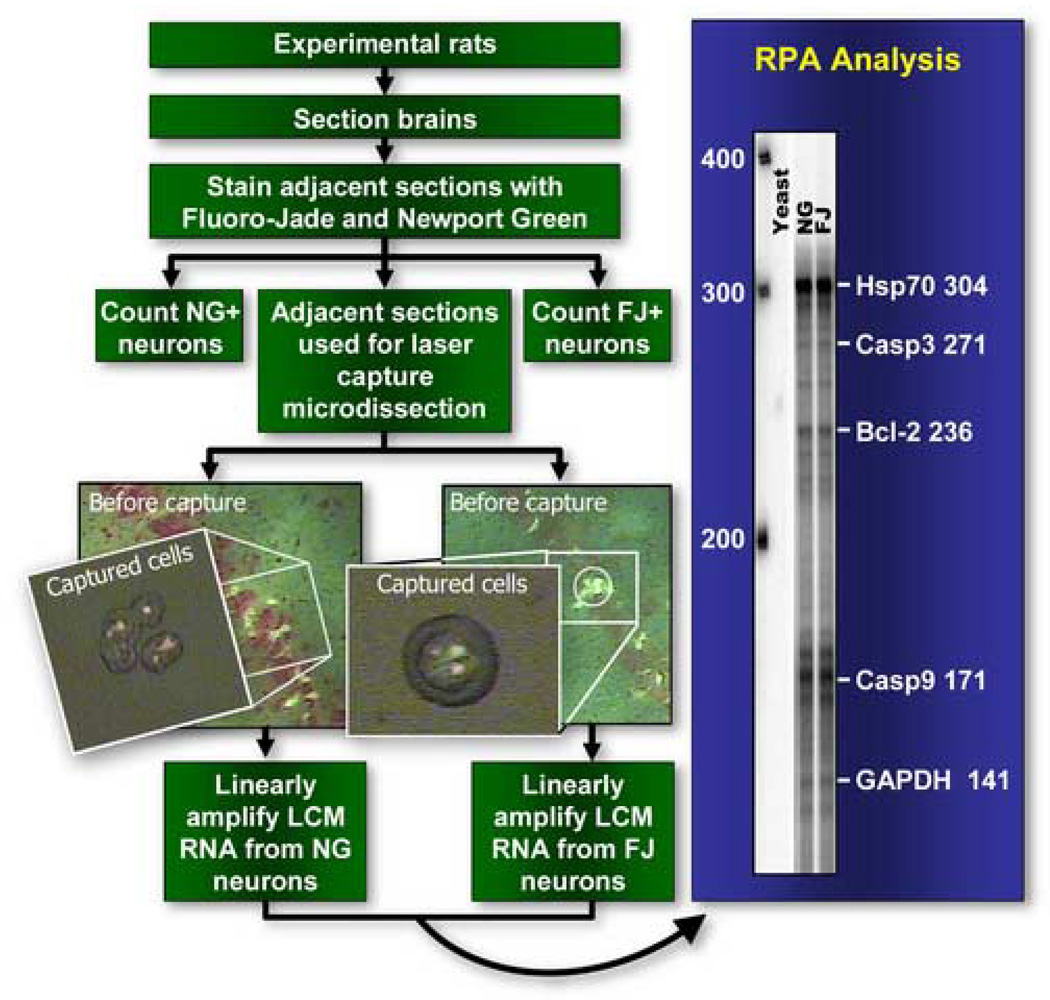
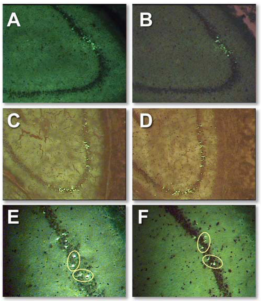
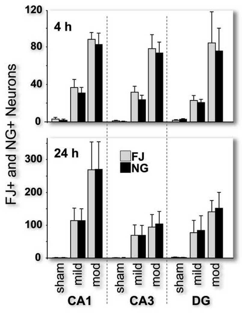
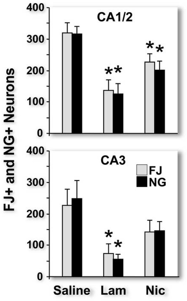
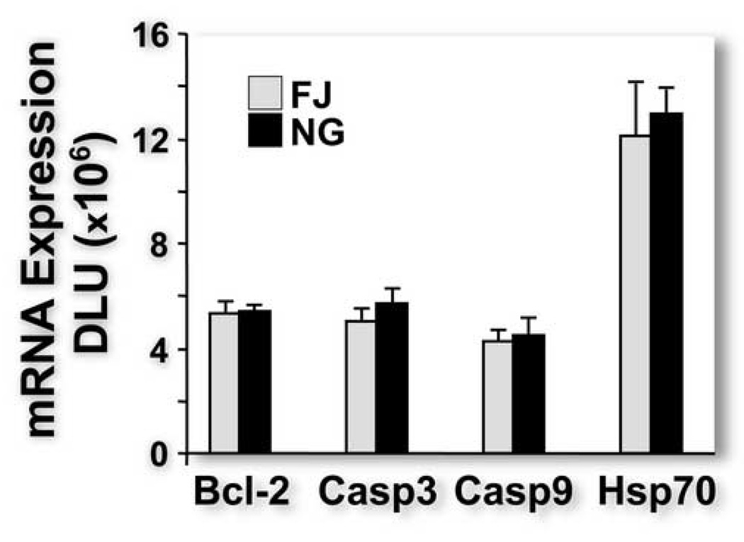
Hippocampal damage contributes to cognitive dysfunction after traumatic brain injury (TBI). We previously showed that Fluoro-Jade, a fluorescent stain that labels injured, degenerating brain neurons, quantifies the extent of hippocampal injury after experimental fluid percussion TBI in rats. Coincidentally, we observed that injured neurons in the rat hippocampus also stained with Newport Green, a fluorescent dye specific for free ionic zinc. Here, we show that, regardless of injury severity or therapeutic intervention, the post-TBI population of injured neurons in rat hippocampal subfields CA1, CA3 and dentate gyrus is indistinguishable, both in numbers and anatomical distribution, from the population of neurons containing high levels of zinc. Treatment with lamotrigine, which inhibits presynaptic release of glutamate and presumably zinc that is co-localized with glutamate, reduced numbers of Fluoro-Jade-positive and Newport Green-positive neurons equally as did treatment with nicardipine, which blocks voltage-gated calcium channels through which zinc enters neurons. To confirm using molecular techniques that Fluoro-Jade and Newport Green-positive neurons are equivalent populations, we isolated total RNA from 25 Fluoro-Jade-positive and 25 Newport Green-positive pyramidal neurons obtained by laser capture microdissection (LCM) from the CA3 subfield, linearly amplified the mRNA and used quantitative ribonuclease protection analysis to demonstrate similar expression of mRNA for selected TBI-induced genes. Our data suggest that therapeutic interventions aimed at reducing neurotoxic zinc levels after TBI may reduce hippocampal neuronal injury.
Figures
Similar articles
-
Traumatic brain injury and hemorrhagic hypotension suppress neuroprotective gene expression in injured hippocampal neurons.Anesthesiology. 2005 Apr;102(4):806-14. doi: 10.1097/00000542-200504000-00017. Anesthesiology. 2005. PMID: 15791111
-
Dose-dependent neuronal injury after traumatic brain injury.Brain Res. 2005 May 24;1044(2):144-54. doi: 10.1016/j.brainres.2005.02.054. Epub 2005 Apr 13. Brain Res. 2005. PMID: 15885213
-
Regional distribution of fluoro-jade B staining in the hippocampus following traumatic brain injury.Exp Neurol. 2005 May;193(1):125-30. doi: 10.1016/j.expneurol.2004.11.025. Exp Neurol. 2005. PMID: 15817271
-
Analysis of long-term gene expression in neurons of the hippocampal subfields following traumatic brain injury in rats.Neuroscience. 2005;131(1):87-97. doi: 10.1016/j.neuroscience.2004.10.041. Neuroscience. 2005. PMID: 15680694
-
Development and application of novel histochemical tracers for localizing brain connectivity and pathology.Brain Res. 2016 Aug 15;1645:31-5. doi: 10.1016/j.brainres.2016.03.053. Epub 2016 May 4. Brain Res. 2016. PMID: 27155454 Review.
Cited by
-
Improving treatments and outcomes: an emerging role for zinc in traumatic brain injury.Nutr Rev. 2012 Jul;70(7):410-3. doi: 10.1111/j.1753-4887.2012.00486.x. Epub 2012 May 22. Nutr Rev. 2012. PMID: 22747843 Free PMC article. Review.
-
The Influence of Heavy Metals and Trace Elements on Comatose Patients with Severe Traumatic Brain Injury in the First Week of Admission.J Toxicol. 2018 Sep 17;2018:7252606. doi: 10.1155/2018/7252606. eCollection 2018. J Toxicol. 2018. PMID: 30310389 Free PMC article.
-
Long-lasting suppression of acoustic startle response after mild traumatic brain injury.J Neurotrauma. 2015 Jun 1;32(11):801-10. doi: 10.1089/neu.2014.3451. Epub 2015 Mar 31. J Neurotrauma. 2015. PMID: 25412226 Free PMC article.
-
Ion channels and zinc: mechanisms of neurotoxicity and neurodegeneration.J Toxicol. 2012;2012:785647. doi: 10.1155/2012/785647. Epub 2012 May 7. J Toxicol. 2012. PMID: 22645609 Free PMC article.
-
Metabolic fate of glucose in rats with traumatic brain injury and pyruvate or glucose treatments: A NMR spectroscopy study.Neurochem Int. 2017 Jan;102:66-78. doi: 10.1016/j.neuint.2016.11.014. Epub 2016 Dec 3. Neurochem Int. 2017. PMID: 27919624 Free PMC article.
References
-
- Ahmad S, Fowler LJ, Whitton PS. Effects of combined lamotrigine and valproate on basal and stimulated extracellular amino acids and monoamines in the hippocampus of freely moving rats. Naunyn Schmiedebergs Arch Pharmacol. 2005;371:1–8. - PubMed
-
- Anderson KJ, Miller KM, Fugaccia I, Scheff SW. Regional distribution of Fluoro-Jade B staining in the hippocampus following traumatic brain injury. Exp Neurol. 2005;193:125–130. - PubMed
-
- Arciniegas D, Adler L, Topkoff J, Cawthra E, Filley CM, Reite M. Attention and memory dysfunction after traumatic brain injury: cholinergic mechanisms, sensory gating, and a hypothesis for further investigation. Brain Inj. 1999;13:1–13. - PubMed
-
- Bacher A, Zornow MH. Lamotrigine inhibits extracellular glutamate accumulation during transient global cerebral ischemia in rabbits. Anesthesiology. 1997;86:459–463. - PubMed
Publication types
MeSH terms
Substances
Grants and funding
LinkOut - more resources
Full Text Sources
Miscellaneous
