Review
. 2007 May 1;74(2):184-95.
doi: 10.1016/j.cardiores.2006.10.002. Epub 2006 Oct 7.

The role of TGF-beta signaling in myocardial infarction and cardiac remodeling

Affiliations
Review

The role of TGF-beta signaling in myocardial infarction and cardiac remodeling

Marcin Bujak et al. Cardiovasc Res. .

Abstract

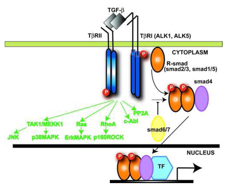
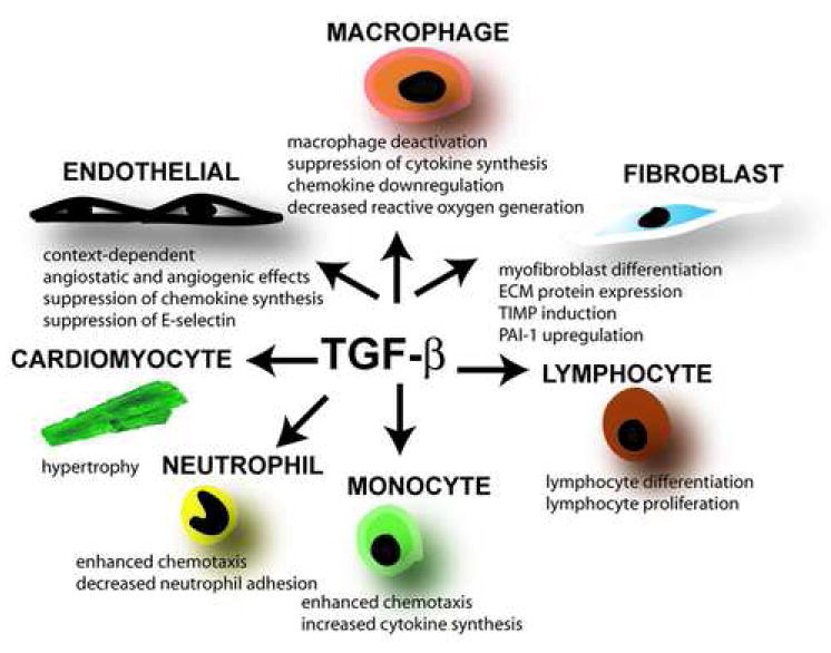
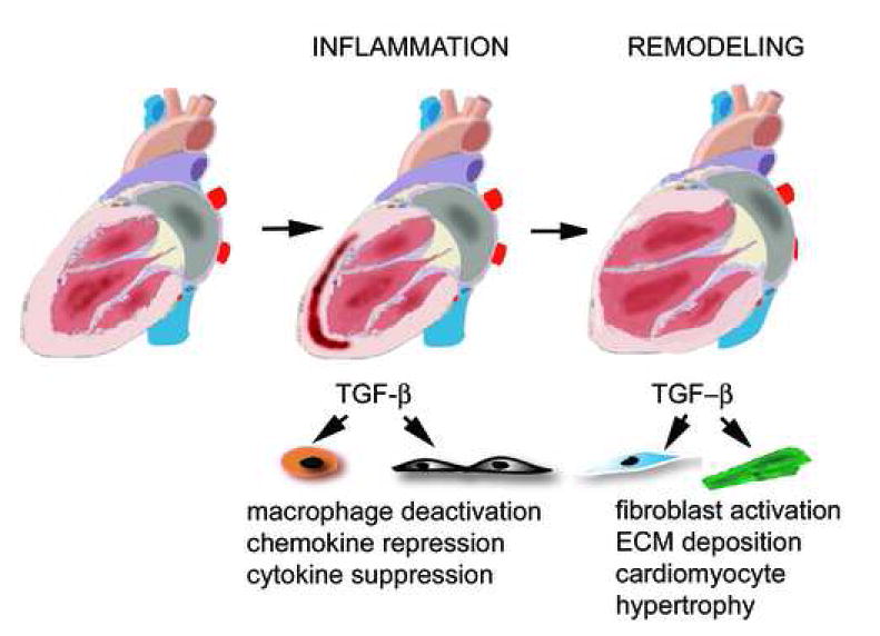
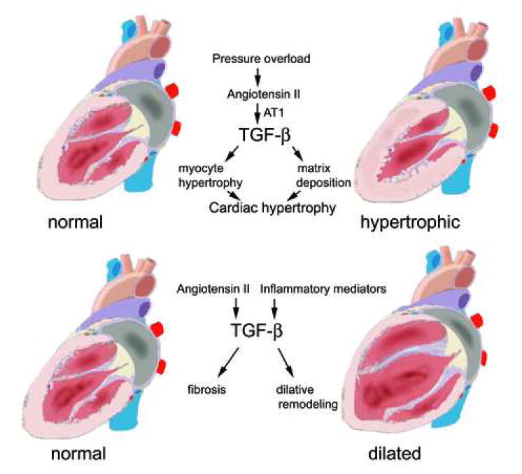
Transforming Growth Factor (TGF)-beta is markedly induced and rapidly activated in the infarcted myocardium. However, understanding of the exact role of TGF-beta signaling in the infarcted and remodeling heart has been hampered by the complex and unusual biology of TGF-beta activation and by the diversity of its effects eliciting multiple, and often opposing cellular responses. Experimental studies suggest that TGF-beta signaling may be crucial for repression of inflammatory gene synthesis in healing infarcts mediating resolution of the inflammatory infiltrate. In addition, TGF-beta may play an important role in modulating fibroblast phenotype and gene expression, promoting extracellular matrix deposition in the infarct by upregulating collagen and fibronectin synthesis and by decreasing matrix degradation through induction of protease inhibitors. TGF-beta is also a key mediator in the pathogenesis of hypertrophic and dilative ventricular remodeling by stimulating cardiomyocyte growth and by inducing interstitial fibrosis. In this review we summarize the current knowledge on the role of TGF-beta in infarct healing and cardiac remodeling.


Figures

Figure 1
Figure 1. Smad-dependent and Smad-independent pathways in TGF-β signaling
TGF-β binds a complex of transmembrane receptor serine/threonine kinases (type I and type II) inducing transphosphorylation of the GS segments in the type I receptor by the type II receptor kinases. The activated type I receptors phosphorylate selected Smads. Receptor-activated Smads (R-Smads) form a complex with the common Smad4. Subsequently the Smad complexes translocate into the nucleus, where they regulate transcription of target genes. The structurally divergent inhibitory Smads (Smad6 and Smad7), negatively regulate TGF-β signaling. In addition, TGF-β signals through Smad-independent cascades (green) activating Erk, JNK, p38MAPK, Protein Phosphatase 2A (PP2A) and RhoA pathways.
Figure 2
Figure 2. The diverse, multifunctional, and pleiotropic effects of TGF-β on cell types involved in infarct healing
During the pro-inflammatory phase of healing TGF-β may induce mononuclear cell chemotaxis and modulate lymphocyte phenotype. The effects of TGF-β on mature macrophages are predominantly suppressive, inhibiting pro-inflammatory cytokine and chemokine synthesis and decreasing reactive oxygen generation. These actions may be important in regulating resolution of the post-infarction inflammatory response. TGF-β also induces acquisition of the myofibroblastic phenotype and promotes extracellular matrix deposition by increasing collagen and fibronectin synthesis, and by inhibiting matrix degradation through TIMP upregulation. Spared cardiomyocytes respond to TGF-β by undergoing hypertrophy. The effects of TGF-β on endothelial cells are complex and context-dependent; their importance in regulating infarct angiogenesis is unknown. TGF-β mediated effects on fibrous tissue deposition and cardiac hypertrophy may be important in the pathogenesis of left ventricular remodeling.
Figure 3
Figure 3. Role of TGF-β signaling in infarct healing and post-infarction remodeling
Myocardial infarction triggers an inflammatory reaction that ultimately results in formation of a scar. Infarct healing is associated with alterations in the geometric characteristics of the ventricle, dilation and hypetrophy; these changes are termed “ventricular remodeling”. In the early phases of infarct healing TGF-β may be important in resolution of the inflammatory response by deactivating macrophages and by suppressing endothelial cell chemokine and cytokine synthesis. At a later stage, TGF-β activates fibrogenic pathways by inducing extracellular matrix deposition and may contribute to the pathogenesis of left ventricular remodeling by promoting fibrosis and hypertrophy of the non-infarcted myocardium.
Figure 4
Figure 4. TGF-β in hypetrophic and dilative remodeling
Evidence suggests a central role for TGF-β in the pathogenesis of hypertrophic cardiac remodeling. Pressure overload activates the Renin Angiotensin System generating angiotensin II. The hypertrophic effects of Angiotensin II are mediated through activation of TGF-β signaling pathways. TGF-β induces both cardiomyocyte hypertrophy and interstitial fibrosis. In contrast, the role of TGF-β in dilated cardiomyopathy is less clearly established. TGF-β induction mediated through Angiotensin II generation or via activation of inflammatory pathways may result in cardiac fibrosis playing a role in dilative remodeling.

References

    1. Schiller M, Javelaud D, Mauviel A. TGF-beta-induced SMAD signaling and gene regulation: consequences for extracellular matrix remodeling and wound healing. J Dermatol Sci. 2004;35:83–92. - PubMed
    1. Letterio JJ, Roberts AB. Regulation of immune responses by TGF-beta. Annu Rev Immunol. 1998;16:137–61. - PubMed
    1. Annes JP, Munger JS, Rifkin DB. Making sense of latent TGFbeta activation. J Cell Sci. 2003;116:217–24. - PubMed
    1. Lyons RM, Keski-Oja J, Moses HL. Proteolytic activation of latent transforming growth factor-beta from fibroblast-conditioned medium. J Cell Biol. 1988;106:1659–65. - PMC - PubMed
    1. Ignotz RA, Massague J. Transforming growth factor-beta stimulates the expression of fibronectin and collagen and their incorporation into the extracellular matrix. J Biol Chem. 1986;261:4337–45. - PubMed

Publication types

MeSH terms

Substances